Abstract
Ascorbic acid (AsA) and gluthathione (GSH) are two key components of the antioxidant machinery of eukaryotic and prokaryotic cells. The cyanobacterium Synechocystis sp. PCC 6803 presents both compounds in different concentrations (AsA, 20-100 μM and GSH, 2-5 mM). Therefore, it is important to have precise and sensitive methods to determine the redox status in the cell and to detect variations in this antioxidants. In this protocol, we describe an improved method to estimate the content of both antioxidants (in their reduced and oxidized forms) from the same sample obtained from liquid cultures of Synechocystis sp. PCC 6803.
Keywords: Cyanobacteria, Antioxidants, Glutathione, Ascorbic acid, Oxidative stress
Background
The redox status in the cell can be altered by multiple factors generating oxidative stress. We used this protocol to quantify GSH and AsA contents in Synechocystis sp. PCC 6803 exposed to heat (50 °C). As described for Arabidopsis thaliana, heat stress resulted in a decline in the content of both antioxidants and triggered cell death by ferroptosis ( Distéfano et al., 2017 ; Aguilera et al., 2019 preprint). Although this protocol was set for Synechocystis sp. PCC 6803, it can be applied to determine GSH and AsA contents in other cyanobacteria. In cyanobacteria AsA contents are very low, uM range in normal conditions and pM under some treatments. For that reason we propose this senstitive method with an improved cell lysate procedure.
Materials and Reagents
Pipette tips: 0.1-10 µl, 1-200 µl and 100-1,000 µl
1.5 ml microcentrifuge tubes
50 ml conical centrifuge tubes
Sterile syringes: 1 ml and 10 ml
Sterile acid-washed glass beads (150-212 µm) (Sigma-Aldrich, catalog number: G1145)
0.22 µm nylon menbrane filters (e.g., GVS, catalog number: FJ13BNPNY002AD01)
Synechocystis PCC 6803 liquid cultures
Ice
Liquid nitrogen
Sterile Mili-Q water
Water for HPLC (Sigma-Aldrich, catalog number 900682)
Bond Elut C18 cartridges (Agilent Technologies, catalog number: 12102028)
TFA (Trifluoroacetic acid) (Carlo Erba, catalog number: 411564)
DTT (Dithiothreitol) (Sigma-Aldrich, catalog number: D0632)
Methanol HPLC-grade (Sigma-Adrich, catalog number: 34860)
Phosphoric acid for HPLC (85%) (Sigma-Adrich, catalog number: 49685)
Potassium phosphate dibasic (Sigma-Aldrich, catalog number: P8281)
Potassium dihydrogen phosphate anhydrous (Sigma-Aldrich, catalog number: 543841)
Ethylenediaminetetraacetic acid (EDTA) (Sigma-Aldrich, catalog number: 431788)
NADPH (β-Nicotinamide adenine dinucleotide 2′-phosphate reduced tetrasodium salt hydrate ≥93% (HPLC) (Sigma-Aldrich, catalog number: N1630)
DTNB (5,5′-Dithiobis(2-nitrobenzoic acid)) (Sigma-Aldrich, catalog number: D8130)
GSSG (L-Glutathione oxidized) (Sigma-Aldrich, catalog number: G4376)
Glutathione Reductase from baker's yeast (S. cerevisiae) (Sigma-Aldrich, catalog number: G3664)
4-Vinylpyridine (Sigma-Aldrich, catalog number: V3204)
L-Glutathione reduced (Sigma-Aldrich, catalog number: G4251)
L-Ascorbic acid (Sigma-Aldrich, catalog number: A5960)
Neubauer counting chamber (BrandTM BlaubrandTM, catalog number: 10350141)
Phosphate buffer 100 mM pH7 (see Recipies)
Phosphate buffer 100 mM pH 7.5 + EDTA 5 mM (see Recipies)
Equipment
Pipettes
Neubauer counting chamber
Refrigerated centrifuge (50 ml conical tubes: Thermo ScientificTM, model: IECTM CL31; 1.5 ml tubes: Thermo ScientificTM, model: SorvallTM LegendTM Micro 17R)
Vortex mixer (Scientific Industries, model: VORTEX-GENIE 2)
Spectrophotometer (Shimadzu, model: UV-160A-Vis)
pH-meter (Milwaukee, model: MI-150, catalogue number: 11861)
-
HPLC system (Shimadzu Scientific Instruments) equipped with:
LC pump (Shimadzu Scientific Instruments, model: LC-10 AT)
UV-VIS detector (Shimadzu Scientific Instruments, model: SPD-10AV)
Microsphere C-18 column (Agilent technologies, SS 100 x 4.6 mm, 3 μm; catalog number: CP28076)
Software
Microsoft Excel (Microsoft Corporation, Available from: https://office.microsoft.com/excel)
Procedure
-
Cell harvest and antioxidants extraction
Place fifty milliliters of exponentially-growing culture (~2.5 × 10⁷ cells ml-1) in 50 ml conical centrifuge tubes.
Harvest cells by centrifugation (9,300 × g for 10 min, 4 °C).
Discard the supernatant and place the pellet with a sterile spatula in a new and sterile 1.5 ml microcentrifuge tube.
Place the tube on ice. Resuspend cells in 1 ml 3% TFA and add approximately 100 μl of sterile acid-washed glass beads (150-212 µm).
-
Lyse the cells by mixing vigorously (vortex, 1 min), freeze with liquid nitrogen and thaw in ice.
Note: Broken cells can be observed under an optical microscope, in order to ensure a correct rupture of the cells.
Repeat Step A5 at least 3 times.
Centrifuge the homogenate (16,000 × g 15 min, 4 °C).
Collect the supernatant in a new 1.5 ml microcentrifuge tube and keep it on ice before use.
-
Activate Bond Elut C18 columns by applying in the following order:
1.5 ml methanol.
1.0 ml water for HPLC.
1.0 ml phosphate buffer 100 mM pH 7.00.
Discard the solution from the washing steps.
Pass 500 μl of the supernantant (Step A8) through a previously activated Bond Elut C18 cartridge using a siringe.
-
Discard flow-through (500 μl) and pass 1.5 ml phosphate buffer 100 mM pH 7.00 to elute (pH of the elution around 2). Collect the filtrate in a new 1.5 ml microcentrifuge tube and mix thoroughly by vortexing for 10-20 s.
This filtrate is used for ascorbic acid and glutation determination.
Note: It is highly recommended to perform the measurements inmediately after obtaining the filtrate. In particular, ascorbic acid determination needs to be done after the preparation. Freezing the samples is not recommended because after thawing a drastic reduction in the antioxidant content is observed. For glutathione measurements, the supernatant can be stored at -80 °C.
-
Reduced ascorbic acid and total ascorbate measurements
-
To determine reduced ascorbic acid mix in a new 1.5 ml microcentrifuge tube:
240 μl of the filtrate obtained in Step A10
240 μl of K2HPO4 100 mM pH 8.5
50 µl of TFA 10%
15 µl of water for HPLC
-
To determine total ascorbate mix in a new 1.5 ml microcentrifuge tube:
240 μl of the filtrate obtained in Step A10
240 μl of K2HPO4 100 mM pH 8.5
15 µl of DTT 100 mM (final concentration: 3 mM)
Incubate for 5 min at at room temperatur and stop the reaction by adding 50 µl of TFA 10%.
-
-
Reduced ascorbic acid and total ascorbate quantification
To quantify the reduced ascorbic acid concentration, filter the solution obtained in Step B1 through a 0.22 µm nylon menbrane filter and inject 10 μl in a HPLC equipped with and a C-18 column (100 × 4.6 mm, 3 μM). As mobile phase use a solution of KH2PO4 100 mM (pH 3.00) in water for HPLC (adjust pH with phosphoric acid 85%) at 0.35 and 0. 5 ml min-1 flow.
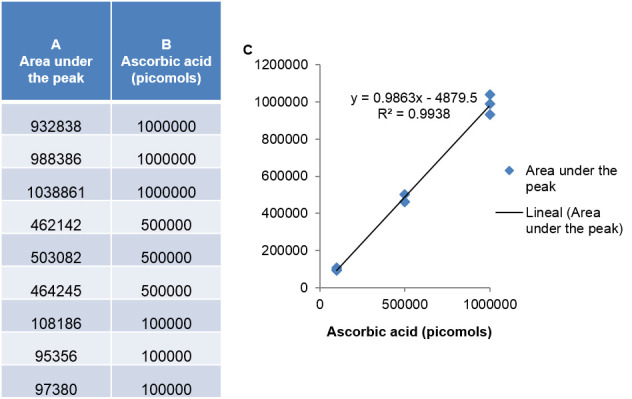
Quantify the area under the peak that displays a maximum at 265 nm (Figure 1).
To quantify total ascorbate concentration filter and inject 10 μl of the second solution obtained in step B2 using the same equipment and conditions described in Steps C1 and C2.
Prepare a calibration curve using a series of standard L-ascorbic acid dilutions (e.g., final concentrations of 0.1, 0.5 and 1 μM) and measure the peak area following the same procedure as described in Step C2 (Figure 1).
-
Adjust the linear function of ascorbic acid concentration vs. peak area and use the slope to calculate the concentration of the unknown samples as described in the formula:
The x2 is included in the formula because cells are resuspended in 1 ml TFA (Step A4) but only half of the supernatant is passed trought the column (Step A9). The x3 is included because in Step A10 one volume of sample is passed through the column (500 μl) and three volumes are collected (1.5 ml).
-
Measurement of glutathione content
The quantitative determination of the total amount of glutathione (GSH + GSSG) employs an enzymatic method. In this, the reduction of DTNB to 5-thio-2-Nitrobenzoato (TNB) by NADPH is quantified spectrophotometrically. This reaction is used to measure the reduction of GSSG to GSH. The rate of the reaction is proportional to the GSH and GSSG concentration.
-
To determine the total glutathione, in a new 1.5 ml microcentrifuge tube add:
650 µl phosphate buffer 100 mM pH 7.5 + EDTA 5 mM
25 µl DNTB (5 mg/ml) (final concentration 0.2 mM)
5 µl NADPH (1 mg/100 µl)
5 µl l glutathione reductase 1:30 (0.5 U)
300 µl of filtrate obtained in Step A10
Note: The blank solution contained all reagents except the filtrate obtained in Step A10.
Mix gently and read the absorbance at 412 nm using a spectrophotometer.
To extract the oxidized gluthathion (GSSG), add 3 µl of Vinylpyridine (95%) to 300 µl of the filtrate obtained in Step A10, gently mix by vortexing and incubate at room temperature for 25 min.
Centrifuge (16,000 × g 10 min, 4 °C) and place the tube on ice
-
In a new 1.5 ml microcentrifuge tube add
365 µl phosphate buffer 100 mM pH 7.5 + EDTA 5 mM.
25 µl DNTB (5 mg/ml).
5 µl NADPH (1 mg/100 μl)
5 µl l glutathione reductase 1:30 (0.5 U)
100 µl of the supernatant obtained in Step D4
Mix gently and read the absorbance at 412 nm.
Quantification of glutathione levels is based on the oxidized form of glutathione (GSSG). To quantify the glutathione levels, prepare a calibration curve with 0-100 µM standard L-Glutathione oxidized solution.
Note: The content of the antioxidant can be expressed either as per mg of fresh or dry weight or as pmol per cell (in the case of of ascorbic acid) and nmol per cell (in the case of glutathione). The content of antioxidants can be correlated with cell number determined with a Neubauer counting chamber.
-
Figure 1. Area under the peak and standard curve to Asa quantification.

Recipes
-
Phosphate buffer 100 mM pH 7
Add 1.071g K2HPO4 and 0.524 g KH2PO4 in 100 ml Sterile Mili-Q water
-
Phosphate buffer 100 mM pH 7.5 + EDTA 5 mM
Solution 1: mix 0.681 g of KH2PO4 and 0.093 g of EDTA in 50 ml sterile Mili-Q water
Solution 2: mix 0.871 g of K2HPO4 and 0.093 g of EDTA in 50ml sterile Mili-Q water
Adjuts the pH of solution 2 by adding solution 1 till reach pH 7.5
Acknowledgments
This research was funded by grants to M.V. Martin from Agencia Nacional de Promoción Científica y Técnica Argentina (PICT 1956 and PICT 0173) and to G.C. Pagnussat from Agencia Nacional de Promoción Científica y Técnica Argentina (PICT-2017-0201).
This protocol was adapted from previously published studies (Griffith, 1980; Bartoli et al., 2006 ; Narainsamy et al., 2016 ).
Competing interests
The authors declare no competing financial interests.
Citation
Readers should cite both the Bio-protocol article and the original research article where this protocol was used.
References
- 1. Aguilera A., Berdun F., Bartoli C., Steelheart C., Alegre M., Salerno G., Pagnussat G. and Martin M. V.(2019). Heat stress induces ferroptosis in a photosynthetic prokaryote. bioRxiv: 828293. [Google Scholar]
- 2. Bartoli C. G., Yu J., Gómez F., Fernández L., McIntosh L. and Foyer C. H.(2006). Inter-relationships between light and respiration in the control of ascorbic acid synthesis and accumulation in Arabidopsis thaliana leaves . J Exp Bot 57(8): 1621-1631. [DOI] [PubMed] [Google Scholar]
- 3. Distéfano A. M., Martin M. V., Córdoba J. P., Bellido A. M., D'Ippólito S., Colman S. L., Soto D., Roldán J. A., Bartoli C. G., Zabaleta E. J., Fiol D. F., Stockwell B. R., Dixon S. J. and Pagnussat G. C.(2017). Heat stress induces ferroptosis-like cell death in plants. J Cell Biol 216(2): 463-476. [DOI] [PMC free article] [PubMed] [Google Scholar]
- 4. Griffith O. W.(1980). Determination of glutathione and glutathione disulfide using glutathione reductase and 2-vinylpyridine. Anal Biochem 106(1): 207-212. [DOI] [PubMed] [Google Scholar]
- 5. Narainsamy K., Farci S., Braun E., Junot C., Cassier-Chauvat C. and Chauvat F.(2016). Oxidative-stress detoxification and signalling in cyanobacteria: the crucial glutathione synthesis pathway supports the production of ergothioneine and ophthalmate. Mol Microbiol 100(1): 15-24. [DOI] [PubMed] [Google Scholar]


