Abstract
Ambiguous genitalia affect 1 in 5,000 live births. Diagnostic procedures can be time-consuming, and often the etiology cannot be established in this group of individuals with differences/disorders of sex development (DSD). We aimed to evaluate the clinical presentation, sex assignment, and diagnostic workup in these patients. In this retrospective observational study, we included infants who presented with ambiguous genitalia from 2006 to 2016 at the Radboudumc (Radboud University Medical Center) DSD expert center. Relevant data were collected from patient records. Sixty-two 46,XY and fourteen 46,XX individuals were included. Sex was assigned in the first days of life and based on the combination of presence or absence of a uterus on ultrasound, AMH level, palpable gonads, and the karyotype (corresponded in 96% of the patients). In 86% of the 46,XX DSD subjects, a diagnosis was made, whereas in only 15/62 (24%) of the 46,XY DSD individuals, etiology was determined. In 52 individuals, genetic testing was performed resulting in a diagnosis in 24 patients (46%). AMH, hCG-stimulated testosterone, and dihydrotestosterone levels contributed to determining etiology, whilst basal testosterone and basal dihydrotestosterone did not. Establishing a diagnosis in infants with ambiguous genitalia is complex and challenging; this study aids to enhance this process and improve current practice.
Keywords: Ambiguous genitalia, Diagnosis, Disorders of sex development, Neonates, Sex determination, Sex differentiation
Ambiguous genitalia affect approximately 1 in 5,000 live births [Sax, 2002; Thyen et al., 2006]. These children form a subgroup of individuals with differences/disorders of sex development (DSD) in whom sex-chromosomal, gonadal, or anatomical sex is atypical [Hughes et al., 2006]. DSD is usually categorized as 46,XX DSD, 46,XY DSD, and sex-chromosomal DSD based on the karyotype and can have a variable etiology. The most common cause in infants with 46,XX DSD is congenital adrenal hyperplasia (CAH), affecting 1 in 14,000-15,000 live births [Pang et al., 1988; Lee et al., 2016]. In 46,XY DSD, the underlying causes are more diverse and can be due to abnormalities in the development of the testes (such as gonadal dysgenesis), disorders in the synthesis of androgens (such as testosterone synthesis deficiency or 5-alpha-reductase-deficiency), or in the actions of androgens (such as androgen receptors problems) [Hughes et al., 2006].
All efforts have to be made in order to establish the underlying condition in a child with ambiguous genitalia as it can cause a lot of concern for the parents and the family, and early sex assignment may help in dealing with these issues [Wisniewski, 2017]. Moreover, early diagnosis is of utmost importance in the case of adrenal DSD in order to start treatment to prevent adrenal crises and complications later in life. The final diagnosis can also help to foresee with which gender the child might identify later on in life. In most patients with 46,XX DSD, the final diagnosis can be determined. In contrast, approximately 20-60% of the patients with 46,XY DSD receive a final diagnosis [Morel et al., 2002; Hughes et al., 2006; De Paula et al., 2016].
In order to find the underlying diagnosis in a newborn with ambiguous genitalia, a variety of diagnostic tests can be performed, such as abdominal and pelvic ultrasound, hormone levels, and cytogenetic studies. A consensus statement on the management of DSD was published in 2006 [Hughes et al., 2006]. However, this was mainly based on expert opinion; the contribution of different tests to sex assignment and the determination of etiology remains unclear [Ogilvy-Stuart and Brain, 2004; Murphy et al., 2011].
Over the past decades, genetic panels are used more commonly in the diagnostic workup of DSD. Progress has been made in genetic research, and genetic analysis has become more accessible and affordable [Eggers et al., 2016; Alhomaidah et al., 2017; Kim et al., 2017]. Although many measurements are available in the diagnostic workup of DSD, it remains unclear which factors actually contribute to sex assignment and final diagnosis [Pang et al., 1988; Bouvattier et al., 2002; Ogilvy-Stuart and Brain, 2004; Bergadá et al., 2006; Hughes et al., 2006; Ahmed and Rodie, 2010; Douglas et al., 2010; Öçal, 2011; Lee et al., 2016]. Current clinical guidelines are mainly based on expert opinion, rather than clinical research. In rare diseases, such as DSD, performing clinical studies remains a major challenge.
Superfluous or unnecessary tests obviously have disadvantages. It can be a physical burden for the infant and psychologically stressful for the parents. A false test result or the lack of reference values can create unnecessary worry or confusion. Furthermore, costs have to be taken into account, and a diagnostic protocol should be efficacious. It is therefore crucial to select a panel of tests that contributes significantly to establishing the final diagnosis.
In this retrospective observational cohort study, we aim to evaluate the clinical presentation and the diagnostic workup in our population of neonates and young children born with ambiguous genitalia. Furthermore, we evaluate the contribution of different diagnostic tests to sex assignment and final diagnosis in order to improve current practice.
Patients and Methods
Patients
We included infants born from 2006 to 2016 who were referred for or presented with suspected ambiguous genitalia at the Radboud University Medical Center (Radboudumc) DSD center, which is 1 of the 3 approved Dutch DSD expert centers. Exclusion criteria were absence of ambiguous genitalia at examination by the Radboudumc experts or lack of data.
Sex Assignment and Final Diagnosis
In our center, there is no predefined protocol for assigning sex and establishing the diagnosis; the results were discussed individually. The experienced multidisciplinary expert team of the Radboudumc DSD center was responsible for these procedures. The team comprises pediatric endocrinologists, pediatric urologists, urologists, gynecologists, radiologists, ethicists, psychologists, sexologists, endocrinologists, specialized nurses, pathologists, clinical geneticists, and a laboratory specialist. In this study, we describe the evaluation of their best practice.
Data Collection
The following data were retrospectively collected from the electronic patient system: information about pregnancy and birth, ethnic background, physical examination, abdominal and/or pelvic ultrasound after birth, all biochemical measurements, results of human chorionic gonadotrophin (hCG) stimulation and genetic tests, the amount of blood samples taken, and the criteria used for sex assignment and final diagnosis. Only samples from venous blood, not cord blood, were included. In addition to these results, all congenital or developmental abnormalities were documented.
Biochemical Analysis
All blood samples were analyzed at the department of laboratory medicine of the Radboudumc, with special expertise in steroid measurement. Dihydrotestosterone (DHT) and dehydroepiandrosterone (DHEA) were analyzed by radioimmunoassay (RIA) after liquid-liquid extraction and chromatography. Testosterone, androstenedione and 17-hydroxyprogesterone (17OHP) were analyzed by RIA after liquid-liquid extraction and chromatography [Swinkels et al., 1990] and from 2014 on by LC-MSMS after protein precipitation and solid phase extraction described in Ter Horst et al. [2016]. Dehydroepiandrosteronesulfate (DHEAS) was measured by immunoassay on an Immulite random access analyzer (Siemens) and from 2015 on an E170 Modular random access analyzer (Roche). Luteinizing hormone (LH) and follicle-stimulating hormone (FSH) were measured by immunoassay using the latter abovementioned analyzer. AMH (gen II) was measured by an immunoassay on an Access random access analyzer (Beckman). Inhibin-B (gen II) was measured by enzyme-linked immunosorbent assay (Beckman).
A subset of patients underwent an hCG stimulation test, which was performed with a single intramuscular injection of 1500 IU hCG. Before and 72 h after the injection, blood samples were taken for testosterone, androstenedione, DHT, 17OHP, DHEAS, and DHEA. ACTH stimulation tests were performed according to local protocol with a single intravenous injection of 250 μg tetracosactide. Blood samples were taken for cortisol, 11-deoxyxcortisol, 17OHP, testosterone, androstenedione, DHEA, and progesterone.
Genetic Analysis
Genetic analysis was performed after a physician counseled the parents. DNA was extracted from peripheral blood using standard procedures. Single-gene analysis was performed by Sanger sequencing. Enrichment was performed using Agilent's SureSelect XT Human All Exon V4 (Agilent Technologies, Santa Clara, CA, USA). Next-generation sequencing was performed using Illumina HiSeq2000TM sequencer at BGI-Europe (Copenhagen, Denmark). Read alignment to the human reference genome (GrCH37/hg19) and variant calling was performed at BGI using BWA and genome analysis toolkit software, respectively. For all cases, variant annotation was performed using a custom designed in-house annotation and variant prioritization pipeline. The analysis of the exome data was divided into 2 steps: the DSD gene panel analysis and the exome analysis. The panel is updated regularly (information is available upon request).
Classification of 46,XY DSD Patients
Based on clinical and biochemical findings, our team made a subclassification of the 46,XY DSD individuals (Table 1). Eight subgroups were defined based on physiological principles and the experience of the team: gonadal dysgenesis, disorders of androgen synthesis, disorders of androgen action (androgen receptor defect), Leydig cell hypoplasia due to LH receptor deficiency, hypogonadotropic hypogonadism, 5-alpha-reductase deficiency, idiopathic when there were no abnormalities in biochemical results, and unclear. The unclear group is characterized as abnormalities in the biochemical results, which cannot be assigned to one of the categories above, or an insufficiently proven diagnosis.
Table 1.
Classification in 46,XY DSD patients
| 1. Gonadal dysgenesis | 2. Disorder of androgen synthesis | 3. Disorder of androgen action (androgen receptor defect) | 4. Leydig cell hypoplasia (LH receptor deficiency) | 5. Hypogonadotropic hypogonadism | 6. 5-Alpha-reductase deficiency | 7. Idiopathic without genetic testing | 8. Idiopathic with genetic testing | 9. Unclear | |
|---|---|---|---|---|---|---|---|---|---|
| AMH | Low | Normal | Normal | Normal | Normal | Normal | Normal | Normal | Abnormalities in the biochemical results, which cannot be explained by one of the previous categories |
| Testosterone | Low | Low | Normal or elevated | Low, precursors also low | Low | Normal or elevated | Normal | Normal | |
| DHT after hCG stimulation | Low | Low | Normal or elevated | Low | Low | Low | Normal | Normal | |
| Testosterone after hCG stimulation | Low | Low or intermediate with elevated precursors | Increased | Low | Increased DHT and testosterone | High ratio of testosterone/DHT | Normal | Normal | |
| Presence of uterus on ultrasound | Yes | No | No | No | No | No | No | No | |
AMH, anti-müllerian hormone; DHT, dihydrotestosterone; hCG, human chorionic gonadotropin.
Statistical Analysis
Data management and analysis was performed using SPSS, version 22.0. Results are solely descriptive and are expressed as counts with percentages or median with interquartile range.
Results
Patient Inclusion and Baseline Characteristics
Seventy-six from a total of 106 identified patients were included: sixty-two 46,XY and fourteen 46,XX DSD patients. Fifteen patients were excluded because they did not have ambiguous genitalia on examination, i.e., normal anatomy in premature born neonates or no anorectal malformation. Fourteen could not be included due to insufficient data, and 1 was excluded because of 49,XXXXY genotype and significant comorbidities. Table 2 shows an overview of the baseline characteristics. About 25% of all patients were born prematurely. 46,XY DSD patients had a lower birth weight compared to 46,XX DSD patients, with 34% having a birth weight <10th percentile. The cause of this phenomenon is unknown but is in concurrence with previous reports [Poyrazoglu et al., 2017]. Consanguinity, ethnicities other than Caucasian, and positive familial history for variations in the external genitalia were more common in the group with 46,XY DSD. Of the 46,XY DSD patients, 26% had additional congenital abnormalities or dysmorphic features versus none in the 46,XX DSD group.
Table 2.
Baseline characteristics and characteristics of sex assignment
| 46,XX (n = 14) | 46,XY (n = 62) | |
|---|---|---|
| Age in years, measured 11–2018, median (IQR) | 6 (5.2–10.7) | 4.5 (2.5–7.5) |
| Gestational age in weeks at birth, median (IQR) | 38 (31–41) | 38 (34–39) |
| Unreported, n (%) | 3 (21) | 10 (16) |
| Birth weight in percentiles, n (%) | ||
| <5 | − | 14 (23) |
| 5–10 | − | 7 (11) |
| 10–50 | 4 (29) | 16 (26) |
| 50–90 | 3 (21) | 9 (14) |
| 90–95 | 2 (14) | 2 (3) |
| >95 | 2 (14) | − |
| Unknown | 3 (21) | 14 (23) |
| Ethnicity, n (%) | ||
| Caucasian | 10 (71) | 28 (45) |
| East-Asian | 1 (7) | 9 (14) |
| West-Asian | 2 (14) | 5 (8) |
| African | − | 3 (5) |
| Mixed | − | 7 (11) |
| Unknown | 1 (8) | 10 (16) |
| Consanguinity of parents − yes, n (%) | − | 6 (10) |
| Other congenital abnormalities or dysmorphic features − yes, n (%) | − | 16 (26) |
| Familial history of abnormalities of the external genitaliaa, n (%) | ||
| Paternal | − | 5 (8) |
| Maternal | 1 (7) | 1 (2) |
| Paternal and maternal | − | 2 (3) |
| No | 8 (57) | 27 (44) |
| Unreported | 5 (36) | 27 (44) |
| Sex assignment postnatal, n (%) | ||
| Female | 13 (93) | 2 (3) |
| Male | 1 (7) | 60 (97) |
| Prader stage, median (IQR) | 4 (3–4) | 4 (3–4) |
| Gonads palpable, n (%) | ||
| Bilateral | − | 43 (70) |
| Unilateral | 1 (7) | 7 (11) |
| None | 13 (93) | 12 (19) |
| Pelvic ultrasonography, n (%) | ||
| Uterus | 11 (79) | 2 (3) |
| Mullerian rests | − | 5 (8) |
| No uterus or müllerian rests | − | 39 (63) |
| No ultrasound performed | 3 (21) | 16 (26) |
| Estimated sex prenatal, n (%) | ||
| Ambiguous genitalia | 3 (21) | 18 (29) |
| Male | − | 8 (13) |
| Female | 1 (7) | 6 (10) |
| Unreported | 10 (71) | 30 (48) |
| Sex assignment, n (%) | ||
| DSD team | 9 (65) | 24 (39) |
| Pediatrician in Radboudumc (not DSD team) | 3 (21) | 7 (11) |
| Resident pediatrician | − | 12 (19) |
| Unreported | 2 (14) | 18 (29) |
| Age at sex assignment in days, median (IQR) | 2 (0.25–4) | 1 (0–2.5) |
| Unreported, n (%) | 6 (43) | 25 (40) |
IQR, interquartile range.
First-, second-, or third-degree relatives of the child.
Performed Tests
The median number and interquartile range (IQR) of biochemical tests performed was 8 (4-19.75) in infants with 46,XX DSD and 17 (12-24.25) in 46,XY DSD; blood sampling was 2 (1-3) in individuals with 46,XX DSD and 3 (2-4) in the 46,XY DSD population. Genetic testing was performed in all 46,XX DSD patients and in 61% of the 46,XY subjects (38/62).
Abdominal ultrasound was performed in 74% of the 46,XY DSD patients and in 79% of the 46,XX DSD patients. A uterus was seen in all 46,XX DSD patients and in two 46,XY DSD patients. In five 46 XY DSD patients, there were possible müllerian rests.
Sex Assignment
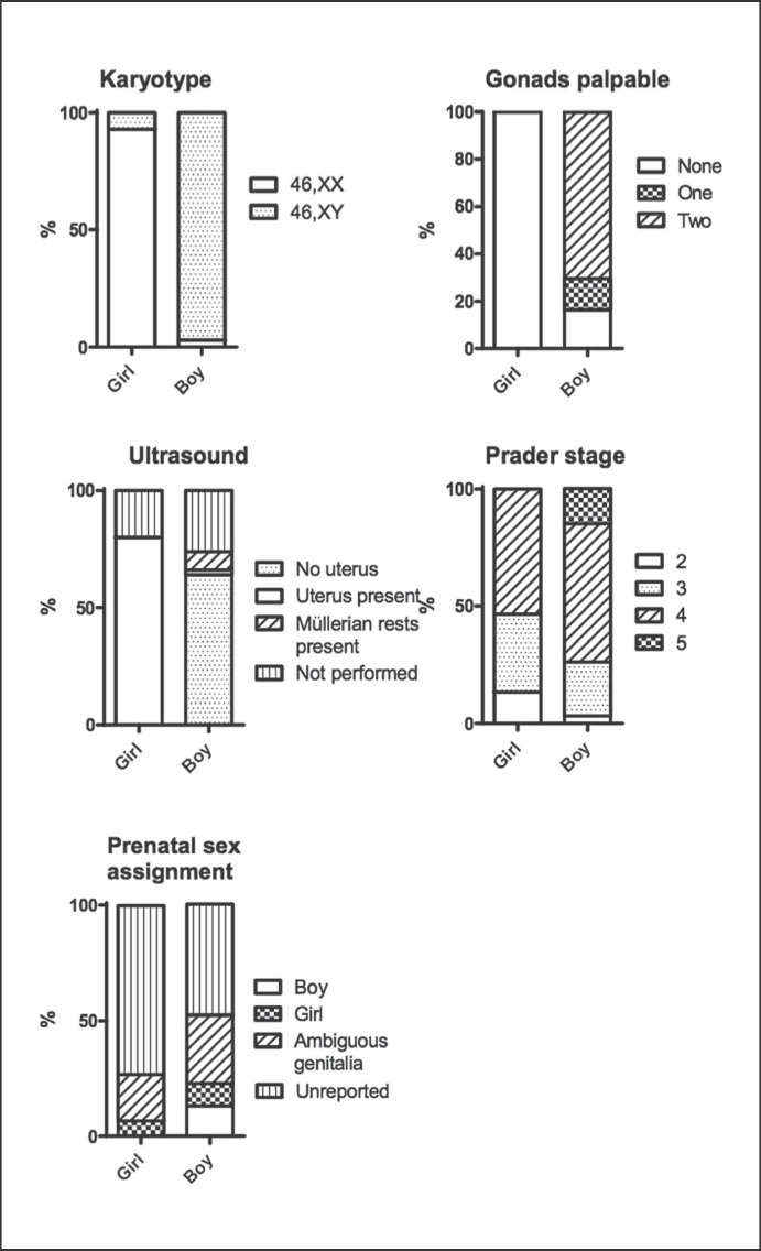
Sex assignment was done by a multidisciplinary team after discussing information from clinical examination, ultrasound, karyotype and biochemical results, including hormone levels. Sixty of the 46,XY DSD patients (97%) were assigned a male sex, thirteen 46,XX DSD patients (93%) were assigned a female sex. In our population, every child in whom 1 or 2 scrotal gonads were palpable received a male sex assignment, and all patients showing a uterus in ultrasound images were assigned females. Assessing internal genitalia on ultrasound can however be challenging and might not always be conclusive [Hughes et al., 2006].]. The sex assignment was mostly in correspondence with the karyotype. Prenatal estimated sex by ultrasound was not predictive for the sex assigned postnatally, and there was no correlation between Prader stage and sex assignment (Fig. 1). Most biochemical tests did not contribute to sex assignment except AMH and testosterone levels after chromatography.
Fig. 1.
Parameters in relation to sex assignment.
In one 46,XX DSD patient with a uterus and only 1 palpable gonad, a male sex had been assigned elsewhere, but sex was later changed because the child expressed her female gender. Therefore, she was included in the female group. Two 46,XY DSD patients were assigned a female sex. One was assigned a female sex at birth and was referred to our clinic at the age of 3 months. A uterus and 2 abdominal gonads were visible on laparoscopic examination. The other patient was also assigned a female sex elsewhere and had a uterus on postnatal ultrasound but also had a high AMH. Genetic analysis showed a mutation in the NR5A1 gene, which affirmed the diagnosis of mixed partial gonadal dysgenesis.
All but 2 patients received sex assignment within one week. One patient received sex assignment at the age of 23 days. This child came to our center at the age of 17 days, after admission in another hospital and initial assignment of a female sex. Her sex was changed to male because of the absence of a uterus on ultrasound and palpable scrotal gonads that produced testosterone. Another child received sex assignment after 8 days because the blood was initially not accessible for analysis.
Final Diagnosis
In our population, 12 out of 14 (86%) 46,XX DSD individuals received a final diagnosis, whereas in only 15/62 (24%) of 46,XY DSD individuals, the etiology could be determined. 46,XX DSD patients were diagnosed within 3 months after birth, whereas children with 46,XY DSD were generally diagnosed later. The definitive diagnoses are summarized in Table 3.
Table 3.
Diagnoses in 46,XX and 46,XY DSD patients
| 46,XX (n = 14) | 46,XY (n = 62) | |
|---|---|---|
| Final diagnosis made − yes, n (%) | 12 (86) | 15 (24) |
| Age in months at diagnosis, median (IQR) | 0 (0–2.25) | 5.5 (5–31.75) |
| No diagnosis made, n (%) | 2 (14) | 47 (76) |
| Diagnosis made, n (%) 46,XX patients | ||
| 21-Hydroxylase deficiency | 10 (83) | |
| Ovotesticular DSD | 1 (8) | |
| POR deficiency | 1 (8) | |
| Unclear | 2 (14) | |
| 46,XY patients | ||
| Gonadal dysgenesis | 1 (2) | |
| Disorders of androgen synthesis | − | |
| Disorders of androgen action | ||
| Partial androgen insensitivity syndrome | 2 (3) | |
| Leydig cell hypoplasia | − | |
| Hypogonadotropic hypogonadism | 5 (8) | |
| Prader-Willi syndrome | 2 (3) | |
| 5-Alpha-reductase deficiency | 5 (8) | |
| Idiopathic without genetic test | 11 (18) | |
| Idiopathic with genetic test | 11 (18) | |
| Unclear | 25 (40) | |
| Others | ||
| Smith-Lemli-Opitz syndrome | 1 (2) | |
| Silver-Russell syndrome | 1 (2) | |
IQR, interquartile range; POR, P450 oxidoreductase deficiency.
The majority of 46,XX DSD patients (10/14) was diagnosed with 21-hydroxylase deficiency. Genetic analysis confirmed the presence of pathogenic mutations in all these infants. Two other 46,XX individuals were diagnosed with P450 oxidoreductase deficiency and ovotesticular DSD, respectively. These diagnoses were based on clinical findings and biochemical tests. In both these patients, genetic analysis did not reveal any mutations (single gene PCR and DSD panel, respectively). In 2 patients, the final diagnosis is not yet made despite whole-exome sequencing.
In 46,XY DSD patients, the most common diagnosis was 5-alpha-reductase deficiency (Table 3). Sixteen 46,XY patients had multiple congenital abnormalities, such as heart defects, patent ductus arteriosus, or periventricular leukomalacia. An etiological diagnosis could be made in 7 patients (44%). In most of the remaining patients (8/9), whole-exome sequencing was performed.
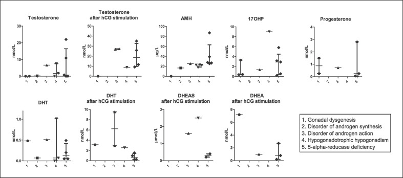
Testosterone and DHT levels after hCG stimulation and AMH were the most useful biochemical tests contributing to the final diagnosis in 46,XY DSD patients. As shown in Figure 2, testosterone after hCG stimulation can better distinguish gonadal dysgenesis and partial androgen insensitivity syndrome (PAIS) from other causes than basal testosterone concentration. DHT after hCG stimulation helps to differentiate between 5-alpha-reductase deficiency and other subgroups. Combining these results led to a likely diagnosis based on physical examination, biochemical evaluation, and abdominal ultrasound which could be confirmed by genetic testing.
Fig. 2.
Laboratory results in 46,XY DSD patients, sorted by etiological groups (median and IQR). Testosterone was measured after chromatography. AMH, anti-müllerian hormone; 17OHP, 17-hydroxyprogesterone; DHT, dihydrotestosterone; DHEAS, dehydroepiandrosteronesulfate; DHEA, dehydroepiandrosterone.
Genetic Testing
In 46,XX individuals, most often single gene analysis was used to confirm 21-hydroxylase deficiency, revealing pathogenic mutations in 10/14 infants (71%). In 46,XY DSD individuals, 38 out of 62 underwent genetic tests, revealing a pathogenic mutation in 37% (14/38). Using single gene analysis, which was performed 17 times, a pathogenic mutation was found in nine 46,XY DSD patients (53%). The DSD panel, which was performed in eighteen 46,XY DSD patients, was used as a primary genetic test or secondary to single gene analysis. It revealed a pathogenic mutation twice and an unclassified variant in another 2 patients. Additional whole-exome sequencing to single-gene analysis or DSD panel, performed in 4 patients, did not reveal any pathogenic mutation. In seven 46,XY patients, whole-exome sequencing was performed as first test, e.g., because of the presence of multiple congenital abnormalities, which revealed a pathogenic mutation in 2 patients.
Discussion
When a neonate with ambiguous genitalia is born, challenging decisions have to be made by the DSD team and the parents concerning sex assignment, diagnostic process, and management. Therefore, a valid and quick diagnostic approach is necessary. In this study, we use a retrospective cohort of DSD patients to describe the practice in our DSD center, assessing different tests to sex assignment and final diagnosis.
Until now, a binary sex assignment (girl or boy) is common practice in most countries, and only a selected number of countries give parents the legal opportunity not to declare sex in official documents. Therefore, sex assignment after birth in neonates born with ambiguous genitalia is still current standard in most countries. There is, however, a growing debate about the need of postnatal sex assignment, and a gender-neutral approach for individuals with DSD conditions is discussed by experts, parents, and human rights groups. The difficulty in establishing a final diagnosis as described in this study contributes to this recent discussion. Reports on a switch in sex and gender dysphoria later in life in individuals with DSD have raised more attention to this question (see https://www.hrw.org/sites/default/files/report_pdf/lgbtintersex0717_web_0.pdf). Furthermore, some groups conceive gender identity as a more fluent (not binary) condition.
In case one wants to perform sex assignment, most experts suggest karyotyping, steroid analysis, MRI, and pelvic ultrasound [Hughes et al., 2006; Ahmed and Rodie, 2010]. In our center, postnatal sex assignment was mainly based on the presence or absence of a uterus on ultrasound, AMH levels, and palpable gonads. Prader stage and prenatal sex determination were not useful as predictive criteria for sex assignment by the multidisciplinary team. Chromosomal sex correlated with sex assignment in most cases, although this was not used as a main criterion. In DSD centers, the diagnostic tests can be performed within 24-48 h; thus, sex assignment can mostly be done within the first 2 days of life to reduce parental stress and concern. In our center, a male sex is currently assigned when gonads are scrotal and ultrasonography does not show a uterus. AMH and testosterone after chromatography are also helpful, although measured values can overlap. Cortisol, ACTH, or DHT did not contribute to sex assignment, but were used for determining final diagnosis, although the small number of female patients in this study limits its conclusive power. Progesterone was superfluous; levels measured in neonates reflect maternal serum levels and hence we do not recommend measurement of progesterone in newborns with DSD. Our study does not provide a defined protocol for sex assignment that can be applied in all circumstances; conditions such as mixed gonadal dysgenesis might need a different method. However, this study does show that multiple factors have to be taken into account, and our data can be used to guide a rational diagnostic approach. Evaluation of the validity of sex assignment in our center remains difficult as most patients are still young and not able to confirm or deny the chosen sex. This limits the power of our conclusions. Therefore, parents need to be counseled about the possibility of changing sex and gender dysphoria later in life, particularly in conditions such as PAIS, 17-beta-HSD deficiency, and 5-alpha-reductase deficiency.
After initial sex assignment, all efforts have to be made to establish a final diagnosis because this might give insight into the need for medical treatment, fertility options, and gender identity. In most 46,XX DSD patients, a final diagnosis can be made in the first weeks of life. The majority of these individuals are diagnosed with 21-hydroxylase deficiency. For 46,XY patients, however, establishing an etiological diagnosis is more difficult. In our group of 46,XY individuals, a final diagnosis could only be made in 24% of the patients, despite excessive diagnostic testing including genetic analysis. The presence of additional congenital abnormalities or developmental delay increases the chance of establishing a diagnosis. This is comparable to numbers reported elsewhere: Baetens et al. [2014] reported a final diagnosis in 20% and 33% in 46,XY neonates and a subgroup with multiple congenital abnormalities or developmental delay, respectively. Parents should be counseled that a final diagnosis cannot be made in all cases.
In our cohort of 46,XY DSD patients, measurement results of basal steroids in the first days of life were highly variable and contributed little to the final diagnosis. It is important to note that the timing of sampling after birth matters for the determination of hormone levels because of so-called mini puberty. There also is a large variation between individuals, and this is of relevance for the interpretation of these values. This emphasizes the importance of hCG stimulation for the evaluation of gonadal function. Because of the small number of subjects in our study, we did not include the different time points in our analysis. The use of hCG-stimulated steroids gave more insight into gonadal function and the underlying diagnosis, especially in 5-alpha-reductase deficiency and PAIS (Fig. 2). Therefore, in our center, we now measure androgens and their precursors preferably 72 h after hCG stimulation. AMH has proven to be a good marker for testicular function [Bergadá et al., 2006; Lee et al., 2016]. It is important to rule out adrenal causes of 46,XY DSD as these children may develop symptoms of cortisol deficiency and need additional medical treatment. A random cortisol is highly variable in the neonatal period and hence does not exclude adrenal insufficiency. It can be expected that testosterone concentration in 46,XY DSD individuals due to adrenal enzymatic defects will be suboptimal even after hCG stimulation. In this case, an ACTH stimulation test should be performed to rule out these defects, such as 3-beta-HSD deficiency, CYP17 deficiency, or Star deficiency, as a cause for ambiguous genitalia.
In addition to biochemical evaluation, it is now customary to confirm a proposed diagnosis by mutation analysis. Also, it is known that DSD is associated with other congenital abnormalities and syndromes. In our cohort, 26% of the 46,XY DSD individuals had additional congenital abnormalities or dysmorphic features. Especially in these patients, a clinical geneticist with experience in the field of DSD should be involved in the diagnostic process. Because of the increasing number of known pathogenic mutations, sequencing a panel of genes is generally the first step when biochemical evaluation does not point in the direction of a specific condition; this can be extended to whole-exome sequencing if no pathogenic mutations are found. Eggers et al. [2016] performed a parallel sequencing targeted DSD panel with 1,031 genes in a large international cohort of 326 children. Both patients with 46,XX and 46,XY DSD were included, but patients with CAH were excluded. They found a genetic diagnosis in 43% of the cases. In our cohort (also excluding CAH children), a pathogenic mutation was found in 14/42 (33%) of the patients by genetic testing. Since most patients underwent only monogenetic analysis in the early years, it is likely that expanding the analysis would increase the number of patients in whom a pathogenic mutation is found.
Our study has several limitations. Since ambiguous genitalia in neonates are rare, the number of patients included in our evaluation is limited, and some data are lacking due to the retrospective design. The possibilities for genetic testing have changed rapidly over the past few years, which limit the evaluation of the diagnostic approach over years. However, we included a representative group of DSD patients and collected a significant amount of data that allows us to draw useful conclusions. Due to the variety of conditions that cause ambiguous genitalia, the subgroups are small. This results in less conclusive power, but also shows the complexity of everyday reality in diagnosing these children.
In conclusion, sex assignment in children with ambiguous genitalia can generally be made in the first days of life based on the karyotype, presence or absence of a uterus on ultrasound, AMH levels, and palpability of gonads. A gender-neutral approach might become more common in the future. hCG-stimulated testosterone, hCG-stimulated DHT, and AMH are the most useful biochemical tests in the evaluation of 46,XY DSD patients. Establishing a final diagnosis can be done in the first weeks of life in 46,XX DSD patients, but it is more complex in 46,XY DSD patients. It can be expected that genetic analysis will speed up this process and increase the number of patients in which a diagnosis can be established in the near future. We suggest an approach of physical examination, ultrasound, genetic testing, and selective biochemical evaluation to avoid unnecessary testing.
Statement of Ethics
The study protocol has been approved by the research institute's committee on human research (CMO 2017-3205).
Disclosure Statement
The authors have no conflicts of interest to declare.
Author Contributions
H.L. Claahsen-van der Grinten and M. van Zoest designed the study, M. van Zoest collected and analyzed the data. E.M. Bijker, H.L. Claahsen-van der Grinten, and M. van Zoest took the lead in writing the manuscript. All authors discussed the results and contributed to the final manuscript.
References
- 1.Ahmed SF, Rodie M. Investigation and initial management of ambiguous genitalia. Best Pract Res Clin Endocrinol Metab. 2010;24:197–218. doi: 10.1016/j.beem.2009.12.001. [DOI] [PubMed] [Google Scholar]
- 2.Alhomaidah D, McGowan R, Ahmed SF. The current state of diagnostic genetics for conditions affecting sex development. Clin Genet. 2017;91:157–162. doi: 10.1111/cge.12912. [DOI] [PubMed] [Google Scholar]
- 3.Baetens D, Mladenov W, Delle Chiaie B, Menten B, Desloovere A, et al. Extensive clinical, hormonal and genetic screening in a large consecutive series of 46,XY neonates and infants with atypical sexual development. Orphanet J Rare Dis. 2014;9:209. doi: 10.1186/s13023-014-0209-2. [DOI] [PMC free article] [PubMed] [Google Scholar]
- 4.Bergadá I, Milani C, Bedecarrás P, Andreone L, Ropelato MG, et al. Time course of the serum gonadotropin surge, inhibins, and anti-Müllerian hormone in normal newborn males during the first month of life. J Clin Endocrinol Metab. 2006;91:4092–4098. doi: 10.1210/jc.2006-1079. [DOI] [PubMed] [Google Scholar]
- 5.Bouvattier C, Carel JC, Lecointre C, David A, Sultan C, et al. Postnatal changes of T, LH, and FSH in 46,XY infants with mutations in the ARgene. J Clin Endocrinol Metab. 2002;87:29–32. doi: 10.1210/jcem.87.1.7923. [DOI] [PubMed] [Google Scholar]
- 6.De Paula GB, Barros BA, Carpini S, Tincani BJ, Mazzola TN, et al. 408 cases of genital ambiguity followed by single multidisciplinary team during 23 years: etiologic diagnosis and sex of rearing. Int J Endocrinol. 2016;2016:4963574. doi: 10.1155/2016/4963574. [DOI] [PMC free article] [PubMed] [Google Scholar]
- 7.Douglas G, Axelrad ME, Brandt ML, Crabtree E, Dietrich JE, et al. Consensus in Guidelines for Evaluation of DSD by the Texas Children's Hospital Multidisciplinary Gender Medicine Team. Int J Pediatr Endocrinol. 2010;2010:919707. doi: 10.1155/2010/919707. [DOI] [PMC free article] [PubMed] [Google Scholar]
- 8.Eggers S, Sadedin S, van den Bergen JA, Robevska G, Ohnesorg T, et al. Disorders of sex development: insights from targeted gene sequencing of a large international patient cohort. Genome Biol. 2016;17:243. doi: 10.1186/s13059-016-1105-y. [DOI] [PMC free article] [PubMed] [Google Scholar]
- 9.Hughes IA, Houk C, Ahmed SF, Lee PA, LWPES Consensus Group. ESPE Consensus Group Consensus statement on management of intersex disorders. Arch Dis Child. 2006;91:554–563. doi: 10.1136/adc.2006.098319. [DOI] [PMC free article] [PubMed] [Google Scholar]
- 10.Kim JH, Kang E, Heo SH, Kim GH, Jang JH, et al. Diagnostic yield of targeted gene panel sequencing to identify the genetic etiology of disorders of sex development. Mol Cell Endocrinol. 2017;444:19–25. doi: 10.1016/j.mce.2017.01.037. [DOI] [PubMed] [Google Scholar]
- 11.Lee PA, Nordenstrom A, Houk CP, Ahmed SF, Auchus R, et al. Global disorders of sex development update since 2006: perceptions, approach and care. Hormone Res Paediatr. 2016;85:158–180. doi: 10.1159/000442975. [DOI] [PubMed] [Google Scholar]
- 12.Morel Y, Rey R, Teinturier C, Nicolino M, Michel-Calemard L, et al. Aetiological diagnosis of male sex ambiguity: a collaborative study. Eur J Pediatr. 2002;161:49–59. doi: 10.1007/s00431-001-0854-z. [DOI] [PubMed] [Google Scholar]
- 13.Murphy C, Allen L, Jamieson MA. Ambiguous genitalia in the newborn: an overview and teaching tool. J Pediatr Adolesc Gynecol. 2011;24:236–250. doi: 10.1016/j.jpag.2011.02.004. [DOI] [PubMed] [Google Scholar]
- 14.Öçal G. Current concepts in disorders of sexual development. J Clin Res Pediatr Endocrinol. 2011;3:105–114. doi: 10.4274/jcrpe.v3i3.22. [DOI] [PMC free article] [PubMed] [Google Scholar]
- 15.Ogilvy-Stuart AL, Brain CE. Early assessment of ambiguous genitalia. Arch Dis Child. 2004;89:401–407. doi: 10.1136/adc.2002.011312. [DOI] [PMC free article] [PubMed] [Google Scholar]
- 16.Pang SY, Wallace MA, Hofman L, Thuline HC, Dorche C, et al. Worldwide experience in newborn screening for classical congenital adrenal hyperplasia due to 21-hydroxylase deficiency. Pediatrics. 1988;81:866–874. [PubMed] [Google Scholar]
- 17.Poyrazoglu S, Darendeliler F, Ahmed SF, Hughes I, Bryce J, et al. Birth weight in different etiologies of disorders of sex development. J Clin Endocrinol Metab. 2017;102:1044–1050. doi: 10.1210/jc.2016-3460. [DOI] [PubMed] [Google Scholar]
- 18.Sax L. How common is intersex? A response to Anne Fausto-Sterling. J Sex Res. 2002;39:174–178. doi: 10.1080/00224490209552139. [DOI] [PubMed] [Google Scholar]
- 19.Swinkels LM, Ross HA, Smals AG, Benraad TJ. Concentrations of total and free dehydroepiandrosterone in plasma and dehydroepiandrosterone in saliva of normal and hirsute women under basal conditions and during administration of dexamethasone/synthetic corticotropin. Clin Chem. 1990;36:2042–2046. [PubMed] [Google Scholar]
- 20.Ter Horst R, Jaeger M, Smeekens SP, Oosting M, Swertz MA, et al. Host and environmental factors influencing individual human cytokine responses. Cell. 2016;167:1111–1124 e13. doi: 10.1016/j.cell.2016.10.018. [DOI] [PMC free article] [PubMed] [Google Scholar]
- 21.Thyen U, Lanz K, Holterhus PM, Hiort O. Epidemiology and initial management of ambiguous genitalia at birth in Germany. Horm Res. 2006;66:195–203. doi: 10.1159/000094782. [DOI] [PubMed] [Google Scholar]
- 22.Wisniewski AB. Psychosocial implications of disorders of sex development treatment for parents. Curr Opin Urol. 2017;27:11–13. doi: 10.1097/MOU.0000000000000344. [DOI] [PMC free article] [PubMed] [Google Scholar]


