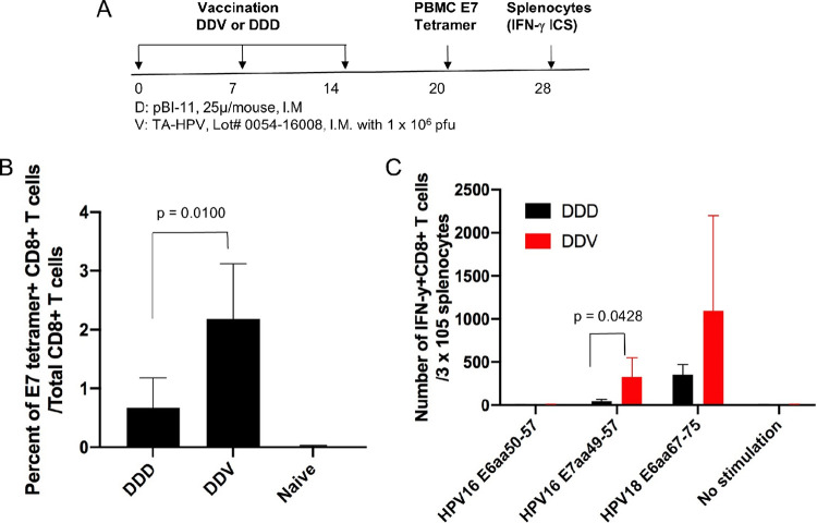
FIG 6.
Characterization of HPV antigen-specific CD8+ T cell-mediated immune responses in mice vaccinated with pBI-11 DNA alone or followed by TA-HPV. Six- to eight-week-old female C57BL/6 mice (5 mice/group) were vaccinated with pBI-11 DNA (25 μg/50 μl/mouse) through i.m. injection. The mice were boosted with the same regimen 7 days later. One week after the second vaccination, one group of the mice was vaccinated with pBI-11 DNA (25 μg/50 μl/mouse) through i.m. injection (DDD). Another group of mice was vaccinated with TA-HPV (1 × 10⁶ PFU/50 μl/mouse) through i.m. injection (DDV). (A) Schema of the experiment. Six days after the last vaccination, peripheral blood was collected from the vaccinated or naive mice for HPV16 E7 tetramer staining. Fourteen days after the last vaccination, splenocytes were prepared from the vaccinated mice and stimulated with either HPV16 E6 (aa 50 to 57), HPV16 E7 (aa 49 to 57), or HPV18 E6 (aa 67 to 75) peptide in the presence of GolgiPlug. Intracellular IFN-γ cytokine staining assay was performed to detect antigen-specific CD8+ T cells. The cells were acquired with a FACSCalibur flow cytometer, and data were analyzed with CellQuest Pro software. (B) Summary of the HPV16 E7 tetramer flow cytometry analysis. (C) Intracellular IFN-γ cytokine staining assay after stimulation with either HPV16 E6 (aa 50 to 57) peptide (5 μg/ml), HPV16 E7 (aa 49 to 57) peptide (1 μg/ml), or HPV18 E6 (aa 67 to 75) peptide (1 μg/ml).

