In this issue, Presti et al.1) reports the protective role of left ventricular (LV) false tendon against LV remodeling and functional mitral regurgitation (MR) after acute myocardial infarction. Unfortunately, the authors did not obtain positive results that false tendon has a protective effect against adverse LV remodeling and ischemic MR. There are many limitations in this study, such as a small study group, a retrospective design, participants with a mild degree of LV systolic dysfunction on baseline echocardiography, low echocardiographic sensitivity to detect the presence of false tendons, and weak consideration of the differences in the anatomical characteristics of false tendon. Therefore, it is difficult to generalize the results of this study. If a new study is carried out with a modified design to address these shortcomings, however, other interesting results might be obtained.
ANATOMIC FEATURES OF LV FALSE TENDON
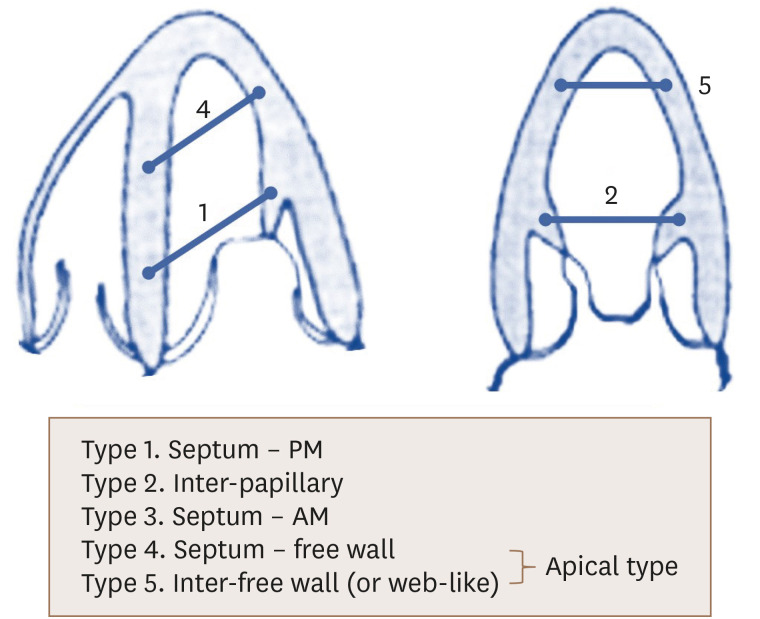
LV false tendon is an interesting normal variant structure. According to autopsy results, false tendons of varying size and location are found in 48%–78% of cardiac specimens.2),3),4) In addition, false tendons show a wide range of individual variations in shape and structure. A false tendon can be a long, greater than 2-mm-thick, well-vascularized, muscular column containing a conduction system. On the other hand, it can be composed of very thin, multi-stranded, avascular fibrous strings. False tendon is frequently classified by its connections (Figure 1). In types 1, 3, and 5, Purkinje fibers are more likely to be contained within the false tendon, which might be related to electrocardiogram abnormalities or arrhythmia.3) Unfortunately, the diagnostic sensitivity of echocardiographic detection of false tendon is 27%–85%, and additional evaluations using variable off-axis windows are required to determine exact sizes and locations.3),4)
Figure 1. Common types of left ventricular false tendon according to connections.
AM: anterior papillary muscle, PM: posterior papillary muscle.
As false tendons become taut in diastole and loose in systole due to blood flow in the cardiac cycle, they vibrate and produce sounds in a manner similar to the Aeolian harp, which resonates with wind.5) These musical chords produced by false tendon are one of the presumed mechanisms of innocent (Still's) murmur.6)
CLINICAL ASSOCIATION OF LV FALSE TENDON
The presence of LV false tendon does not differ between the normal population and patients with congenital heart disease,2) and it is not associated with increased mortality.7) Occasionally, false tendons contain conductive tissue, and an association between false tendons and arrhythmic disease has been recognized. In some cases of idiopathic LV tachycardia, the arrhythmic origin can be either papillary muscle or false tendon.8),9) In false tendon types 1 and 3, in which the false tendon connects the ventricular septum to either of the papillary muscles (Figure 1), it can be expressed as terminal notching or slurring of the QRS complex (J wave) to the inferior leads on electrocardiography.10) In cases of premature ventricular contraction originating from false tendon, a premature beat might have a right-bundle branch block and downward axis morphology according to the direction of the Purkinje fibers.10)
POTENTIAL PROTECTIVE ROLE OF FALSE TENDON AGAINST LV REMODELING AND MR
False tendons might be effective in preventing secondary MR by limiting LV dilatation. In the past when mitral valve replacement surgery included resection of papillary muscles, the LV progressively dilated, resulting in poor long-term outcomes. On the other hand, in chronic MR caused by LV remodeling after posterolateral myocardial infarction, a chord-like device connecting the LV walls to each other, similar to false tendon, had an effect of limiting LV remodeling.11) The device shifted end-systolic and end-diastolic pressure-volume relationships to the left, decreased myofiber stress throughout the cardiac cycle, and decreased mitral regurgitant volume without a change of pump function.
In conclusion, false tendon is a structure whose function is not fully understood. Along with the moderate band of the right ventricle, the pericardium, and the papillary muscles of the LV, false tendon might be a natural safety device that prevents adverse remodeling and mechanistic inefficiency of the heart in response to volume changes. Hopefully, this question will be resolved in future research.
Footnotes
Conflict of Interest: The author has no financial conflicts of interest.
References
- 1.Presti SL, Baruqui DL, Perez J, et al. The role of false tendons in left ventricular remodeling and secondary mitral regurgitation after acute myocardial infarction. J Cardiovasc Imaging. 2021;29:46–56. doi: 10.4250/jcvi.2020.0112. [DOI] [PMC free article] [PubMed] [Google Scholar]
- 2.Gerlis LM, Wright HM, Wilson N, Erzengin F, Dickinson DF. Left ventricular bands. A normal anatomical feature. Br Heart J. 1984;52:641–647. doi: 10.1136/hrt.52.6.641. [DOI] [PMC free article] [PubMed] [Google Scholar]
- 3.Loukas M, Louis RG, Jr, Black B, Pham D, Fudalej M, Sharkees M. False tendons: an endoscopic cadaveric approach. Clin Anat. 2007;20:163–169. doi: 10.1002/ca.20347. [DOI] [PubMed] [Google Scholar]
- 4.Philip S, Cherian KM, Wu MH, Lue HC. Left ventricular false tendons: echocardiographic, morphologic, and histopathologic studies and review of the literature. Pediatr Neonatol. 2011;52:279–286. doi: 10.1016/j.pedneo.2011.06.007. [DOI] [PubMed] [Google Scholar]
- 5.Silbiger JJ. Left ventricular false tendons: anatomic, echocardiographic, and pathophysiologic insights. J Am Soc Echocardiogr. 2013;26:582–588. doi: 10.1016/j.echo.2013.03.005. [DOI] [PubMed] [Google Scholar]
- 6.Nishimura T, Kondo M, Umadome H, Shimono Y. Echocardiographic features of the false tendons in the left ventricle. Am J Cardiol. 1981;48:177–183. doi: 10.1016/0002-9149(81)90588-9. [DOI] [PubMed] [Google Scholar]
- 7.Kenchaiah S, Benjamin EJ, Evans JC, Aragam J, Vasan RS. Epidemiology of left ventricular false tendons: clinical correlates in the Framingham Heart Study. J Am Soc Echocardiogr. 2009;22:739–745. doi: 10.1016/j.echo.2009.03.008. [DOI] [PMC free article] [PubMed] [Google Scholar]
- 8.Yamada T, Doppalapudi H, McElderry HT, et al. Electrocardiographic and electrophysiological characteristics in idiopathic ventricular arrhythmias originating from the papillary muscles in the left ventricle: relevance for catheter ablation. Circ Arrhythm Electrophysiol. 2010;3:324–331. doi: 10.1161/CIRCEP.109.922310. [DOI] [PubMed] [Google Scholar]
- 9.Thakur RK, Klein GJ, Sivaram CA, et al. Anatomic substrate for idiopathic left ventricular tachycardia. Circulation. 1996;93:497–501. doi: 10.1161/01.cir.93.3.497. [DOI] [PubMed] [Google Scholar]
- 10.Nakagawa M, Ezaki K, Miyazaki H, et al. Electrocardiographic characteristics of patients with false tendon: possible association of false tendon with J waves. Heart Rhythm. 2012;9:782–788. doi: 10.1016/j.hrthm.2011.12.022. [DOI] [PubMed] [Google Scholar]
- 11.Carrick R, Ge L, Lee LC, et al. Patient-specific finite element-based analysis of ventricular myofiber stress after Coapsys: importance of residual stress. Ann Thorac Surg. 2012;93:1964–1971. doi: 10.1016/j.athoracsur.2012.03.001. [DOI] [PMC free article] [PubMed] [Google Scholar]


