Abstract
Background
Modified Dunn procedure has become popular for the treatment of severe cases of slipped capital femoral epiphysis (SCFE). We assessed the outcomes in a consecutive series of thirty Indian adolescents treated by the modified Dunn procedure.
Materials and methods
All patients treated by the modified Dunn procedure by a single senior Paediatric Orthopaedic surgeon over six years were retrospectively reviewed. Only moderate and severe slips undergoing modified Dunn procedure were included. Clinical records and radiographs were reviewed to obtain demographic information; to classify the slips by duration of symptoms, severity and physeal stability; and to assess the outcomes by Harris Hip Score, radiological changes and rate of complications.
Results
Thirty consecutive hips with 19 stable and 11 unstable slips were included. Mean age was 13.05 years, 25 boys and 5 girls; six were acute slips, six chronic and eighteen acute-on-chronic. There were 20 moderate and 10 severe slips. Slip angle correction was on average 43.63° ± 8.42° (p < 0.001). At a mean follow-up of 25.36 months, the slip angle averaged 9.9° ± 3.78°, and alpha angle was 33.63° ± 4.14. The average Harris Hip Score was 81.833 ± 7.12 points, with six excellent, 17 good, six fair and one poor result. Osteonecrosis occurred in two hips (6.6%). One hip had post-operative subluxation which was corrected.
Conclusion
This study adds to the evidence that the modified Dunn procedure is safe, reliable and reproducible. It should be the first choice for the treatment of moderate and severe SCFE.
Keywords: Slipped capital femoral epiphysis, Modified dunn procedure, Capital realignment, Indian adolescents, Safe surgical dislocation
Introduction
Slipped capital femoral epiphyses (SCFE) have traditionally been classified morphologically as mild, moderate and severe, according to the Southwick method [1]. Higher-grade slips are associated with poorer outcomes. The ideal treatment of these moderate and severe slips has been debatable with two main schools of thought. For years, in situ fixation had been advocated as the treatment of choice for all types of slips [2, 3]. Carney et al. [3] proposed that even in the moderate and severe slips, the proximal femur would remodel sufficiently over time.
However, other authors noted a more guarded long-term prognosis after in situ pinning [4] and a number of osteotomies were described to restore the proximal femoral anatomy. These include the sub-capital (Dunn [5] and Fish [6]), cervical (Kramer [7] and Barmada [8]) and inter-trochanteric (Southwick [1] and Imhauser [9]) osteotomies. Osteotomies closer to the site of deformity produced the best anatomical correction, but carried the highest risk of avascular necrosis (AVN). The subtrochanteric and intertrochanteric osteotomies did not address the cam impingement, and hence, they did not always prevent degenerative arthritis.
Dunn [5, 10] described a technique for performing a sub-capital osteotomy which shortened the femoral neck to preserve blood supply to the femoral head. However, other authors were unable to reproduce Dunn’s results and reported high rates of complications with this technique [11–14]. In 2009, Ziebarth et al. [15] proposed a combination of the Dunn sub-capital osteotomy with the Ganz safe surgical hip dislocation. In a series of 40 moderate to severe slips, they were able to restore the proximal femoral alignment while preserving the blood supply to the femoral capital epiphysis, in all cases.
There have been very few studies on the results of the modified Dunn procedure in Indian adolescents [16]. We present our experience using this technique in the management of moderate and severe slips in this population subset. Our aim was to quantify the radiological correction, functional outcome and complication rate using this technique in Indian adolescents.
Materials and Methods
The clinical records and imaging studies of 30 patients treated using the modified Dunn procedure by a single senior surgeon, between 2013 and 2018, were retrospectively reviewed. Demographic details collected for each case included age, gender, side involved, time between symptom onset and surgery. Slips were classified temporally (acute, chronic, acute on chronic) as well as on the basis of physeal stability and slip severity. Only patients with moderate and severe slips (Southwick angle ≥ 30°) were treated using the modified Dunn procedure and, thus, included in the study. Patients with underlying endocrine or metabolic disorders were also included. Patients with mild slips who underwent in situ pinning, and those who had undergone surgery prior to presentation at our center were excluded. The diagnosis was made on the basis on clinical findings and plain radiographs of both hips. CT scan was done in every patient to exactly quantify the degree of slip and for planning. Informed consent was obtained from the parents of all individual participants included in the study.
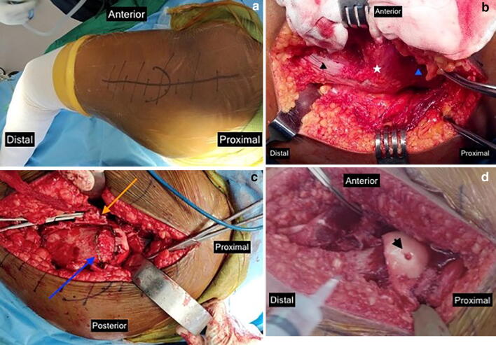
The modified Dunn procedure was performed (Fig. 1a–d) as per the technique described by Leunig et al. [17]. Briefly, with the patient in the lateral decubitus position, surgical hip dislocation was performed after a digastric trochanteric osteotomy. While dislocating the hip, care was taken to temporarily fix the femoral epiphysis in the slipped position with two thick Kirschner wires to prevent further slipping during the dislocation maneuver. After careful preparation of the extended retinacular flaps, the epiphysis was carefully separated from the metaphysis, fully exposing the neck. Next, the posteromedial callus was excised and the femoral neck trimmed judiciously so as to allow capital realignment without tension on the retinacular vessels. At this stage, vascularity of the femoral head was confirmed by drilling a small hole using a Kirschner wire and checking for active pulsatile bleeding. The author’s preferred method of fixation was with one 6.5- or 7-mm cannulated cancellous screw supplemented with one or two Kirschner wires. The hip joint was reduced, and the trochanter fixed slightly distally using 3.5 mm self-tapping cortical screws. From the operative notes, details regarding operative time, estimated blood loss and post-reduction epiphyseal vascularity were noted for each case.
Fig. 1.
Intra-operative photographs of modified Dunn osteotomy performed on a left hip. a Incision centered over greater trochanter. b Deep exposure, just prior to digastric osteotomy; star—greater trochanter, black triangle—vastus lateralis, blue triangle—abductors. c Extensile retinacular flaps raised (yellow arrow), Dunn osteotomy being planned (blue arrow). d Slip reduced, active bleeding seen from epiphysis after reduction (arrow head)
Post-operatively, the child remained nil weight bearing for four weeks followed by protected weight bearing with crutches for four more weeks or till full union of the trochanteric osteotomy was confirmed, after which unrestricted weight-bearing was allowed. Subsequent follow-up was at six monthly intervals. At each follow-up, clinical findings (range of motion, limb lengths) were noted and functional outcomes were assessed using the Harris Hip Score [18]. Radiological outcomes were quantified in terms of slip angle correction, and lateral view alpha angle. Details of any complications were also recorded.
Data were analyzed using SPSS software. Differences in pre- and post-operative slip angles were determined using a paired t-test.
Results
We have presented the mean 25.36 months (range 13–60 months) follow-up of 30 hips in 30 patients treated by the modified Dunn procedure by a single surgeon between 2013 and 2018. Results are summarized in Table 1.
Table 1.
Summary of results of our study
| Parameter | Mean | Range |
|---|---|---|
| Patient age at surgery | 13.05 ± 1.41 years | 11–15.5 years |
| Time from symptom onset till surgery | 25.86 days | 2–60 days |
| Duration of surgery | 146.16 ± 19.48 min | 120–200 min |
| Estimated blood loss | 239 ± 79.03 ml | 150–400 ml |
| Pre-operative slip angle | 53.53 ± 7.37° | 45°–65° |
| Post-operative slip angle | 9.9° ± 3.78° | 5°–15° |
| Slip angle correction | 43.63° ± 8.42° | 23°–55° |
| Post-operative alpha angle | 33.63° ± 4.14 | 28°–45° |
| Harris Hip Score | 81.833 ± 7.12 points | 55–90 points |
| Duration of follow-up | 25.36 months | 13–60 months |
The average age at surgery was 13.05 ± 1.41 years (range 11–15.5 years). There were 25 boys and 5 girls in our series. Right hip was involved in 16, left hip in 14. There were 19 stable slips, and 11 unstable slips, (Loder et al. [19]). As per the Fahey and O’Brian system [20], six, six, and 18 slips were acute, chronic and acute-on-chronic, respectively. In terms of slip angle, there were 20 moderate slips and 10 severe slips.
Average time from symptom onset till surgery was 25.86 days (range 2–60 days). Average duration of surgery was 146.16 ± 19.48 min and blood loss averaged 239 ± 79.03 ml. In 12 cases, there was hemorrhagic fluid on opening the joint capsule, indicative of an unstable slip. In ten hips (all unstable), there was no bleeding on the femoral head immediately after dislocation, which re-appeared in all but two hips, post-reduction.
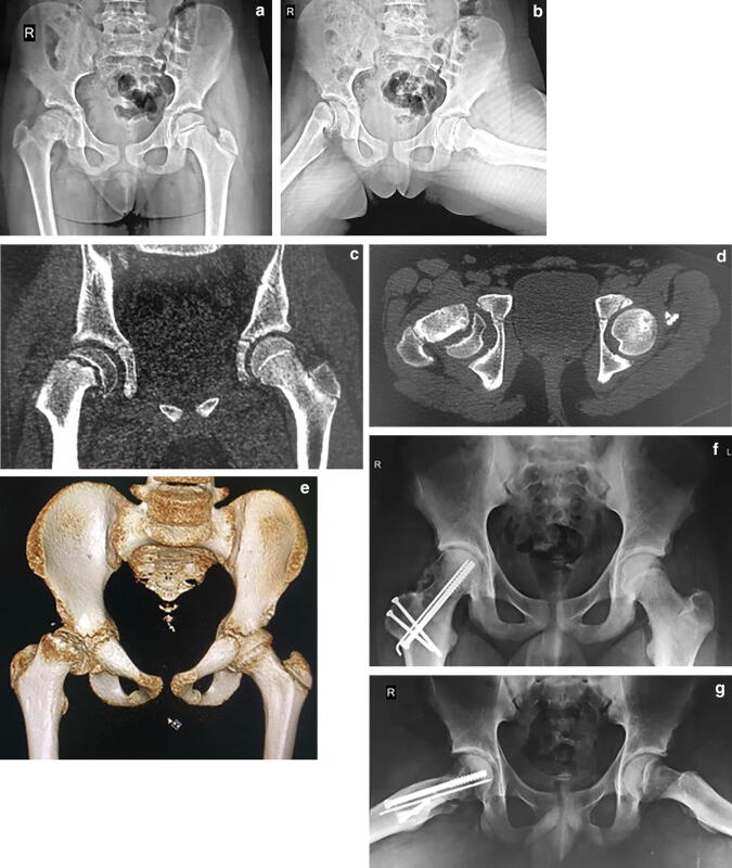
Mean slip angle was corrected from 53.53 ± 7.37° (range 45°–65°) to 9.9° ± 3.78° (range 5°–15°) (p < 0.001). Average correction of slip angle was 43.63° ± 8.42° (range 23°–55°). Mean post-operative alpha angle was 33.63° ± 4.14° (range 28°–45°). Pre- and post-operative imaging of a patient treated by the modified Dunn procedure for a stable slip of moderate severity are given in Fig. 2a–g.
Fig. 2.
A thirteen-year-old boy with right-sided, acute on chronic, stable slip. Pre-operative X-ray pelvis with both hip joints, a anteroposterior and b frog leg lateral views showing a moderate slip (52°). c Coronal, d transverse, and e three-dimensional reconstruction computerized tomography images prior to surgery. f and g Radiographs at 2.5-year follow-up after capital realignment using modified Dunn procedure (final slip angle 3°, alpha angle 26°)
Functional outcome was assessed using the Harris Hip Score [18], whereby results were graded as excellent (≥ 90), good (80–90), fair (70–80) and poor (< 70). Results were excellent in six cases, good in 17, fair in six and poor in one. The average Harris Hip Score was 81.833 ± 7.12 points (range 55–90). Hip range of motion was restored to near normal in all cases. Mean hip flexion, flexed internal rotation and flexed external rotation at final follow-up were 101.66° ± 7.71° (range 80°–110°), 23.16° ± 6.89° (range 0°–30°) and 31.33 °± 7.63° (range 20–50°), respectively. Eighteen patients had minor limb length discrepancy, with mean shortening of 0.55 cm (0.5–1.5 cm) on the operated side.
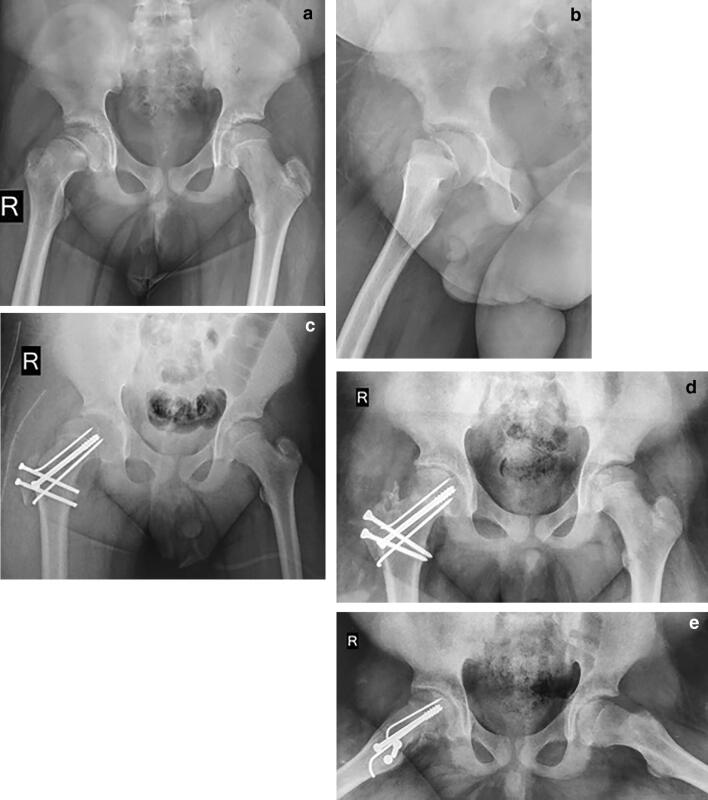
Three complications were encountered in our series. Osteonecrosis occurred in two hips. Both the hips had unstable, severe, acute on chronic slips. One child had mild radiological AVN which was asymptomatic and was asked to be kept on regular follow-up; while, the other child had severe, symptomatic AVN at around 18 months of follow-up. The child was almost skeletally mature and was advised regarding total hip replacement. One patient suffered a post-operative hip subluxation (Fig. 3a–c), which on re-exploration was found to be due to capsular interposition. Following revision surgery, the hip was stable and the child went on to have a good outcome (Fig. 3d, e).
Fig. 3.
A thirteen-year-old boy operated for right-sided acute, unstable slip. X-ray pelvis with both hip joints a anteroposterior and b cross table lateral views at presentation. c Immediate post-operative anteroposterior radiograph showing subluxation. d Anteroposterior and e frog leg lateral radiographs at 18 months after revision surgery (removal of interposed capsular tissue and the trochanteric screws exchange). Note the concentric joint reduction and absence of AVN
Discussion
The chief goals of treatment of SCFE are, first, prevention of slip progression by obliteration of the physis; and second, prevention of degenerative joint disease by providing near anatomic proximal femoral anatomy, while maintaining the vascularity of the capital femoral epiphysis. The modified Dunn procedure allows the surgeon to achieve both these goals. The surgical dislocation of the hip allows complete exposure of the head and neck of the femur allowing precise capital realignment; while the creation of extended periosteal flaps and trimming of the neck allow realignment to be done without stressing the retinacular vessels, thus preserving vascularity of the femoral head.
Since first described in 2009 [15], the modified Dunn procedure has increasingly been used for the treatment of moderate to severe SCFE. Our study was able to reproduce the results of previous series using the modified Dunn technique to a large extent. In 30 hips with a mean pre-operative slip angle of 53°, proximal femoral anatomy was restored in all cases. Mean slip angle and alpha angle at the final follow-up were 9.9° and 33.63°, respectively, which is comparable to that reported in previous series (Table 2). The Harris Hip Score at the final follow-up averaged 81.8 points. This is slightly lower than that reported by most studies (Table 2), which is likely due to the larger number of children with chronic slips, and children presenting to us late after symptom onset.
Table 2.
Comparison of results of the modified Dunn procedure in different studies
| Author (year) | Pre-operative slip angle (°) | Post-operative slip angle (°) | Post-operative alpha angle (°) | Post-operative harris hip score (points) |
|---|---|---|---|---|
| Ziebarth (2009) [15] | 56.6 | 8.6 | 40.6 | 99.6 |
| Slongo (2010) [21] | 47.6 | 4.6 | 37.5 | 99 |
| Huber (2011) [22] | 44.9 | 5.2 | 41.4 | 97.1 |
| Masse (2012) [24] | 50.65 | 9.45 | 43.11 | 98.2 |
| Madan (2013) [31] | 59 | 8 | – | 89.1 |
| Novais (2015) [34] | 65 | 16 | 44 | –þ |
| Elmarghany (2017) [36] | 52.5 | 5.6 | 51.15 | 96.16 |
| Javier (2017) [23] | 59.1 | 5.4 | 40.8 | 76.3 |
| Lerch (2019) [28] | 64 | 7 | 38 | 94 |
| Our study (2020) | 53.53 | 9.9 | 33.63o | 81.8 |
We had a low rate of complications in our series. There were two cases of AVN (6.66%), both of which occurred in unstable slips. None of the stable slips in our series went on to develop AVN. Previous studies have reported AVN rates with the modified Dunn procedure ranging from zero to 66.7% (Table 3) [21–36]. There are many reasons for this wide range. Studies with a higher ratio of unstable vs stable slips tended to have higher AVN rates [23, 26, 29, 30, 32]. This may be due to the natural course of unstable slips; a recent meta-analysis [37] found the cumulative rate of osteonecrosis in unstable SCFE to be 23.9% (range 0–58%), irrespective of mode of treatment. Upasani et al. [32] found an inverse correlation between surgeon experience and complication rate with the modified Dunn procedure. Many of the reports on the modified Dunn procedure are multicenter studies with surgeons having varying levels of experience with the procedure. The steep learning curve with this technically demanding procedure may account for some of the variability in AVN rates between different studies. Our study compares favorably to the rates of AVN described in the literature.
Table 3.
Comparison of AVN rates of the modified Dunn procedure in different studies
| Author (year) | Total hips | Stable | Unstable | Total AVN | AVN in stable slips* | AVN in unstable slips** |
|---|---|---|---|---|---|---|
| Ziebarth (2009) [15] | 40 | 70% | 30% | 0 | – | – |
| Slongo (2010) [21] | 23 | 87% | 13% | 4% | 1/1 | Nil |
| Madan (2013) [31] | 28 | 39% | 61% | 7.1% | Nil | 2/2 |
| Souder (2014) [33] | 17 | 59% | 41% | 24% | 2/4 | 2/4 |
| Novais (2015) [34] | 15 | 100% | – | 6.6% | 1/1 | – |
| Javier (2017) [23] | 21 | 29% | 71% | 47.6% | 2/10 | 8/10 |
| Novais (2018) [26] | 27 | – | 100% | 26% | – | 7/7 |
| Ebert (2019) [27] | 15 | 100% | – | 26% | 4/4 | – |
| Lerch (2019) [28] | 46 | 70% | 30% | 5% | 2/2 | Nil |
| Our study (2020) | 30 | 63% | 37% | 6.6% | Nil | 2/2 |
*Cases of AVN in stable slips / Total cases of AVN in the study
**Cases of AVN in unstable slips / Total cases of AVN in the study
We also had one case of hip instability following the procedure (Fig. 3). Iatrogenic hip instability is a devastating but rare complication of the modified Dunn procedure which has recently received greater attention. A multicenter review [38] identified 17 cases of iatrogenic hip instability out of 406 modified Dunn procedures (4%) performed across eight institutions over the course of seven years. Factors that were believed to contribute to the occurrence of this complication include: the external rotation contracture and attenuation of the anterior hip capsule in patients with chronic SCFE, weakening of the already compromised capsule by the extensive capsulotomy, dislocation of the hip for performing the capital realignment, excessive trimming of the femoral neck, and loose capsular repair to prevent constriction of the retinacular vessels. Most patients with hip instability in the aforementioned study had poor outcomes, with the majority developing AVN and three having undergone arthroplasty within a short follow-up of just 2 years. The one case of post-operative hip instability encountered in our series was found to have occurred due to interposition of in-folded capsule which was preventing concentric reduction of the joint. Upon removal, joint stability was restored, and the child went on to have an uneventful recovery, with no evidence of AVN at 18-month follow-up.
We did not encounter any implant failures in our series. Previous studies have reported implant breakage in up to 15% of cases [15, 22, 30]. We believe the absence of this complication in our series is due to the practice of using heavy ≥ 6.5 mm screws for capital fixation in all cases. Similar findings were reported by Sankar et al. [30] who had implant breakage of 15% in their series, all of which occurred in cases where either two solid 4.5 mm screws or multiple threaded wires had been used for epiphyseal fixation. None of their patients treated with 6.5-mm cannulated screws had implant failures.
Studies regarding the safety and reproducibility of the modified Dunn procedure in the Indian population are scarce. Arora et al. [16] analyzed the outcomes and disease associations of SCFE at a tertiary care center in India. In a series of 30 slips, eight underwent the modified Dunn procedure, and the rest were pinned in situ. All eight were unstable slips, with a mean pre-operative slip angle of 72°, corrected to a mean of 4.8° post-operatively. Two cases were complicated by AVN. The authors found that the modified Dunn procedure had good early results in their series, and spared the need for secondary procedures as after in situ pinning. They found high BMI, Vitamin D deficiency and endocrine disorders to be risk factors for SCFE in Indian adolescents.
Our study has some limitations. This was a small cohort of 30 patients. However, SCFE being relatively uncommon in Indian adolescents, this is probably the largest series of hips treated by the modified Dunn procedure in this population, to be published. Second, we did not have a control group of patients treated by another modality, for example, in situ pinning. However, given the mounting evidence demonstrating the association between cam type of FAI in moderate and severe slips pinned in situ with early onset of degenerative osteoarthritis, we felt that in situ pinning was not justified in these patients when there existed an alternative treatment option. Finally, this was a retrospective study with a short-term follow-up. Longer-term prospective studies are necessary to study the behavior of these hips in the future.
Despite its limitations, this study reports the outcomes of the modified Dunn procedure in a series of 30 Indian adolescents. The radiological and functional outcomes and the complication rates with this procedure were found to be comparable to those previously suggested by the literature. It is important to note, however, that this is a technically demanding procedure and should preferably be undertaken by those with prior experience in performing safe surgical dislocation.
Conclusion
Our study adds to the evidence that the modified Dunn osteotomy procedure is safe, reliable and reproducible. It should be the first choice for management for moderate and severe slipped capital femoral epiphysis.
Author contributions
MVA: Concepts, design, definition of intellectual content, literature search, clinical studies, experimental studies, data acquisition, data analysis, statistical analysis, manuscript preparation, manuscript editing, manuscript review, guarantor. DAP: design, literature search, manuscript preparation, manuscript editing, manuscript review. SVV: concept, design, definition of intellectual content, clinical studies, data acquisition, data analysis, manuscript preparation, manuscript editing.
Funding
None.
Compliance with Ethical Standards
Conflict of interest
None.
Patient declaration statement
“The authors certify that they have obtained all appropriate patient consent forms. In the form the patient(s) has/have given his/her/their consent for his/her/their images and other clinical information to be reported in the journal. The patients understand that their names and initials will not be published and due efforts will be made to conceal their identity, but anonymity cannot be guaranteed.”
Ethical standard statement
All the procedures performed in this study were in accordance with the ethical standards of the National research committee and with the 1964 Helsinki declaration and its later amendments or comparable ethical standards.
Informed consent
Since it was a retrospective study, a formal consent was not necessary.
Footnotes
Publisher's Note
Springer Nature remains neutral with regard to jurisdictional claims in published maps and institutional affiliations.
Contributor Information
Mandar V. Agashe, Email: mandarortho@gmail.com
Deepika A. Pinto, Email: deepupinto@gmail.com
Sandeep Vaidya, Email: drsvvaidya@gmail.com.
References
- 1.Southwick WO. Osteotomy through the lesser trochanter for slipped capital femoral epiphysis. Journal of Bone and Joint Surgery. American Volume. 1967;49(5):807–835. doi: 10.2106/00004623-196749050-00001. [DOI] [PubMed] [Google Scholar]
- 2.Boyer DW, Mickelson MR, Ponseti IV. Slipped capital femoral epiphysis. Long-term follow-up study of one hundred and twenty-one patients. J Bone Jt Surg—Ser A. 1981;63(1):85–95. doi: 10.2106/00004623-198163010-00011. [DOI] [PubMed] [Google Scholar]
- 3.Carney BT, Weinstein SL, Noble J. Long-term follow-up of slipped capital femoral epiphysis. J Bone Jt Surg. 1991;73(5):667–674. doi: 10.2106/00004623-199173050-00004. [DOI] [PubMed] [Google Scholar]
- 4.Ross PM, Lyne ED, Morawa LG. Slipped capital femoral epiphysis long-term results after 10–38 years. Clinical Orthopaedics and Related Research. 1979;141:176–180. [PubMed] [Google Scholar]
- 5.Dunn DM. The treatment of adolescent slipping of the upper femoral epiphysis. Journal of Bone and Joint Surgery. British Volume. 1964;46:621–629. doi: 10.1302/0301-620X.46B4.621. [DOI] [PubMed] [Google Scholar]
- 6.Fish JB. Cuneiform osteotomy of the femoral neck in the treatment of slipped capital femoral epiphysis. Journal of Bone and Joint Surgery. American Volume. 1984;66(8):1153–1168. doi: 10.2106/00004623-198466080-00002. [DOI] [PubMed] [Google Scholar]
- 7.Kramer W, Craig W, Noel S. Compensating osteotomy at the base of the femoral neck for slipped capital femoral epiphysis. Journal of Bone and Joint Surgery. 1976;58(6):796–800. doi: 10.2106/00004623-197658060-00009. [DOI] [PubMed] [Google Scholar]
- 8.Barmada R, Bruch RF, Gimbel JS, Ray RD. Base of the neck extracapsular osteotomy for correction of deformity in slipped capital femoral epiphysis. Clinical Orthopaedics and Related Research. 1978;132(132):98–101. [PubMed] [Google Scholar]
- 9.Imhauser G. Pathogenesis and therapy of hip dislocation in youth. Zeitschrift fur Orthopadie und Ihre Grenzgebiete. 1956;88(1):3–41. [PubMed] [Google Scholar]
- 10.Dunn DM, Angel JC. Replacement of the femoral head by open operation in severe adolescent slipping of the upper femoral epiphysis. Journal of Bone and Joint Surgery Series B. 1978;60B(3):394–403. doi: 10.1302/0301-620X.60B3.681417. [DOI] [PubMed] [Google Scholar]
- 11.Rostoucher P, Bensahel H, Pennecot GF, Kaewpornsawan K, Mazda K. Slipped capital femoral epiphysis: evaluation of different modes of treatment. Journal of Pediatric Orthopedics Part B. 1996;5(2):96–101. doi: 10.1097/01202412-199605020-00008. [DOI] [PubMed] [Google Scholar]
- 12.Velasco R, Schai PA, Exner GU. Slipped capital femoral epiphysis: a long-term follow-up study after open reduction of the femoral head combined with subcapital wedge resection. Journal of Pediatric Orthopedics Part B. 1998;7(1):43–52. doi: 10.1097/01202412-199801000-00008. [DOI] [PubMed] [Google Scholar]
- 13.Jerre R, Hansson G, Wallin J, Karlsson J. Long-term results after realignment operations for slipped upper femoral epiphysis. Journal of Bone and Joint Surgery Series B. 1996;78(5):745–750. doi: 10.1302/0301-620X.78B5.0780745. [DOI] [PubMed] [Google Scholar]
- 14.Fron D, Forgues D, Mayrargue E, Halimi P, Herbaux B. Follow-up study of severe slipped capital femoral epiphysis treated with Dunn’s osteotomy. Journal of Pediatric Orthopedics. 2000;20(3):320–325. [PubMed] [Google Scholar]
- 15.Ziebarth K, Zilkens C, Spencer S, Leunig M, Ganz R, Kim YJ. Capital realignment for moderate and severe SCFE using a modified dunn procedure. Clinical Orthopaedics and Related Research. 2009;467(3):704–716. doi: 10.1007/s11999-008-0687-4. [DOI] [PMC free article] [PubMed] [Google Scholar]
- 16.Arora S, Dutt V, Palocaren T, Madhuri V. Slipped upper femoral epiphysis: Outcome after in situ fixation and capital realignment technique. Indian J Orthop. 2013;47(3):264–271. doi: 10.4103/0019-5413.111492. [DOI] [PMC free article] [PubMed] [Google Scholar]
- 17.Leunig M, Slongo T, Ganz R. Subcapital realignment in slipped capital femoral epiphysis: surgical hip dislocation and trimming of the stable trochanter to protect the perfusion of the epiphysis. Instructional Course Lectures. 2008;57:499–507. [PubMed] [Google Scholar]
- 18.Harris WH. Traumatic arthritis of the hip after dislocation and acetabular fractures: treatment by mold arthroplasty. An end-result study using a new method of result evaluation. Journal of Bone and Joint Surgery America. 1969;51(4):737–755. doi: 10.2106/00004623-196951040-00012. [DOI] [PubMed] [Google Scholar]
- 19.Loder RT, Richards BS, Shapiro PS, Reznick LR, Aronson DD. Acute slipped capital femoral epiphysis: the importance of physeal stability. Journal of Bone and Joint Surgery. American Volume. 1993;75(8):1134–1140. doi: 10.2106/00004623-199308000-00002. [DOI] [PubMed] [Google Scholar]
- 20.Fahey JJ, O’Brien ET. Acute slipped capital femoral epiphysis: Review of the literature and report of ten cases. Journal of Bone and Joint Surgery. American Volume. 1965;47:1105–1127. doi: 10.2106/00004623-196547060-00001. [DOI] [PubMed] [Google Scholar]
- 21.Slongo T, Kakaty D, Krause F, Ziebarth K. Treatment of slipped capital femoral epiphysis with a modified Dunn procedure. Journal of Bone and Joint Surgery—Series A. 2010;92(18):2898–2908. doi: 10.2106/JBJS.I.01385. [DOI] [PubMed] [Google Scholar]
- 22.Huber H, Dora C, Ramseier LE, Buck F, Dierauer S. Adolescent slipped capital femoral epiphysis treated by a modified Dunn osteotomy with surgical hip dislocation. Journal of Bone and Joint Surgery—Series B. 2011;93B(6):833–838. doi: 10.1302/0301-620X.93B6.25849. [DOI] [PubMed] [Google Scholar]
- 23.Masquijo JJ, Allende V, D’Elia M, Miranda G, Fernández CA. Treatment of slipped capital femoral epiphysis with the modified dunn procedure: a multicenter study. Journal of Pediatric Orthopedics. 2019;39(2):71–76. doi: 10.1097/BPO.0000000000000936. [DOI] [PubMed] [Google Scholar]
- 24.Massè A, Aprato A, Grappiolo G, Turchetto L, Campacci A, Ganz R. Surgical hip dislocation for anatomic reorientation of slipped capital femoral epiphysis: preliminary results. Hip Int. 2012;22(2):137–144. doi: 10.5301/HIP.2012.9208. [DOI] [PubMed] [Google Scholar]
- 25.Cosma D, Vasilescu DE, Corbu A, Valeanu M, Vasilescu D. The modified dunn procedure for slipped capital femoral epiphysis does not reduce the length of the femoral neck. Pakistan J Med Sci. 2016;32(2):379–384. doi: 10.12669/pjms.322.8638. [DOI] [PMC free article] [PubMed] [Google Scholar]
- 26.Novais EN, Maranho DA, Heare T, Sink E, Carry PM, O’Donnel C. The modified Dunn procedure provides superior short-term outcomes in the treatment of the unstable slipped capital femoral epiphysis as compared to the inadvertent closed reduction and percutaneous pinning: a comparative clinical study. International Orthopaedics. 2019;43(3):669–675. doi: 10.1007/s00264-018-3993-9. [DOI] [PubMed] [Google Scholar]
- 27.Ebert N, Rupprecht M, Stuecker R, Breyer S, Stiel N, Priemel MH, et al. Outcome of the modified Dunn procedure in severe chronic or acute on chronic slipped capital femoral epiphysis. J Orthop Surg Res. 2019;14(1):1–7. doi: 10.1186/s13018-019-1433-1. [DOI] [PMC free article] [PubMed] [Google Scholar]
- 28.Lerch TD, Vuilleumier S, Schmaranzer F, Ziebarth K, Steppacher SD, Tannast M, et al. Patients with severe slipped capital femoral epiphysis treated by the modified Dunn procedure have low rates of avascular necrosis, good outcomes, and little osteoarthritis at long-term follow-up. Bone Joint J. 2019;101-B(4):403–14. [DOI] [PubMed]
- 29.Alves C, Steele M, Narayanan U, Howard A, Alman B, Wright JG. Open reduction and internal fixation of unstable slipped capital femoral epiphysis by means of surgical dislocation does not decrease the rate of avascular necrosis: a preliminary study. Journal of Children's Orthopaedics. 2012;6(4):277–283. doi: 10.1007/s11832-012-0423-1. [DOI] [PMC free article] [PubMed] [Google Scholar]
- 30.Sankar WN, Vanderhave K, Matheney TH, Herrera-Soto J, Karlen J. The modified dunn procedure for unstable. J Bone Jt Surg Am. 2013;95:585–591. doi: 10.2106/JBJS.L.00203. [DOI] [PubMed] [Google Scholar]
- 31.Madan SS, Cooper AP, Davies AG, Fernandes JA. The treatment of severe slipped capital femoral epiphysis via the Ganz surgical dislocation and anatomical reduction. Bone Joint J. 2013;95-B(3):424–429. doi: 10.1302/0301-620X.95B3.30113. [DOI] [PubMed] [Google Scholar]
- 32.Upasani VV, Matheney TH, Spencer SA, Kim YJ, Millis MB, Kasser JR. Complications after modified dunn osteotomy for the treatment of adolescent slipped capital femoral epiphysis. Journal of Pediatric Orthopedics. 2014;34(7):661–667. doi: 10.1097/BPO.0000000000000161. [DOI] [PubMed] [Google Scholar]
- 33.Souder CD, Bomar JD, Wenger DR. The role of capital realignment versus in situ stabilization for the treatment of slipped capital femoral epiphysis. Journal of Pediatric Orthopedics. 2014;34(8):791–798. doi: 10.1097/BPO.0000000000000193. [DOI] [PubMed] [Google Scholar]
- 34.Novais EN, Hill MK, Carry PM, Heare TC, Sink EL. Modified Dunn Procedure is Superior to In Situ Pinning for Short-term Clinical and Radiographic Improvement in Severe Stable SCFE. Clinical Orthopaedics and Related Research. 2015;473(6):2108–2117. doi: 10.1007/s11999-014-4100-1. [DOI] [PMC free article] [PubMed] [Google Scholar]
- 35.Davis RL, Samora WP, Persinger F, Klingele KE. Treatment of unstable versus stable slipped capital femoral epiphysis using the modified dunn procedure. Journal of Pediatric Orthopedics. 2019;39(8):411–415. doi: 10.1097/BPO.0000000000000975. [DOI] [PubMed] [Google Scholar]
- 36.Elmarghany M, Abd El-Ghaffar TM, Seddik M, Akar A, Gad Y, Ragheb E, et al. Surgical hip dislocation in treatment of slipped capital femoral epiphysis. Sicot-J. 2017;3:10. doi: 10.1051/sicotj/2016047. [DOI] [PMC free article] [PubMed] [Google Scholar]
- 37.Zaltz I, Baca G, Clohisy JC. Unstable SCFE: Review of treatment modalities and prevalence of osteonecrosis hip. Clinical Orthopaedics and Related Research. 2013;471(7):2192–2198. doi: 10.1007/s11999-012-2765-x. [DOI] [PMC free article] [PubMed] [Google Scholar]
- 38.Upasani VV, Birke O, Klingele KE, Millis MB. Iatrogenic hip instability is a devastating complication after the modified dunn procedure for severe slipped capital femoral epiphysis. Clinical Orthopaedics and Related Research. 2017;475(4):1229–1235. doi: 10.1007/s11999-016-5094-7. [DOI] [PMC free article] [PubMed] [Google Scholar]


