Abstract
Stimulant-use disorders have been associated with lower availability of dopamine type-2 receptors (D2R) and greater availability of type-3 receptors (D3R). Links between D2R levels, cognitive performance, and suppression of the default mode network (DMN) during executive functioning have been observed in healthy and addicted populations; however, there is limited evidence regarding a potential role of elevated D3R in influencing cognitive control processes in groups with and without addictions. Sixteen individuals with cocaine-use disorder (CUD) and 16 healthy comparison (HC) participants completed [11C]-(+)-PHNO PET imaging of D2R and D3R availability and fMRI during a Stroop task of cognitive control. Independent component analysis was performed on fMRI data to assess DMN suppression during Stroop performance. In HC individuals, lower D2R-related binding in the dorsal putamen was associated with improved task performance and greater DMN suppression. By comparison, in individuals with CUD, greater D3R-related binding in the substantia nigra was associated with improved performance and greater DMN suppression. Exploratory moderated-mediation analyses indicated that DMN suppression was associated with Stroop performance indirectly through D2R in HC and D3R in CUD participants, and these indirect effects were different between groups. To our knowledge, this is the first evidence of a dissociative and potentially beneficial role of elevated D3R availability in executive functioning in cocaine-use disorder.
Subject terms: Addiction, Cognitive control
Introduction
Links between stimulant use and alterations in dopaminergic systems are well established [1, 2]. Regarding dopamine receptor levels in individuals with a stimulant-use disorder, evidence indicates bidirectional differences in the dopamine type-2 family of receptors, with lower availability of type-2 receptors (D2R) in striatal regions and higher availability of type-3 receptors (D3R) in midbrain regions [3–7]. Given some distinctions in regional distributions of these receptor subtypes, they have been hypothesized to contribute differently to cognitive mechanisms in addictions. D2R-related circuitry is proposed to contribute to compulsivity and habitual behavior and D3R-related circuitry to impulsivity and reward sensitivity [7–11]. However, investigations dissociating potential D2R and D3R relationships with cognitive processes in individuals with and without a cocaine-use disorder (CUD) are limited.
Beyond contributions to reinforcement processing, dopaminergic processes have also been implicated in executive functions including working memory, attentional control, and response processing [12, 13]. Dual-state models of dopaminergic roles in executive functioning propose that the type-2 family of receptors is related to cognitive flexibility and adaptability (e.g., set-shifting and reversal learning) while cognitive stability and maintenance (e.g., persistence of working memory) are associated with the type-1 family of receptors [14]. Evidence of neurocognitive impairments in individuals with a stimulant-use disorder are mostly consistent with deficient D2R function according to these models (i.e., greater perseverance, impaired attentional shifting, and poor response inhibition) [15]. However, the extent to which increased D3R-related functioning in CUD may compensate or subjugate D2R-related roles in executive functions is unclear. While both decreased D2R and increased D3R have been separately linked to greater impulsivity and cognitive-control impairments [16], their potentially dissociative influences are unknown. Furthermore, individual differences in D2R and D3R alterations, which may not be correlated in individuals with a stimulant-use disorder [6, 7], may support evidence of differing patterns and degrees of neurocognitive deficits in stimulant additions [17, 18].
The default mode network (DMN), integrating activity in the posterior cingulate with regions of the ventromedial frontal and lateral parietal cortices, is an established resting-state functional brain network broadly associated with non-goal-directed cognition [19, 20]. Alterations in DMN functioning have been noted in a range of psychiatric conditions including addictions, depression, schizophrenia, and attention-deficit/hyperactivity disorder [21–24]. Suppression of the DMN may be a marker of the global functioning of externally oriented cognitive systems [25, 26] and has been linked to dopaminergic functioning [27–30]. In healthy populations, greater suppression of the DMN during visual attention has been associated with reduced dopamine transporter availability (i.e., greater synaptic dopamine levels) in the striatum [31]. Greater connectivity between nodes of the DMN during working memory has been linked to greater D2R-related, but not D3R-related, availability [30]. Functional impairments of the DMN have been noted across substance-use disorders [21]. In individuals with a stimulant-use disorder, greater DMN suppression during cue-reactivity is associated with greater D2R-related binding [32]. Furthermore, methylphenidate, which increases synaptic dopamine levels, increases suppression of the DMN during response inhibition in individuals who use cocaine [33] and youth with attention-deficit/hyperactivity disorder [24].
The current study investigated potential relationships between D2R/D3R availability and global executive functioning during cognitive control in individuals with CUD relative to healthy comparison (HC) individuals. Relationships were examined between D2R and D3R availability (assessed with [11C]-(+)-PHNO PET), DMN functioning (assessed using independent component analysis of fMRI during a Stroop task), and Stroop-related cognitive-control performance between and within groups. We hypothesized that across all participants, greater availability of D2R would be associated with better task performance (i.e., reduced errors and interference delays) and greater DMN suppression during Stroop performance. In CUD, we expected higher availability of D3R, proposed to reflect increased appetitive (nonexecutive) processing [34], would be associated with lower DMN suppression, and poorer Stroop performance.
Methods
Participants
Sixteen nontreatment-seeking individuals with CUD and 16 age- and gender-matched HC participants were recruited from the local community. Physical exams with medical history, routine laboratory studies, pregnancy tests, and electrocardiograms were performed to assess medical eligibility. Urine toxicology screening for cocaine, amphetamines, marijuana, opiates, benzodiazepines, and barbiturates (Integrated EZ Split Key Cup; Redwood Toxicology Laboratories, Santa Rosa, CA, USA) was performed to confirm cocaine-use status in CUD participants and the absence of other recent drug use in both CUD and HC participants. Participants were assessed for DSM-IV diagnoses using the Structured Clinical Interview for DSM-IV [35]. All CUD participants met criteria for cocaine dependence (comparable to DSM-5 criteria for a CUD of at least moderate severity) and were admitted to an inpatient research facility to monitor abstinence prior to completing imaging procedures. Exclusion criteria included the presence or history of a general medical (e.g., cardiovascular, diabetic/metabolic) or neurological (e.g., cerebrovascular, seizures, traumatic brain injury) illness or psychotic disorder, or met criteria for any current Axis I psychiatric diagnosis (other than cocaine- or tobacco-use disorders), pregnancy or breast-feeding, or any condition that would interfere with PET or MRI participation (e.g., claustrophobia, metallic implants). Intelligence was estimated using the Shipley Institute of Living Scale [36]. Tobacco-using CUD participants were allowed regular smoke-breaks during inpatient residency, and no smoking was allowed within an hour prior to PET/MRI scanning to limit potential influences of acute tobacco use [37, 38]. Individuals from a prior PET report [6] completing Stroop fMRI procedures were included in the current analysis. All study procedures were approved by the Yale Human Investigation, Yale University Radiation Safety, Yale-New Haven Hospital Radioactive Drug Research, and Yale MRI Safety Committees, and participants provided written informed consent.
[11C](+)PHNO PET
[11C]-(+)-PHNO was prepared as previously described [39, 40], and details of PET imaging, performed on a Siemens high-resolution research tomograph (Siemens/CTI, Knoxville, TN, USA), are provided as Supplementary Material. Average [11C]-(+)-PHNO binding potential (BPND) values were computed from smoothed parametric images for two regions of interest (ROI), the dorsal putamen (DPU) [41], and substantia nigra (SN) [39], as binding in these regions largely reflect D2R- and D3R-related availability, respectively [42–44].
Stroop fMRI
The event-related Stroop color-word interference task has been shown previously to activate brain regions and functional networks underlying cognitive control [45, 46]. Briefly, participants completed six 3-min runs consisting of congruent (e.g., “red” displayed in red font) and incongruent stimuli (e.g., “red” displayed in blue font) during which they were instructed to respond silently, an approach that has been shown to produce activation equivalent to overt response methods [47]. Acquisition and spatial processing of functional images, collected on a Siemens 3T Trio system (Siemens Medical Solutions, Malvern, PA), are provided as Supplementary Material.
Independent component analysis
ICA was performed on the fMRI time series using the Group ICA Toolbox (GroupICAT v4.0b; https://trendscenter.org/software/gift/) [48]. Minimum description length criterion [49] estimated that a mean of 25 maximally independent components was present in each functional run. Data from all participants were concatenated into a single group, reduced through a principal component analysis, and 25 components were extracted from this group aggregate using the InfoMax algorithm [50]. ICA was iterated 20 times using ICASSO to assess stability and consistency of extracted components [51]. Component time courses and corresponding spatial source maps were reconstructed and scaled to percent BOLD signal change for each participant to facilitate comparisons.
The DMN was visually selected a priori from the set of 25 identified components. Task-relatedness of the DMN was assessed using multiple regression analyses of the component time course with the time courses of incongruent and congruent stimuli convolved with canonical hemodynamic activity and including motion parameters from spatial processing as nuisance regressors. The resulting β-weights were averaged for each stimulus type across runs for each participant as a measure of DMN “engagement,” with more negative engagement indicating greater DMN suppression.
Stroop behavioral performance
Participants completed two runs of the task aloud prior to scanning and a maximum of five runs immediately following scanning to assess behavioral performance. Vocal responses and reaction times were collected using presentation (Neurobehavioral Systems, Inc., Berkeley, CA). Preliminary analyses indicated no differences in performance between runs completed before and after scanning; thus, Stroop interference delays (i.e., incongruent minus congruent vocal reaction time) and percent incongruent errors were averaged across all runs completed outside of the scanner.
Statistical analyses
Initial analyses were performed within modality (i.e., PET, fMRI, and behavioral performance, separately) using linear mixed models with a between-subjects factor of group (HC, CUD) in SPSS 26.0 (IBM Corporation, Armonk, NY). [11C]-(+)-PHNO BPND was investigated using a within-subjects factor of ROI (DPU and SN). DMN engagements were similarly tested in a second mixed model using a within-subjects factor of stimulus type (i.e., incongruent and congruent). A third mixed model was used to investigate Stroop performance using a within-subjects factor of behavior (interference delay and incongruent error percentage). Main effects and interactions of the unimodal mixed models were assessed at Bonferroni-corrected PBonferroni < 0.05 (alpha = 0.05, n = 3; P < 0.017) and post hoc tests included models to investigate within-group effects. These three within-modality models were each repeated with inclusion of covariates for tobacco-smoking status across the entire sample and cocaine-use measures (days of past-month cocaine use, years of lifetime cocaine use, and duration of abstinence (at PET scan), modeled together) within the CUD group.
Bimodal analyses were then performed. Primary hypotheses regarding relationships with D2R- and D3R-related binding were tested by repeating the models of DMN engagement and Stroop performance separately with the inclusion of two covariates for BPND (DPU, SN) together. To examine relationships between performance and DMN engagement, the model of Stroop performance was repeated with a single covariate of the average (mean of congruent and incongruent) DMN engagement. Main effects and interactions of these three models examining bimodal relationships were assessed at PBonferroni < 0.05 (alpha = 0.05, n = 3; P < 0.017) and post hoc tests included models to investigate within-group effects.
Following bimodal testing, exploratory analyses were performed to examine potential moderated-mediation relationships across all three modalities (D2R/D3R, DMN suppression, and Stroop interference delays). Analyses were performed using PROCESS [52] for SPSS with 5000 bootstrap resamples to handle the limited sample sizes. Models tested whether group moderated potential mediations by D2R- and D3R-related availability, modeled separately, in associations between DMN engagement and Stroop interference delays. Associations and conditional indirect effects were calculated at 95% CI and within-group post hoc mediation models were performed. 95% CIs that did not include zero determined significant mediations and moderated mediations.
Results
Participant characteristics and D2/3R availability
There were no group differences in age, gender, IQ, body mass, alcohol use, or radiotracer injection parameters (Table 1). Demographic characteristics did not interact with group on [11C]-(+)-PHNO BPND, Stroop performance, or DMN engagement (P > 0.1). Consistent with clinical profiles, CUD participants were more likely to smoke tobacco daily than were HC participants; however, there was no effect of daily smoking on [11C]-(+)-PHNO BPND, Stroop performance, or network engagement measures (P > 0.1). There was a group difference in the time between PET and fMRI scans, with CUD participants completing scans ~1 week apart, while scans averaged 1 month apart in HC participants.
Table 1.
Participant characteristics and injection details.
| Variable | HC (N = 16) | CUD (N = 16) | P value |
|---|---|---|---|
| Participant characteristics | |||
| Age, years (SD) | 42.3 (6.2) | 43.4 (5.3) | 0.59 |
| Gender, F (%) | 4 (25.0) | 2 (12.5) | 0.37 |
| Estimated IQ, total (SD) | 99.7 (15.2) | 95.6 (11.7) | 0.40 |
| Body-mass, BMI (SD) | 30.4 (6.5) | 29.8 (7.0) | 0.79 |
| Daily tobacco user, N (%) | 1 (6.3) | 13 (81.3) | <0.001 |
| Weekly alcohol use, drinks (SD) | 1.3 (2.2) | 7.3 (17.9) | 0.20 |
| Cocaine-use frequency, days/month (SD) | – | 18.4 (7.0) | – |
| CUD chronicity, years (SD) | – | 19.8 (6.1) | – |
| Abstinence from cocaine, days (SD) | – | 7.4 (5.4) | – |
| Time between PET/MRI, days (SD) | 30.9 (29.7) | 9.1 (6.5) | 0.01 |
| [11C]-(+)-PHNO injection | |||
| Radioactive dose, MBq (SD) | 430 (144) | 408 (157) | 0.68 |
| Injected mass, μg/kg (SD) | 0.024 (0.007) | 0.022 (0.007) | 0.53 |
| Specific activity, MBq/nmol (SD) | 56.3 (22.9) | 55.6 (18.1) | 0.92 |
CUD cocaine-use disorder, HC healthy comparison, SD standard deviation.
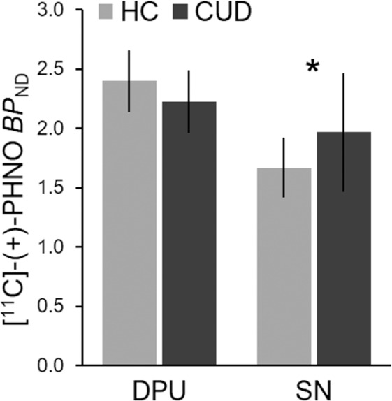
Consistent with prior reports [4–7], CUD and HC differed in [11C]-(+)-PHNO BPND across regions (group-by-ROI interaction: F1,30 = 10.38, P = 0.003; PBonferroni = 0.009). CUD participants had 17% greater BPND in the D3R-rich SN (t30 = 2.13, P = 0.042), and 7% lower BPND in the D2R-rich DPU that did not reach statistical significance (t30 = 1.85, P = 0.074) (Fig. 1). BPND in other regions commonly reported in [11C]-(+)-PHNO research is provided in Supplementary Table S1. BPND values in the SN and DPU were positively correlated in HC (r = 0.55, P = 0.026) but not in CUD (r = 0.15, P = 0.58) participants. There was no interaction of cocaine-use measures on ROI BPND in CUD (P’s > 0.07).
Fig. 1. [11C]-(+)-PHNO BPND in regions of interest.

Individuals with cocaine-use disorder (CUD) had higher BPND than healthy comparison (HC) participants in the substantia nigra (SN), reflecting D3R-related binding differences (P = 0.042). Lower BPND, the dorsal putamen (DPU), reflecting D2R-related binding, did not reach significance (P = 0.074). Analyses were performed on smoothed parametric images. Error bars indicate standard deviation. *P < 0.05.
Default mode network
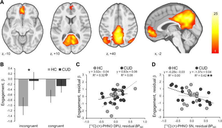
On average across participants and stimuli, the DMN (Fig. 2a) was significantly suppressed (displaying negative engagement associated with task events) during Stroop performance (main effect of stimuli: F1,30 = 7.06, P = 0.013; PBonferroni = 0.04). There was no difference in DMN suppression between incongruent and congruent events (within-subjects effect of stimulus type: F1,30 = 0.27, P = 0.61). There was no difference between CUD and HC in average DMN engagement (main effect of group: F1,30 = 3.12, P = 0.09) and no group-by-stimulus interaction (F1,30 = 2.76, P = 0.11) on DMN suppression. Within groups, DMN suppression was greater in relation to incongruent compared to congruent stimuli in HC (t15 = 2.91, P = 0.011) but not CUD (t15 = 0.22, P = 0.83) participants (Fig. 2b). In CUD participants, DMN engagement was not associated with cocaine-use measures (P > 0.5).
Fig. 2. The default mode network (DMN) identified by independent component analysis of Stroop fMRI data and relationships with [11C]-(+)-PHNO BPND.
a Spatial pattern of the visually selected DMN displayed at voxel-wise PFWE < 0.001 and cluster extent >200 contiguous voxels. b Suppression of the DMN in response to high-conflict incongruent events was greater in healthy comparison (HC) as compared to cocaine-use disorder (CUD) individuals; error bars indicate standard deviation. c In HC participants, lower D2R-related binding in the DPU was associated with greater DMN suppression (DPU and engagement residuals from regressions with SN BPND). d In CUD participants, greater D3R-related binding in the SN was associated with greater DMN suppression (SN and engagement residuals from regressions with DPU BPND). °P < 0.10; *P < 0.05; ** P < 0.01.
Differences in DMN engagement related to incongruent and congruent stimuli were not associated with [11C]-(+)-PHNO BPND values across participants or within groups (i.e., there were no interactions of DPU or SN covariates on stimulus or stimulus-by-group effects; P > 0.3). The time between PET and fMRI scans did not interact with covariates of D2R/D3R-related availability on DMN engagement between or within groups (P’s > 0.4). Lower D2R-related BPND in the DPU was associated with greater average (i.e., mean incongruent and congruent) DMN suppression but did not survive family-wise correction (main effect of DPU: F1,26 = 5.33, P = 0.029; PBonferroni = 0.087). This relationship did not differ between groups (group-by-DPU interaction: F1,26 = 1.89, P = 0.18), did not achieve significance within HC participants (F1,13 = 4.48, P = 0.054) and was not present in CUD participants (F1,13 = 0.75, P = 0.40) (Fig. 2c). An association between higher D3R-related BPND in the SN and greater average DMN suppression was not significant across participants (main effect of SN BPND: F1,26 = 3.07, P = 0.09) or different between groups (group-by-SN interaction: F1,26 = 0.38, P = 0.54); however, it was significant in CUD (F1,13 = 13.01, P = 0.003) and not present within HC participants (F1,13 = 0.30, P = 0.60) (Fig. 2d). Exploratory correlations between BPND in other regions commonly reported in [11C]-(+)-PHNO research and DMN engagement are provided in Supplementary Table S2.
Stroop performance
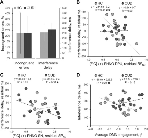
On average, participants committed errors on 22.7% (SD = 16.6) of incongruent trials, with an average interference delay of 271 ms (SD = 77). There were no differences between CUD and HC participants on Stroop performance (F1,30 = 1.07, P = 0.31) or group-by-behavior interaction (F1,30 = 1.07, P = 0.31) (Fig. 3a). In CUD, Stroop performance was not associated with cocaine-use measures (P > 0.06).
Fig. 3. Stroop performance and relationships with [11C]-(+)-PHNO BPND and DMN engagement.

a Incongruent errors and interference delays (incongruent minus congruent reaction times) did not differ between cocaine-use disorder (CUD) and healthy comparison (HC) participants. Error bars indicate standard deviation. b In HC participants, higher D2R-related binding in the dorsal putamen (DPU) was associated with shorter interference delays (DPU and delay residuals from regressions with BPND in the substantia nigra (SN) to reflect results of mixed model analyses). c In CUD, higher D3R-related binding in the SN was associated with shorter interference delays (SN and delay residuals from regressions with BPND in the DPU to reflect results of mixed model analyses). d In HC participants, greater average DMN engagement (i.e., less DMN suppression) was associated with reduced interference delays. *P < 0.05; **P < 0.01.
Across participants, D2R-related binding was associated with interference delays and not error rates (behavior-by-DPU interaction: F1,26 = 7.48, P = 0.011; PBonferroni = 0.033). CUD and HC participants differed in this association (group-by-behavior-by-DPU interaction: F1,26 = 8.54, P = 0.007; PBonferroni = 0.021), with greater D2R-related binding associated with shorter interference delays in HC participants (F1,13 = 11.97, P = 0.004) and no relation to delays in CUD participants (F1,13 = 0.25, P = 0.088) (Fig. 3b). There was no association between D3R-related binding and difference between Stroop performance across participants (behavior-by-SN interaction: F1,26 = 0.02, P = 0.90) or between groups (group-by-behavior-by-SN interaction: F1,26 = 3.64, P = 0.068). However, within groups, greater D3R-related binding was associated with shorter interference delays in CUD (F1,13 = 7.99, P = 0.014) but not in HC (F1,13 = 0.81, P = 0.38) participants (Fig. 3c). Additional exploratory correlations between BPND in other regions commonly reported in [11C]-(+)-PHNO research and interference engagement are provided in Supplementary Table S2. No relationships between D2R/D3R and incongruent error rates were observed across participants or within groups (P’s > 0.1).
There was no association between average DMN suppression and differences between Stroop performance measures across participants (behavior-by-DMN interaction: F1,26 = 0.26, P = 0.62), though a group difference in DMN associations with performance was significant (group-by-behavior-by-DMN interaction: F1,26 = 6.86, P = 0.014; PBonferroni = 0.042). Greater DMN suppression was associated with longer interference delays in HC (F1,14 = 4.60, P = 0.050) but not CUD (F1,14 = 2.49, P = 0.14) participants (Fig. 3d). No relationships between DMN suppression and incongruent error rates were observed across participants or within groups (P’s > 0.1).
Exploratory moderated mediation
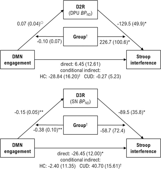
Moderated-mediation results are summarized in Fig. 4 and detailed in Supplementary Table S3. Results of post hoc within-group mediation models are shown in Supplementary Fig. S1. The model of D2R mediation yielded conditional effects such that DMN engagement had a significant indirect effect, through D2R, on Stroop interference delays only in HC (Β = −28.85, SE = 16.20, 95% CI = −70.36, −6.62). A significant index of moderated mediation (index = 28.57, SE = 16.53, 95% CI = 6.75, 71.81) indicated the D2R-related mediating effect between DMN and Stroop performance in HC differed from CUD participants. The model of D3R mediation revealed that DMN engagement had a significant indirect effect on Stroop performance only in CUD (Β = 40.70; SE = 15.61; 95%CI = 12.95, 74.37), and the index of moderated mediation (index = 43.10, SE = 16.84, 95% CI = 11.05, 77.03) indicated this significantly differed from HC participants.
Fig. 4. Moderated-mediation models.

The D2R-related mediation model (top) indicated a significant index of moderated mediation (index = 28.57, SE = 16.53, 95% CI = 6.75, 71.81) in which a conditional indirect effect of average engagement of the default mode network (DMN) on Stroop interference reaction times through D2R was significant in healthy comparison participants (HC) but not in individuals with a cocaine-use disorder (CUD). The D3R-related mediation model (bottom) indicated a significant moderated mediation (index = 43.10, SE = 16.84, 95%CI = 11.05, 77.03) in which a conditional indirect effect of DMN engagement on Stroop performance through D3R was significant in CUD but not HC individuals. Unstandardized coefficient values and standard error (Β(SE)) are shown for the moderated-mediation models. °P < 0.10, *P < 0.05; **P < 0.01. †95% confidence intervals of moderated mediation and conditional indirect effects indirect do not include zero, indicating a significant effect.
Discussion
The current study investigated relationships between D2R/D3R availability, DMN suppression, and behavioral performance during cognitive control in CUD. Consistent with prior [11C]-(+)-PHNO research in similarly sized samples of individuals using stimulants, CUD relative to HC participants had higher D3R-related availability in the SN while lower D2R-related availability in the DPU did not reach statistical significance [4, 5, 7]. Contrary to hypotheses, greater D2R-related availability was associated with less DMN suppression. However, consistent with hypotheses, greater D2R-related availability was associated with better Stroop performance (i.e., shorter interference delays) in HC individuals. In CUD participants, there were no relationships between D2R-related availability, DMN suppression, and behavioral performance. However, greater D3R availability in CUD was associated with greater DMN suppression and better Stroop performance. Exploratory moderated-mediation analyses indicated that D2R-related availability mediated the relationship between DMN suppression and interference delays in HC differently than CUD participants, in whom D3R-related availability mediated the relationship between DMN suppression and performance. Findings suggest that greater D3R-related availability may reflect activity through alternative functional mechanisms that compensate for deficient D2R-related signaling in CUD within the context of cognitive control.
D2R/D3R and DMN suppression during cognitive control
The DMN was suppressed on average during Stroop performance; however, suppression was significant within HC but not CUD individuals. Contrary to hypotheses and prior reports [30, 31], greater DMN suppression was linked to lower D2R-related availability across the study sample, and this relationship appeared strongest within the HC group (though did not achieve statistical significance). Research has suggested that the positive associations between D2R availability and increased brain activity during executive functioning may be dependent on subjective task difficulty [53]. Thus, one potential interpretation, consistent with behavioral finding of lower D2R-related binding related to longer interference delays in HC, is that low-D2R individuals experienced the Stroop task as more cognitively demanding in a manner linked to greater DMN suppression.
In CUD individuals, DMN suppression appeared to be generally blunted during the Stroop task, though only significantly differed from HC in response to the high-conflict incongruent events. Coordination between the DMN and executive function networks, typically indicated by greater DMN suppression under cognitive demands, may be dysregulated in CUD [54]. Thus, the lack of DMN suppression in CUD in the current study, in absence of performance impairments, may reflect this de-coupling between functional systems. However, associations between greater D3R-related binding and greater DMN suppression may also indicate increased activity through alternate processes to compensate for this de-coupling. Consistent with this interpretation is the observation that greater D3R-releated binding was also associated with shorter interference delays in CUD.
Stroop performance and D2R/D3R
Although there were no behavioral differences between CUD and HC in the current study, a dissociative relationship was observed between D2R and D3R availability and interference delays. In HC individuals, greater D2R-related binding was associated with shorter interference delays, consistent with the hypotheses. While research into the influence of dopaminergic agents on Stroop performance in healthy populations are mixed [55–57], greater availability of striatal D2R has been associated with improved cognitive performance more broadly [58]. In the current study, this association between D2R binding and cognitive performance was not present in CUD participants. Rather, in CUD individuals, shorter interference delays were related to greater D3R-related availability. Limited evidence of the influences of elevated D3R in CUD has indicated potential associations with greater impulsivity and more risky decision-making [7]. While this finding suggests a role for increased D3R reflecting a compensatory functional response to declining D2R in CUD, this relationship was observed in the absence of an association with D2R in CUD. One speculative interpretation is that the increases in D3R may reflect neuroadaptive changes of alternate functional mechanisms to compensate for losses in D2R-related functioning. This would be consistent with our prior findings using this task that despite equivalent performance in CUD and HC individuals, response times were differentially associated with distinct functional networks in each group [46]. However, it remains to be determined whether D3R-related support of cognitive-control performance in CUD occurs through appropriation of D2R-related processes or through distinct mechanisms.
D2R/D3R, DMN, and performance relationships
Exploratory moderated-mediation analyses indicated a dissociative role of D2R- and D3R-related intermediary functional processes in the pathway between DMN suppression and Stroop-related cognitive control in CUD and HC. In HC, associations between DMN suppression and interference delays were mediated through D2R-related binding as a potential indicator of greater DMN-anti-correlated (“task-on”) functional systems linked to D2R. In CUD, associations between DMN suppression and interference delays were mediated through D3R-related functional systems. Preclinical and human research have indicated that losses in D2R and gains in D3R may occur over the course of CUD [5, 59]; however, neither D2R- or D3R-related binding was related to CUD chronicity in the current study, and implications of higher D3R availability reflecting increased functioning of compensatory mechanisms remain speculative. Similarly, whether functional systems linked to D2R in HC individuals are appropriated by D3R signaling or circumvented by D3R-related mechanisms warrants further research.
To our knowledge, this is the first demonstration of dissociative D2R- and D3R-related mediation of links between DMN suppression and cognitive-control performance in individuals with CUD compared to healthy individuals. The mechanisms of D2R/D3R influence on cognitive control likely involve complex interactions with other neuromodulatory systems (e.g., serotonin and acetylcholine) throughout subcortical and cortical regions [60–65]. Similarly, the phasic activation and deactivation of dopamine receptors may have nonlinear effects on cognitive processes relative to baseline tonic dopamine functioning [66]. This complexity is demonstrated by evidence that both D3R agonism and antagonism may improve cognitive functioning, including in individuals with stimulant-use disorders [67–71]. The current findings lend some insight into these mechanisms, providing evidence linking greater D3R availability, DMN suppression, and cognitive-control performance in individuals with CUD.
Limitations
The small sample sizes in this multimodal study, while within the range of previous [11C]-(+)-PHNO research of stimulant-use disorders, may limit detection of fMRI-related differences. The use of [11C]-(+)-PHNO limited investigation to subcortical regions with relatively high concentrations of D2R and D3R. While this radiotracer allows dissociation of D2R- and D3R-related binding signals, evidence using a high-affinity, nonselective D2R/D3R radiotracers indicates alterations in extrastriatal and cortical regions in individuals with CUD [72, 73] that may also be influencing DMN suppression and cognitive performance. The Stroop task employed used silent responding during fMRI scanning; thus, performance during scanning was not directly assessed. Current analyses employed linear statistics to examine D2R/D3R relationships, and possible nonlinear relationships may exist between dopaminergic activity and cognitive and behavioral performance [66]. Prior research using the Stroop and other neurocognitive tasks suggests performance improvements over repeated testing may be linked to adaptions of neural processes to optimize behavior [74, 75], and future studies should examine potential D2R/D3R relationships with engagement of additional large-scale brain networks. Furthermore, links between dopamine D2R/D3R availability and task fatigue during Stroop performance have been previously reported [76], but were not directly examined in the current study. The current CUD sample represents a somewhat homogenous population with respect to disease severity (e.g., a history of at least 7 years, and at least twice-weekly use) and were all assessed following <20 days of abstinence, limiting generalizability to individuals at different stages of disease and recovery. Similarly, while dopaminergic alterations appear to be robust to different stimulants of abuse (e.g., cocaine, methamphetamine; [1]), research is required to investigate the generalizability of these functional implications in people with non-cocaine stimulant use. PET and fMRI scans were performed closer in time in individuals with CUD, and while analyses indicate that this did not influence current results, future multimodal research should be cautious to balance timing of scans. This investigation examined relationships between dopaminergic receptors, DMN suppression, and behavioral performance under cognitive-control demands using a simple event-related Stroop task. Further research using a range of cognitive-control-related tasks of varying difficulties (that may detect group differences in performance) is required to replicate and extend these findings within this domain of interest [77].
Conclusions
To our knowledge, this is the first report linking increased D3R-related receptor availability to improved neurocognitive performance in CUD. During performance of a Stroop task, greater D3R-related [11C]-(+)-PHNO binding in the SN was associated with greater DMN suppression and shorter interference delays in individuals with CUD. This relationship of increased availability, improved cognitive performance, and greater DMN suppression has been reported with respect to D2R in healthy populations, suggesting increases in D3R may reflect a potential functional compensation for deficient D2R-related processes in CUD. These findings provide additional insight into mixed neurocognitive profiles in individuals with stimulant-use disorders and indicate a potential mechanism of preclinical evidence that D3R partial agonists may have some efficacy in treating CUD [78, 79]. Research examining concurrent D2R and D3R links to additional cognitive domains implicated in addictions (e.g., reward processing), as well as potential relationships with other large-scale functional networks, may provide further insights into individual variability and pharmacological challenges for CUD.
Funding and disclosure
This research was funded by the National Institute on Drug Abuse (NIDA) (P20-DA027844, R03-DA027456, K01-DA042998, R01-DA039136). Additional financial support was provided by the State of Connecticut Department of Mental Health and Addiction Services and the National Center on Addiction and Substance Abuse. This publication was also made possible by CTSA Grant Number UL1 TR000142 from the National Center for Advancing Translational Science (NCATS), a component of NIH. Its contents are solely the responsibility of the authors and do not necessarily represent the official view of the funding agencies. MNP has received financial support or compensation for the following: MNP has consulted for and advised RiverMend Health, Opiant Pharmaceuticals, Idorsia, the Addiction Policy Forum and AXA; has received research support from the Mohegan Sun Casino and the National Center for Responsible Gaming; has participated in surveys, mailings or telephone consultations related to addictive disorders or other health topics; has consulted for or advised law offices and gambling entities on issues related to addictive disorders and behaviors; has provided clinical care in the Connecticut Department of Mental Health and Addiction Services Problem Gambling Services Program; has performed grant reviews; has edited journals and journal sections; has given academic lectures in grand rounds, CME events and other clinical or scientific venues; and has generated books or book chapters for publishers of mental health texts. MNP has participated in World Health Organization meetings relating to considerations on gaming and gambling. MNP has participated in research meetings and groups focused on problematic internet use including with respect to gaming with support from European and Asian funding agencies. REC has received research funding from Astellas, Astra Zeneca, BMS, Lilly, Pfizer, Taisho, and UCB. The authors declare no competing interests.
Supplementary information
Author contributions
All authors contributed to: the acquisition, analysis, or interpretation of data; drafting the work or revising it critically for important intellectual content; and provided final approval of the version to be published. PDW, RTM, REC, and MNP contributed substantially to the conception or design of the work and agree to be accountable for all aspects of the work in ensuring that questions related to the accuracy or integrity of any part of the work are appropriately investigated and resolved.
Footnotes
Publisher’s note Springer Nature remains neutral with regard to jurisdictional claims in published maps and institutional affiliations.
Supplementary information
Supplementary Information accompanies this paper at (10.1038/s41386-020-00874-7).
References
- 1.Nutt DJ, Lingford-Hughes A, Erritzoe D, Stokes PR. The dopamine theory of addiction: 40 years of highs and lows. Nat Rev Neurosci. 2015;16:305–12. doi: 10.1038/nrn3939. [DOI] [PubMed] [Google Scholar]
- 2.Volkow ND, Fowler JS, Wang GJ, Baler R, Telang F. Imaging dopamine’s role in drug abuse and addiction. Neuropharmacology. 2009;56:3–8. doi: 10.1016/j.neuropharm.2008.05.022. [DOI] [PMC free article] [PubMed] [Google Scholar]
- 3.Volkow N, Wang G-J, Fowler J, Logan J, Gatley S, Hitzemann R, et al. Decreased striatal dopaminergic responsiveness in detoxified cocaine-dependent subjects. Nature. 1997;386:830–33. doi: 10.1038/386830a0. [DOI] [PubMed] [Google Scholar]
- 4.Boileau I, Payer D, Houle S, Behzadi A, Rusjan PM, Tong J, et al. Higher binding of the dopamine D3 receptor-preferring ligand [11C]-(+)-propyl-hexahydro-naphtho-oxazin in methamphetamine polydrug users: a positron emission tomography study. J Neurosci. 2012;32:1353–59. doi: 10.1523/JNEUROSCI.4371-11.2012. [DOI] [PMC free article] [PubMed] [Google Scholar]
- 5.Matuskey D, Gallezot J-D, Pittman B, Williams W, Wanyiri J, Gaiser E, et al. Dopamine D3 receptor alterations in cocaine-dependent humans imaged with [11C](+)PHNO. Drug Alcohol Depend. 2014;139:100–5. doi: 10.1016/j.drugalcdep.2014.03.013. [DOI] [PMC free article] [PubMed] [Google Scholar]
- 6.Worhunsky PD, Matuskey D, Gallezot J-D, Gaiser EC, Nabulsi N, Angarita GA, et al. Regional and source-based patterns of [11C]-(+)-PHNO binding potential reveal concurrent alterations in dopamine D2 and D3 receptor availability in cocaine-use disorder. Neuroimage. 2017;148:343–51. doi: 10.1016/j.neuroimage.2017.01.045. [DOI] [PMC free article] [PubMed] [Google Scholar]
- 7.Payer DE, Behzadi A, Kish SJ, Houle S, Wilson AA, Rusjan PM, et al. Heightened D3 dopamine receptor levels in cocaine dependence and contributions to the addiction behavioral phenotype: a positron emission tomography study with [11C]-+-PHNO. Neuropsychopharmacology. 2014;39:321–28. doi: 10.1038/npp.2013.192. [DOI] [PMC free article] [PubMed] [Google Scholar]
- 8.Buckholtz JW, Treadway MT, Cowan RL, Woodward ND, Li R, Ansari MS, et al. Dopaminergic network differences in human impulsivity. Science. 2010;329:532–32. doi: 10.1126/science.1185778. [DOI] [PMC free article] [PubMed] [Google Scholar]
- 9.Seeman P. Parkinson’s disease treatment may cause impulse–control disorder via dopamine D3 receptors. Synapse. 2015;69:183–89. doi: 10.1002/syn.21805. [DOI] [PubMed] [Google Scholar]
- 10.Volkow ND, Wang G-J, Telang F, Fowler JS, Logan J, Childress A-R, et al. Cocaine cues and dopamine in dorsal striatum: mechanism of craving in cocaine addiction. J Neurosci. 2006;26:6583–88. doi: 10.1523/JNEUROSCI.1544-06.2006. [DOI] [PMC free article] [PubMed] [Google Scholar]
- 11.Heidbreder CA, Gardner EL, Xi Z-X, Thanos PK, Mugnaini M, Hagan JJ, et al. The role of central dopamine D3 receptors in drug addiction: a review of pharmacological evidence. Brain Res Rev. 2005;49:77–105. doi: 10.1016/j.brainresrev.2004.12.033. [DOI] [PMC free article] [PubMed] [Google Scholar]
- 12.Cropley VL, Fujita M, Innis RB, Nathan PJ. Molecular imaging of the dopaminergic system and its association with human cognitive function. Biol Psychiatry. 2006;59:898–907. doi: 10.1016/j.biopsych.2006.03.004. [DOI] [PubMed] [Google Scholar]
- 13.Ott T, Nieder A. Dopamine and cognitive control in prefrontal cortex. Trends Cogn Sci. 2019;23:213–34. doi: 10.1016/j.tics.2018.12.006. [DOI] [PubMed] [Google Scholar]
- 14.Durstewitz D, Seamans JK. The dual-state theory of prefrontal cortex dopamine function with relevance to catechol-o-methyltransferase genotypes and schizophrenia. Biol Psychiatry. 2008;64:739–49. doi: 10.1016/j.biopsych.2008.05.015. [DOI] [PubMed] [Google Scholar]
- 15.Spronk DB, van Wel JH, Ramaekers JG, Verkes RJ. Characterizing the cognitive effects of cocaine: a comprehensive review. Neurosci Biobehav Rev. 2013;37:1838–59. doi: 10.1016/j.neubiorev.2013.07.003. [DOI] [PubMed] [Google Scholar]
- 16.Lee B, London ED, Poldrack RA, Farahi J, Nacca A, Monterosso JR, et al. Striatal dopamine d2/d3 receptor availability is reduced in methamphetamine dependence and is linked to impulsivity. J Neurosci. 2009;29:14734–40. doi: 10.1523/JNEUROSCI.3765-09.2009. [DOI] [PMC free article] [PubMed] [Google Scholar]
- 17.Frazer KM, Richards Q, Keith DR. The long-term effects of cocaine use on cognitive functioning: a systematic critical review. Behav Brain Res. 2018;348:241–62. doi: 10.1016/j.bbr.2018.04.005. [DOI] [PubMed] [Google Scholar]
- 18.Frazer KM, Manly JJ, Downey G, Hart CL. Assessing cognitive functioning in individuals with cocaine use disorder. J Clin Exp Neuropsychol. 2018;40:619–32. doi: 10.1080/13803395.2017.1403569. [DOI] [PubMed] [Google Scholar]
- 19.Andrews-Hanna JR. The brain’s default network and its adaptive role in internal mentation. Neuroscientist. 2012;18:251–70. doi: 10.1177/1073858411403316. [DOI] [PMC free article] [PubMed] [Google Scholar]
- 20.Buckner RL, Andrews-Hanna JR, Schacter DL. The brain’s default network: anatomy, function, and relevance to disease. Ann NY Acad Sci. 2008;1124:138. doi: 10.1196/annals.1440.011. [DOI] [PubMed] [Google Scholar]
- 21.Zhang R, Volkow ND. Brain default-mode network dysfunction in addiction. Neuroimage. 2019;200:313–31. doi: 10.1016/j.neuroimage.2019.06.036. [DOI] [PubMed] [Google Scholar]
- 22.Sheline YI, Barch DM, Price JL, Rundle MM, Vaishnavi SN, Snyder AZ, et al. The default mode network and self-referential processes in depression. Proc Natl Acad Sci USA. 2009;106:1942–47. doi: 10.1073/pnas.0812686106. [DOI] [PMC free article] [PubMed] [Google Scholar]
- 23.Garrity AG, Pearlson GD, McKiernan K, Lloyd D, Kiehl KA, Calhoun VD. Aberrant “default mode” functional connectivity in schizophrenia. Am J Psychiatry. 2007;164:450–57. doi: 10.1176/ajp.2007.164.3.450. [DOI] [PubMed] [Google Scholar]
- 24.Peterson B, Potenza M, Wang Z, Zhu H, Martin A, Marsh R, et al. A functional MRI study of the effects of psychostimulants on default-mode processing during performance of the word-color stroop task in youth with ADHD. Am J Psychiatry. 2009;166:1286–94. doi: 10.1176/appi.ajp.2009.08050724. [DOI] [PMC free article] [PubMed] [Google Scholar]
- 25.Anticevic A, Cole MW, Murray JD, Corlett PR, Wang X-J, Krystal JH. The role of default network deactivation in cognition and disease. Trends Cogn Sci. 2012;16:584–92. doi: 10.1016/j.tics.2012.10.008. [DOI] [PMC free article] [PubMed] [Google Scholar]
- 26.Binder JR. Task-induced deactivation and the” resting” state. Neuroimage. 2012;62:1086–91. doi: 10.1016/j.neuroimage.2011.09.026. [DOI] [PMC free article] [PubMed] [Google Scholar]
- 27.Nagano‐Saito A, Lissemore JI, Gravel P, Leyton M, Carbonell F, Benkelfat C. Posterior dopamine D2/3 receptors and brain network functional connectivity. Synapse. 2017;71:e21993. doi: 10.1002/syn.21993. [DOI] [PubMed] [Google Scholar]
- 28.Nagano-Saito A, Liu J, Doyon J, Dagher A. Dopamine modulates default mode network deactivation in elderly individuals during the Tower of London task. Neurosci Lett. 2009;458:1–5. doi: 10.1016/j.neulet.2009.04.025. [DOI] [PubMed] [Google Scholar]
- 29.Dang LC, Donde A, Madison C, O’Neil JP, Jagust WJ. Striatal dopamine influences the default mode network to affect shifting between object features. J Cogn Neurosci. 2012;24:1960–70. doi: 10.1162/jocn_a_00252. [DOI] [PMC free article] [PubMed] [Google Scholar]
- 30.Nour MM, Dahoun T, McCutcheon RA, Adams RA, Wall MB, Howes OD. Task-induced functional brain connectivity mediates the relationship between striatal D2/3 receptors and working memory. Elife. 2019;8:e45045. doi: 10.7554/eLife.45045. [DOI] [PMC free article] [PubMed] [Google Scholar]
- 31.Tomasi D, Volkow ND, Wang R, Telang F, Wang G-J, Chang L, et al. Dopamine transporters in striatum correlate with deactivation in the default mode network during visuospatial attention. PloS One. 2009;4:e6102. doi: 10.1371/journal.pone.0006102. [DOI] [PMC free article] [PubMed] [Google Scholar]
- 32.Tomasi D, Wang GJ, Wang R, Caparelli EC, Logan J, Volkow ND. Overlapping patterns of brain activation to food and cocaine cues in cocaine abusers: association to striatal D2/D3 receptors. Hum Brain Mapp. 2015;36:120–36. doi: 10.1002/hbm.22617. [DOI] [PMC free article] [PubMed] [Google Scholar]
- 33.Matuskey D, Luo X, Zhang S, Morgan PT, Abdelghany O, Malison RT, et al. Methylphenidate remediates error-preceding activation of the default mode brain regions in cocaine-addicted individuals. Psychiatry Res Neuroimaging. 2013;214:116–21. doi: 10.1016/j.pscychresns.2013.06.009. [DOI] [PMC free article] [PubMed] [Google Scholar]
- 34.Payer D, Balasubramaniam G, Boileau I. What is the role of the D3 receptor in addiction? A mini review of PET studies with [11C]-(+)-PHNO. Prog Neuropsychopharmacol Biol Psychiatry. 2014;52:4–8. doi: 10.1016/j.pnpbp.2013.08.012. [DOI] [PubMed] [Google Scholar]
- 35.First MB, Spitzer RL, Miriam G, Williams JBW. Structured clinical interview for DSM-IV-TR axis I disorders, research version, patient edition. (SCID-I/P). New York: Biometrics Research, New York State Psychiatric Institute; 2002.
- 36.Zachary RA, Shipley WC. hipley Institute of Living Scale: revised manual. Los Angeles: Western Psychological Services; 1986. [Google Scholar]
- 37.Le Foll B, Guranda M, Wilson AA, Houle S, Rusjan PM, Wing VC, et al. Elevation of dopamine induced by cigarette smoking: novel insights from a [11 C]-(+)-PHNO PET study in humans. Neuropsychopharmacology. 2014;39:415–24. doi: 10.1038/npp.2013.209. [DOI] [PMC free article] [PubMed] [Google Scholar]
- 38.Tanabe J, Nyberg E, Martin LF, Martin J, Cordes D, Kronberg E, et al. Nicotine effects on default mode network during resting state. Psychopharmacology. 2011;216:287–95. doi: 10.1007/s00213-011-2221-8. [DOI] [PMC free article] [PubMed] [Google Scholar]
- 39.Gallezot J-D, Zheng M-Q, Lim K, Lin S-F, Labaree D, Matuskey D, et al. Lin S-f, Labaree D, Matuskey D, et al. Parametric imaging and test–retest variability of 11C-(+)-PHNO binding to D2/D3 dopamine receptors in humans on the high-resolution research tomograph PET Scanner. J Nucl Med. 2014;55:960–66. doi: 10.2967/jnumed.113.132928. [DOI] [PMC free article] [PubMed] [Google Scholar]
- 40.Wilson AA, McCormick P, Kapur S, Willeit M, Garcia A, Hussey D, et al. Radiosynthesis and evaluation of [11C]-(+)-4-Propyl-3, 4, 4a, 5, 6, 10b-hexahydro-2 H-naphtho [1, 2-b][1, 4] oxazin-9-ol as a potential radiotracer for in vivo imaging of the dopamine D2 high-affinity state with positron emission tomography. J Med Chem. 2005;48:4153–60. doi: 10.1021/jm050155n. [DOI] [PubMed] [Google Scholar]
- 41.Mawlawi O, Martinez D, Slifstein M, Broft A, Chatterjee R, Hwang D-R, et al. Imaging human mesolimbic dopamine transmission with positron emission tomography: I. accuracy and precision of D2 receptor parameter measurements in ventral striatum. J Cereb Blood Flow Metab. 2001;21:1034–57. doi: 10.1097/00004647-200109000-00002. [DOI] [PubMed] [Google Scholar]
- 42.Searle G, Beaver JD, Comley RA, Bani M, Tziortzi A, Slifstein M, et al. Imaging dopamine D3 receptors in the human brain with positron emission tomography,[11C]PHNO, and a selective D3 receptor antagonist. Biol Psychiatry. 2010;68:392–99. doi: 10.1016/j.biopsych.2010.04.038. [DOI] [PubMed] [Google Scholar]
- 43.Searle GE, Beaver JD, Tziortzi A, Comley RA, Bani M, Ghibellini G, et al. Mathematical modelling of [11C]-(+)-PHNO human competition studies. Neuroimage. 2013;68:119–32. doi: 10.1016/j.neuroimage.2012.11.033. [DOI] [PubMed] [Google Scholar]
- 44.Tziortzi AC, Searle GE, Tzimopoulou S, Salinas C, Beaver JD, Jenkinson M, et al. Imaging dopamine receptors in humans with [11C]-(+)-PHNO: dissection of D3 signal and anatomy. Neuroimage. 2011;54:264–77. doi: 10.1016/j.neuroimage.2010.06.044. [DOI] [PubMed] [Google Scholar]
- 45.Leung H-C, Skudlarski P, Gatenby JC, Peterson BS, Gore JC. An event-related functional MRI study of the Stroop color word interference task. Cereb Cortex. 2000;10:552–60. doi: 10.1093/cercor/10.6.552. [DOI] [PubMed] [Google Scholar]
- 46.Worhunsky PD, Stevens MC, Carroll KM, Rounsaville BJ, Calhoun VD, Pearlson GD, et al. Functional brain networks associated with cognitive control, cocaine dependence, and treatment outcome. Psychol Addict Behav. 2013;27:477. doi: 10.1037/a0029092. [DOI] [PMC free article] [PubMed] [Google Scholar]
- 47.Brown GG, Kindermann SS, Siegle GJ, Granholm E, Wong EC, Buxton RB. Brain activation and pupil response during covert performance of the Stroop Color Word task. J Int Neuropsychological Soc. 1999;5:308–19. doi: 10.1017/S1355617799544020. [DOI] [PubMed] [Google Scholar]
- 48.Calhoun VD, Adali T, Pearlson GD, Pekar JJ. A method for making group inferences from functional MRI data using independent component analysis. Hum Brain Mapp. 2001;14:140–51. doi: 10.1002/hbm.1048. [DOI] [PMC free article] [PubMed] [Google Scholar]
- 49.Li YO, Adali T, Calhoun VD. Estimating the number of independent components for functional magnetic resonance imaging data. Hum Brain Mapp. 2007;28:1251–66. doi: 10.1002/hbm.20359. [DOI] [PMC free article] [PubMed] [Google Scholar]
- 50.Bell AJ, Sejnowski TJ. An information-maximization approach to blind separation and blind deconvolution. Neural Comput. 1995;7:1129–59. doi: 10.1162/neco.1995.7.6.1129. [DOI] [PubMed] [Google Scholar]
- 51.Himberg J, Hyvärinen A, Esposito F. Validating the independent components of neuroimaging time series via clustering and visualization. NeuroImage. 2004;22:1214–22. doi: 10.1016/j.neuroimage.2004.03.027. [DOI] [PubMed] [Google Scholar]
- 52.Hayes AF. Introduction to mediation, moderation, and conditional process analysis: a regression-based approach. New York, NY: Guilford Publications; 2017. [Google Scholar]
- 53.Salami A, Garrett DD, Wåhlin A, Rieckmann A, Papenberg G, Karalija N, et al. Dopamine D2/3 binding potential modulates neural signatures of working memory in a load-dependent fashion. J Neurosci. 2019;39:537–47. doi: 10.1523/JNEUROSCI.1493-18.2018. [DOI] [PMC free article] [PubMed] [Google Scholar]
- 54.Liang X, He Y, Salmeron BJ, Gu H, Stein EA, Yang Y. Interactions between the salience and default-mode networks are disrupted in cocaine addiction. J Neurosci. 2015;35:8081–90. doi: 10.1523/JNEUROSCI.3188-14.2015. [DOI] [PMC free article] [PubMed] [Google Scholar]
- 55.Scholes KE, Harrison BJ, O’Neill BV, Leung S, Croft RJ, Pipingas A, et al. Acute serotonin and dopamine depletion improves attentional control: findings from the stroop task. Neuropsychopharmacology. 2007;32:1600. doi: 10.1038/sj.npp.1301262. [DOI] [PubMed] [Google Scholar]
- 56.Curley LE, Kydd RR, Kirk IJ, Russell BR. Using fMRI to compare the effects of benzylpiperazine with dexamphetamine—their differences during the Stroop paradigm. J Integr Neurosci. 2016;15:109–22. doi: 10.1142/S0219635216500084. [DOI] [PubMed] [Google Scholar]
- 57.Roesch-Ely D, Scheffel H, Weiland S, Schwaninger M, Hundemer H-P, Kolter T, et al. Differential dopaminergic modulation of executive control in healthy subjects. Psychopharmacology. 2005;178:420–30. doi: 10.1007/s00213-004-2027-z. [DOI] [PubMed] [Google Scholar]
- 58.Volkow ND, Gur RC, Wang G-J, Fowler JS, Moberg PJ, Ding Y-S, et al. Association between decline in brain dopamine activity with age and cognitive and motor impairment in healthy individuals. Am J Psychiatry. 1998;155:344–49. doi: 10.1176/ajp.155.10.1325. [DOI] [PubMed] [Google Scholar]
- 59.Nader MA, Morgan D, Gage HD, Nader SH, Calhoun TL, Buchheimer N, et al. PET imaging of dopamine D2 receptors during chronic cocaine self-administration in monkeys. Nat Neurosci. 2006;9:1050–56. doi: 10.1038/nn1737. [DOI] [PubMed] [Google Scholar]
- 60.Daw ND, Kakade S, Dayan P. Opponent interactions between serotonin and dopamine. Neural Netw. 2002;15:603–16. doi: 10.1016/S0893-6080(02)00052-7. [DOI] [PubMed] [Google Scholar]
- 61.Fujishiro H, Umegaki H, Suzuki Y, Oohara-Kurotani S, Yamaguchi Y, Iguchi A. Dopamine D2 receptor plays a role in memory function: implications of dopamine–acetylcholine interaction in the ventral hippocampus. Psychopharmacology. 2005;182:253–61. doi: 10.1007/s00213-005-0072-x. [DOI] [PubMed] [Google Scholar]
- 62.Calabresi P, Picconi B, Parnetti L, Di, Filippo M. A convergent model for cognitive dysfunctions in Parkinson’s disease: the critical dopamine–acetylcholine synaptic balance. Lancet Neurol. 2006;5:974–83. doi: 10.1016/S1474-4422(06)70600-7. [DOI] [PubMed] [Google Scholar]
- 63.Briand LA, Gritton H, Howe WM, Young DA, Sarter M. Modulators in concert for cognition: modulator interactions in the prefrontal cortex. Prog Neurobiol. 2007;83:69–91. doi: 10.1016/j.pneurobio.2007.06.007. [DOI] [PMC free article] [PubMed] [Google Scholar]
- 64.Cools R. Dopaminergic control of the striatum for high-level cognition. Curr Opin Neurobiol. 2011;21:402–07. doi: 10.1016/j.conb.2011.04.002. [DOI] [PubMed] [Google Scholar]
- 65.Di Giovanni G, Di Matteo V, Pierucci M, Esposito E. Serotonin–dopamine interaction: electrophysiological evidence. Prog Brain Res. 2008;172:45–71. doi: 10.1016/S0079-6123(08)00903-5. [DOI] [PubMed] [Google Scholar]
- 66.Cools R, D’Esposito M. Inverted-U–shaped dopamine actions on human working memory and cognitive control. Biol Psychiatry. 2011;69:e113–25. doi: 10.1016/j.biopsych.2011.03.028. [DOI] [PMC free article] [PubMed] [Google Scholar]
- 67.Sokoloff P, Le, Foll B. The dopamine D3 receptor, a quarter century later. Eur J Neurosci. 2017;45:2–19. doi: 10.1111/ejn.13390. [DOI] [PubMed] [Google Scholar]
- 68.Ersche KD, Roiser JP, Lucas M, Domenici E, Robbins TW, Bullmore ET. Peripheral biomarkers of cognitive response to dopamine receptor agonist treatment. Psychopharmacology. 2011;214:779–89. doi: 10.1007/s00213-010-2087-1. [DOI] [PMC free article] [PubMed] [Google Scholar]
- 69.Nakajima S, Gerretsen P, Takeuchi H, Caravaggio F, Chow T, Le Foll B, et al. The potential role of dopamine D3 receptor neurotransmission in cognition. Eur Neuropsychopharmacol. 2013;23:799–813. doi: 10.1016/j.euroneuro.2013.05.006. [DOI] [PMC free article] [PubMed] [Google Scholar]
- 70.Keck TM, John WS, Czoty PW, Nader MA, Newman AH. Identifying medication targets for psychostimulant addiction: unraveling the dopamine D3 receptor hypothesis. J Med Chem. 2015;58:5361–80. doi: 10.1021/jm501512b. [DOI] [PMC free article] [PubMed] [Google Scholar]
- 71.Moeller SJ, Honorio J, Tomasi D, Parvaz MA, Woicik PA, Volkow ND, et al. Methylphenidate enhances executive function and optimizes prefrontal function in both health and cocaine addiction. Cereb Cortex. 2014;24:643–53. doi: 10.1093/cercor/bhs345. [DOI] [PMC free article] [PubMed] [Google Scholar]
- 72.Narendran R, Mason NS, Himes ML, Frankle WG. Imaging cortical dopamine transmission in cocaine dependence: a [11C] FLB 457-amphetamine positron emission tomography (PET) study. Biol Psychiatry. 2020. 10.1016/j.biopsych.2020.04.001. [Epub ahead of print]. [DOI] [PMC free article] [PubMed]
- 73.Fotros A, Casey KF, Larcher K, Verhaeghe JA, Cox SM, Gravel P, et al. Cocaine cue-induced dopamine release in amygdala and hippocampus: a high-resolution PET [18 F] fallypride study in cocaine dependent participants. Neuropsychopharmacology. 2013;38:1780–88. doi: 10.1038/npp.2013.77. [DOI] [PMC free article] [PubMed] [Google Scholar]
- 74.Chen Z, Lei X, Ding C, Li H, Chen A. The neural mechanisms of semantic and response conflicts: an fMRI study of practice-related effects in the Stroop task. Neuroimage. 2013;66:577–84. doi: 10.1016/j.neuroimage.2012.10.028. [DOI] [PubMed] [Google Scholar]
- 75.Kelly AC, Garavan H. Human functional neuroimaging of brain changes associated with practice. Cereb Cortex. 2004;15:1089–102. doi: 10.1093/cercor/bhi005. [DOI] [PubMed] [Google Scholar]
- 76.Moeller S, Tomasi D, Honorio J, Volkow N, Goldstein R. Dopaminergic involvement during mental fatigue in health and cocaine addiction. Transl Psychiatry. 2012;2:e176. doi: 10.1038/tp.2012.110. [DOI] [PMC free article] [PubMed] [Google Scholar]
- 77.Tasks NAMHCWo. Criteria MfRD. Bethesda, MD: National Institute of Mental Health; 2016.
- 78.Powell GL, Bonadonna JP, Vannan A, Xu K, Mach RH, Luedtke RR, et al. Dopamine D3 receptor partial agonist LS-3-134 attenuates cocaine-motivated behaviors. Pharmacol Biochem Behav. 2018;175:123–29. doi: 10.1016/j.pbb.2018.10.002. [DOI] [PubMed] [Google Scholar]
- 79.Roman V, Gyertyan I, Saghy K, Kiss B, Szombathelyi Z. Cariprazine (RGH-188), a D 3-preferring dopamine D 3/D 2 receptor partial agonist antipsychotic candidate demonstrates anti-abuse potential in rats. Psychopharmacology. 2013;226:285–93. doi: 10.1007/s00213-012-2906-7. [DOI] [PubMed] [Google Scholar]
Associated Data
This section collects any data citations, data availability statements, or supplementary materials included in this article.


