



Abstract
Rationale and Objectives:
Mobile mammography units provide preventive health care to patients facing barriers to annual screening. This study reviews the outcomes of a mobile mammography service during a recent five-year period.
Materials and Methods:
This retrospective study analyzed the examinations by mobile mammography during a five-year period (9,327 examinations). The patients recalled, biopsies performed, and cancers detected were tallied. The race, age, breast cancer size, lymph node involvement, and metastases were recorded. The PPV and CDR metrics were calculated as outlined by the ACR BI-RADS Atlas.
Results:
The program identified cancer in 14 cases (CDR = 1.5 per 1,000 examinations [95% CI, 0.9-2.5]) with 11 being invasive. The majority of these cancers were small and of low stage. Lymph node status was determined in 11 of the 14 cases (1 as N1mi, 5 as N0, 4 as N1,1 as N2a). Abnormalities led to 1,686 examinations recalled (RR = 17.8%; PPV 1 = 0.8% [95% CI, 0.5-1.4%]). 101 were recommended for biopsy (PPV 2 = 13.9% [95% CI, 8.4-21.9%]) and 98 pursued biopsy (PPV 3 = 14.3% [95% CI, 8.7-22.6%]). Patient age ranged from 41 to 67 years with an average of 50.6 years.
Conclusions:
The program detected many cancers in an asymptomatic population facing barriers to breast cancer screening. These findings are underscored by the cancers detected at an early stage with a favorable prognosis and support the need for the development of similar programs.
Keywords: Breast cancer, breast cancer screening, mobile mammography
Introduction:
Breast cancer is the second most common cancer in the world, the most frequent cancer-related cause of death in women within less developed regions of the world, and the second most frequent cancer-related cause of death in the remaining developed regions [1]. Within the United States, breast cancer accounts for nearly one in three cancers diagnosed in women [2].
Screening for breast cancer remains a key component of women’s health as screening mammography reduces breast cancer mortality [3–6]. Lack of participation in screening mammography is linked to social disparities such as lower income levels, lack of transportation, unemployment, less access to care, and less expensive housing [7]. These considerations are components of overall socioeconomic status. Decreased socioeconomic status is attributed to a lower number of women who will receive screening mammography and potential follow-up diagnostic imaging [8–11]. A patient’s socioeconomic status plays a critical role in the ability of a patient to seek preventive care, and socioeconomic status serves as an independent predictor of breast cancer stage at diagnosis [12]. Mobile mammography is one of the best strategies to target women of lower socioeconomic status to increase access [10, 13–18]. Mobile mammography is also associated with an increased participation rate as women are more likely to undergo mammography within three months if offered a mobile option and health education [17, 19]. Encouraging participation with screening is critical to maximizing the chance of detecting breast cancer while it is still at a low stage.
Several reports have demonstrated the value of mobile mammography to increase screening participation and highlight demographics that particularly benefit from its use [10, 13–17, 19]. However, not much information is available concerning the Breast Imaging Reporting and Data System (BI-RADS) audit outcomes or the pathological outcomes of these screened patients [20]. It is the hope of the authors that this review will contribute toward that goal.
Project VALET (Valuable Area Life-Saving Exams in Town) is a mobile mammography screening service provided by a tertiary cancer center. This program operates on an annual budget of 1.6-2 million dollars and targets underserved women in the greater Houston metropolitan area who face barriers to breast cancer screening. These financial barriers relate to the cost of travel, navigation, insurance, appointment availability, and the costs of screening and diagnostic procedures. The patients in some instances have also encountered communication barriers that stem from their primary language or changes in their addresses. This study examines Project VALET’s outcomes over a recent five-year period.
Materials and Methods:
This was an institutional review board-approved, Health Insurance Portability and Accountability Act (HIPAA) compliant retrospective case review. The primary objective was to determine the number of breast cancers detected by screening mammography and to examine the stage of the detected cancers. The secondary objective was to determine the recall rate and to identify patient characteristics that may be associated with benefits from screening mammography. Patients of all ages and of any race/ethnicity who participated in the Project VALET program during a recent five-year period spanning October 1st, 2012 to September 30th, 2017 were identified. Clinicians at health clinics within the greater Houston metropolitan area referred these participating patients. The majority of the referring clinicians were physicians but occasionally a referring clinic designated a nurse practitioner or physician assistant as a referring clinician, according to their workflow. If a patient reported a symptomatic complaint such as focal pain, a palpable abnormality, nipple discharge, or skin changes, the patient became ineligible for Project VALET and was instead referred to another facility for a diagnostic mammography examination. The only exception to this guideline was if the patient was experiencing generalized soreness in conjunction with her menstrual cycle that had been previously established by the clinic and the patient.
On the screening appointment day, the patients first met with a representative from the mammography screening program inside their health clinics. The representative was bilingual to assist in gathering a pertinent history, obtaining consent, and signing appropriate release forms. The representative established if the visit was a baseline screening examination. If the patient had prior mammography examinations then a release form was obtained to acquire the comparison images. Up to 14 business days were allowed for older images to arrive before the current images were submitted for interpretation.
Full field digital mammography was acquired on two separate mobile mammography vans. Each mobile van utilized a single Hologic Selenia S digital mammographic unit with R2 Cernova™ computer-aided detection (CAD), accredited by the American College of Radiology (ACR) (Selenia, Hologic, Marlborough, MA) (Fig. 1). Mammography technologists in conjunction with the appropriate staff from the department of imaging physics performed quality control on the digital units. Image interpretation was performed by 19 radiologists with fellowship training in breast imaging.
Figure 1:
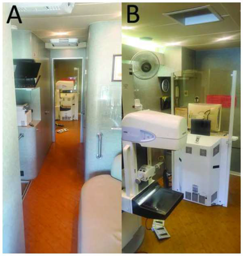
Photographs from the interior of a mobile mammography van. A, View of the patient waiting area, corridor, and mammography suite. B, View of the mammography suite with the breast imaging unit (Selenia, Hologic, Marlborough, MA).
The referring clinicians and patients were sent result letters between 2 and 14 days after screening. The clinicians then contacted the patients if screening mammography identified findings requiring additional diagnostic work-ups. Each facility participating in the program referred diagnostic work-ups to a facility within the greater metropolitan area, based on considerations such as location and funding. Patients sought diagnostic mammograms at four additional facilities within the local area based on their preferences. Several medical oncologists affiliated with the main tertiary cancer center also staff a local county health organization. Due to this affiliation, this local county health organization was included in the institutional review board approval to assist in gathering pathologic outcomes but clinicians from the local county health organization did not interpret screening mammograms for this project.
The number of patients who underwent screening mammography and the number of patients recalled due to possible abnormalities identified during screening mammography were tallied. Written reports from subsequent biopsies which demonstrated malignancy were obtained to track patient outcomes. The number of biopsies performed and the number of cancers detected from these biopsies were recorded. The positive predictive value (PPV) metrics and cancer detection rate (CDR) were calculated as outlined by the ACR BI-RADS [20]. These metrics were also calculated for the same set of radiologists interpreting screening mammography at a permanent site during the same time period to evaluate for potential differences in radiologist performance with respect to mobile mammography.
The medical records and the mammographic images of the patients diagnosed with cancer were retrospectively reviewed. The patients’ race/ethnicity, and age at the time of the cancer diagnosis were recorded. Breast cancer size was recorded by imaging. Regional lymph node involvement and distant metastases were reviewed and the stage was recorded, when available. The tumor histopathologic profile, nuclear grade, estrogen receptor (ER) status, progesterone receptor (PR) status, and human epidermal growth factor receptor 2 (HER2/neu) overexpression were recorded, when available.
Results:
A total of 9,327 screening mammography examinations were performed during the five year period from October 1st, 2012 to September 30th, 2017. This pool of examinations examined 7,391 unique women. Of these patients, 5,871 (79.4%) of the women received a single screening exam during the five year time period. Patients who received two examinations, three examinations, four examinations, five examinations, and six examinations accounted for 1,190 (16.1%) patients, 254 (3.4%) patients, 68 (0.9%) patients, 6 (0.1%) patients, and 2 (less than 0.1%) patients, respectively.
The age and the race/ethnicity of the patients were recorded. Patients between 40-44 years of age made up 32.7% of the patients screened. Patients between 45-49 years of age comprised 26.9% of the screened population. Patients over the age of 50 years accounted for 39.9% of the patients screened. The remaining patients less than 40 years of age made up 0.5% of the population. Patients of Hispanic, Black, Asian, White, and other race/ethnicity comprised 76%, 10%, 8%, 4% and 2% of the total population, respectively.
Mammography screening led to 1,686 examinations recalled due to mammographic abnormalities with a recall rate of 17.8%. The number of patients lost to follow-up is not available for the first year of the study. However, an average of 44 patients were lost to follow-up for the remaining four years. Of these patients, 101 women were recommended for biopsy and 98 of these women elected to pursue biopsy. Biopsy demonstrated evidence of malignancy in 14 cases with 11 of the malignancies found to be invasive (Table 1). The cancer detection rate was 1.5 cancers per 1,000 screening mammograms (95% confidence interval [CI], 0.9-2.5). The positive predictive value of abnormal interpretation (PPV1) was 14 cancers detected / 1686 patients recalled (0.8%) (95% CI, 0.5-1.4%). The positive predictive value of recommendations for tissue diagnosis (PPV2) was 14 cancers detected / 101 biopsies recommended (13.9%) (95% CI, 8.4-21.9%). The positive predictive value of biopsies performed (PPV3) was 14 cancers detected / 98 biopsies performed (14.3%) (95% CI, 8.7-22.6%).
Table 1:
Screen-Detected Breast Cancer Patients
| Age | Race | Cancer Type | Size (cm) | Grade | ER | PR | HER2 | T | N | M | Stage |
|---|---|---|---|---|---|---|---|---|---|---|---|
| 40 | Hispanic | IDC, DCIS | 1.3 | 2 | + | + | − | pT1c | N0 | M0 | IA |
| 41 | Black | IDC, DCIS | 0.5 | 2 | + | + | − | pT1c | N1mi | M0 | IB |
| 41 | Black | IDC, DCIS | 1.5 | 1 | N/A | N/A | N/A | pT1mi | N0 | M0 | IA |
| 44 | Hispanic | IDC, DCIS | 1.5 | 2 | + | + | − | pT1c | N1a | M0 | IIA |
| 44 | Hispanic | IDC, DCIS | 1.8 | 2 | + | − | + | pT1a | N2a | M0 | IIIA |
| 48 | Hispanic | DCIS | 4.4 | 3 | + | + | N/A | pTis | Nx | M0 | 0 |
| 50 | Hispanic | IDC, DCIS | 1.7 | 2 | + | + | − | cT1c | N0 | M0 | IA |
| 53 | Hispanic | ILC | 1.4 | 2 | + | − | − | pT1c | N0 | M0 | IA |
| 54 | Hispanic | IDC | 0.6 | 2 | N/A | N/A | N/A | cT1b | N/A | N/A | N/A |
| 54 | Hispanic | IDC | 2.1 | 3 | + | + | − | cT2 | N1 | N/A | N/A |
| 55 | Hispanic | ILC | 9.1 | 1 | + | + | − | pT1a | N0 | M0 | IA |
| 57 | Asian | DCIS | 2.7 | 2 | N/A | N/A | N/A | cTis | N/A | N/A | N/A |
| 60 | Black | IDC, DCIS | 3.0 | 3 | + | + | − | cT2 | N1 | M0 | IIB |
| 67 | Hispanic | DCIS | 0.5 | 1 | N/A | N/A | N/A | cTis | N1 | N/A | N/A |
Age is reported in years. Size is reported by imaging. ER = Estrogen receptor. PR = Progesterone receptor. HER2 = Human epidermal growth factor receptor 2. T = Primary tumor. N = Regional lymph nodes. M = Distant metastasis. IDC = Invasive ductal carcinoma. DCIS = Ductal carcinoma in situ. ILC = Invasive lobular carcinoma. N/A = Information was unavailable from outside facility.
Pathology in nine (64.3%) of the screen-detected breast cancers demonstrated invasive ductal carcinoma with and without in situ disease (Fig. 2). Ductal carcinoma in situ without invasive disease was identified in three cases (21.4%) (Fig. 3). One of these three cases with evidence of ductal carcinoma in situ was later found to have N1 nodal disease, implying evidence of invasive cancer. This case was lost to follow-up as the patient moved to another country. The remaining two invasive cancers (14.3%) demonstrated invasive lobular carcinoma.
Figure 2:
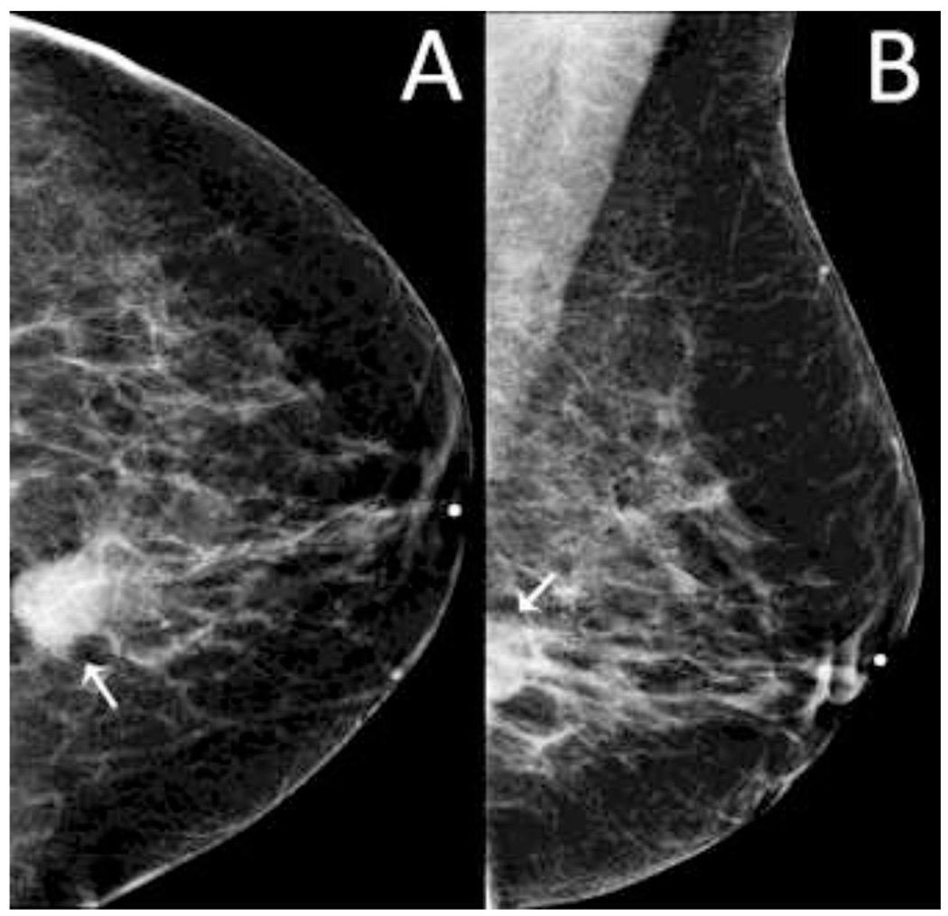
A 60-year-old woman presented for screening mammography. A, Left craniocaudal mammogram demonstrates a round mass in the posterior central left breast (arrow). B, Left mediolateral oblique mammogram demonstrates the same mass (arrow). Ultrasound-guided biopsy demonstrated evidence of high grade invasive ductal carcinoma (ER+, PR+, HER2−). A metastatic axillary lymph node was found without additional metastases, resulting in an overall stage of IIB.
Figure 3:
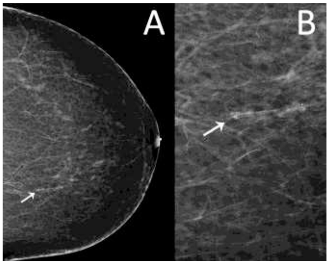
A 57-year-old woman presented for baseline screening mammography. A, Left craniocadual mammogram demonstrates coarse heterogeneous calcifications at 11 o’clock, 13 centimeters from the nipple (arrow). B, Digital zoomed view of the left mediolateral oblique mammogram demonstrates the morphology of the calcifications in greater detail (arrows). Stereotactic biopsy revealed evidence of ductal carcinoma in situ. Further information was unavailable as the patient sought follow-up care in another country.
The age of the patients with screen-detected breast carcinoma ranged from 41 to 67 years. The average age of diagnosis was 50.6 years. Patients over the age of 50 years accounted for eight (57.1%) of the 14 malignancies. Of these eight cancers in patients over 50 years of age, six cancers demonstrated an invasive component (75%). Patients between 40 and 49 years of age accounted for the remaining six screen-detected cancers. Of these six cancers, five (83.3%) demonstrated an invasive component and were all found in patients 40-44 years of age.
The patient’s race/ethnicity was documented for all 14 malignancies. Ten of the cancers were associated with Hispanic women. Three of the cancers were associated with Non-Hispanic Black women. A single malignancy was associated with an Asian woman.
The size of the imaging abnormality identified by screening mammography which prompted diagnostic work-up was recorded for all 14 malignancies. The imaging abnormalities ranged in size from 0.5 cm to 9.1 cm. The mean size was 2.3 cm and the median size was 1.6 cm.
The final tumor size (T) by pathology was obtained from the treating facilities for seven of the 11 invasive malignancies. All seven cases were identified as T1 with one case as pT1mi, two cases as pT1a, and four cases as pT1c. The clinical stage of the remaining four cases were determined with one case as cT1b, one case as cT1c, and two cases as cT2.
Regional lymph node (N) status was determined in 11 of the 14 cases. A single case (9.1%) was identified as N1mi. Five of the cases (45.5%) were determined to be N0, four of the cases (36.4%) were N1, and a single case (9.1%) was N2a.
Distant metastases (M) was recorded for 10 of the 14 cases. No distant metastases were identified in any of the 10 cases. Final stage was determined in 10 of the 14 cases. Five of the cases were stage IA. Single cases were determined to be stage 0, IB, IIA, IIB, and IIIA.
Receptor status was able to be determined in nine of the 11 invasive cancers. Seven (77.8%) of these cases demonstrated ER+, PR+, HER2− receptor status. A single case (11.1%) demonstrated ER+, PR−, HER2+ receptor status. A single case (11.1%) demonstrated ER+, PR−, HER2− receptor status. The receptor status information was unavailable for the remaining two cases as the patients sought care in another country.
Discussion:
This study demonstrated the ability for mobile mammography to identify small invasive breast cancers of low grade in a vulnerable population facing barriers to breast cancer screening. The majority of breast cancers identified demonstrated ER+, PR+, HER2− receptor status. This receptor status allows the malignancy to be targeted by hormonal therapy and is associated with a lower risk of local or regional recurrence [21]. This favorable receptor status was also demonstrated in patients of 40-49 years of age and the majority of these cancers were early stage (stage 0 or I). These cases of invasive disease highlight the importance of mammographic screening for a patient population that may not have access to screening. The cancers detected may have progressed to a later stage with worse outcomes and these results support continued mammographic screening for breast cancer in women aged 40-49 years old [22–24]. An argument could be made that breast cancer screening is unnecessary if small, early stage cancers are primarily detected. If these small, invasive cancers are allowed to progress then the cost of treatment at a later stage can be more expensive. To this end, Rim et al has recently demonstrated the cost-effectiveness of screening underserved women aged 40-64 years of age for breast cancer which is a similar patient demographic to the patients examined in this study [25]. These results are concordant with other studies examining the cost-effectiveness of breast cancer screening in other health care systems [26, 27].
The recall rate observed during the study period is higher than national benchmarks but similar to other mobile mammography programs [28, 29]. The cancer detection rate and the positive predictive value metrics were also noted to be lower than national benchmarks [28]. However, prior studies noted that mobile programs may not achieve all of the established benchmarks when compared to permanent sites [29]. The deviation of the mobile screening mammography benchmarks is thought to be secondary to multiple factors.
First, the composition of our screened population would be expected to yield a smaller number of malignancies due to the amount of patients aged less than 50 years. More than 60% of the underserved women screened were less than 50 years old, with approximately one-third less than 45 years old. The fewer detected malignancies results in lower CDR and PPV metrics. Furthermore, decreased PPVs are often seen in younger populations due to increased breast density and a lack of comparison examinations [22, 30]. The incidence data released by the National Cancer Institute’s Surveillance, Epidemiology, and End Results (SEER) program also demonstrates a lower incidence of in situ and invasive breast cancer in women younger than 50 years compared to older women [23].
Second, the race/ethnicity composition of the study population contributed to a smaller number of malignancies detected. In this study population, 76% of the patients identified themselves as Hispanic. The SEER incidence data highlights the lower incidence of breast cancer observed in Hispanic women as compared to other women. During a similar time period of this review and excluding American Indians and Alaska Natives, SEER incidence data from 2012-2016 reported an incidence of breast cancer in Hispanic women of 97 per 100,000 persons compared to 100-137 per 100,000 persons in other women. Using the same criteria, SEER reported an incidence of in situ breast cancer in Hispanic women of 25 per 100,000 persons compared to 25-35 per 100,000 persons in other women [23].
Screening examinations from the mobile mammography program often lacked comparison studies. A lack of comparison studies can increase recall rates as stability cannot be established for low suspicion findings. In this study population, 80% of the patients received only a single mammogram and 96% of the patients received two or fewer screening examinations. This trend of fewer comparison examinations being available to a mobile mammography service relative to a permanent site has also previously been observed [31]. Project VALET currently obtains the release form needed to acquire comparison examinations on the day of screening and allows up to 14 business days for the prior images to be retrieved. There is a risk that the prior images will never arrive, particularly in a population that faces challenges with travel and communication. This timeframe has worked well without risking a long delay in reporting the current examinations.
The possibility that radiologists interpreting the mobile mammography screening examinations were contributing to the reported metrics was examined. During the study time period, these examinations performed at a permanent site yielded a CDR of 5.8, a PPV1 of 5.9%, a PPV2 of 25.7%, a PPV3 of 31.9%, and a recall rate of 9.8%. These results are within the expected performance range for screening mammography, suggesting radiologist performance was not a major contributing factor to the difference in performance metrics for the mobile mammography service.
One of the limitations of this study extends from the difficulty of obtaining results from the outside facilities where the patients pursued their diagnostic work-ups, surgeries, and treatments. This barrier often limited the investigators from obtaining diagnostic imaging, pathology results, and treatment information. Additionally, patients served by the mobile program often faced financial difficulty that resulted in a change of a home address or a disconnected phone line which complicated communication between the patients and the mobile mammography staff. These issues reinforce the need for close communication and developing partnerships between a mobile mammography service and regional health clinics.
Digital breast tomosynthesis (DBT) allows for increased CDR, increased PPV, and a decreased recall rate [32, 33]. This technology was not available on the mobile mammography vans until after the reviewed time period, and DBT would likely have provided benefits to our patients. Another limitation of this study stems from comparing examinations and subsequent metrics with DBT at a permanent site with examinations performed by mobile mammography vans without DBT. While the availability of DBT would likely have improved audit metrics of the mobile mammography service, the discrepancies in the metrics between the permanent and the mobile screening programs are most likely a combination of multiple factors, including the availability of comparison examinations, differences in population demographics, and the availability of DBT.
Project VALET is continually examining existing methods and new technologies to improve. In addition to DBT, automated whole breast ultrasound has been examined as an additional capability. At this time, the program has not found an economically feasible solution due to restraints on cost, time, and space.
Project VALET has seen benefit through hiring technologists capable of directing patients in multiple languages. This allows improved efficiency, reduces errors related to patient history, and provides a more comfortable patient experience. Each mobile mammography van is staffed by a project service coordinator and a mammography technologist, which streamlines the different phases of a patient’s appointment. Project VALET also connects patients with navigation services and issues reminders, which reduce the number of missed or late appointments. Another way the program has reduced overall downtime is by implementing a cleaning and preventive maintenance day each week. This allows for thorough cleaning and inspection for any problems prior to an issue becoming catastrophic and removing the van from service.
An additional way in which Project VALET provides benefit to the community is by covering the cost of all diagnostic procedures up to the point of biopsy. Some patients and clinical providers had previously expressed concerns about the utility of mammographic screening if the patient was unable to afford diagnostic evaluation.
Conclusion:
Project VALET is an example of a mobile mammography screening program serving an underserved metropolitan community. The program has resulted in 14 breast cancers recently detected in this asymptomatic population. The years of life potentially saved are underscored by the number of breast cancers detected at a low stage, without nodal involvement or distant metastases. These findings support the need for the development of similar programs in other areas in the United States with a mission to care for the underserved community. Additionally, further investigation into mobile mammography programs would be helpful to identify patient factors, logistics, and technology, including DBT, that might prove beneficial.
Acknowledgments:
We wish to thank Lucy Balderas, Diane Benson, Ada Lo, Olga Lukyanchenko, Cindy Marquez, Monica Oliveros, and Lizbeth Pineda for their contributions to this manuscript. Supported by the NIH/NCI under award number P30 CA016672.
Footnotes
Publisher's Disclaimer: This is a PDF file of an unedited manuscript that has been accepted for publication. As a service to our customers we are providing this early version of the manuscript. The manuscript will undergo copyediting, typesetting, and review of the resulting proof before it is published in its final form. Please note that during the production process errors may be discovered which could affect the content, and all legal disclaimers that apply to the journal pertain.
Financial Disclosures: The spouse of an author is an employee of STERIS Corporation. The authors declare that they have no other potential competing financial interest to disclose.
References:
- 1.Ferlay J, Soerjomataram I, Dikshit R, et al. Cancer incidence and mortality worldwide: sources, methods and major patterns in GLOBOCAN 2012. Int J Cancer 2015; 136:E359–386 [DOI] [PubMed] [Google Scholar]
- 2.DeSantis CE, Fedewa SA, Goding Sauer A, Kramer JL, Smith RA, Jemal A. Breast cancer statistics, 2015: Convergence of incidence rates between black and white women. CA Cancer J Clin 2016; 66:31–42 [DOI] [PubMed] [Google Scholar]
- 3.Tabar L, Yen M-F, Vitak B, Chen H-HT, Smith RA, Duffy SW. Mammography service screening and mortality in breast cancer patients: 20-year follow-up before and after introduction of screening. Lancet 2003; 361:1405–1410 [DOI] [PubMed] [Google Scholar]
- 4.Tabar L, Vitak B, Chen TH, et al. Swedish two-county trial: impact of mammographic screening on breast cancer mortality during 3 decades. Radiology 2011; 260:658–663 [DOI] [PubMed] [Google Scholar]
- 5.Hendrick RE, Helvie MA. Mammography screening: a new estimate of number needed to screen to prevent one breast cancer death. AJR Am J Roentgenol 2012; 198:723–728 [DOI] [PubMed] [Google Scholar]
- 6.Berry DA, Cronin KA, Plevritis SK, et al. Effect of screening and adjuvant therapy on mortality from breast cancer. N Engl J Med 2005; 353:1784–1792 [DOI] [PubMed] [Google Scholar]
- 7.Jensen LF, Pedersen AF, Andersen B, Vedsted P. Identifying specific non-attending groups in breast cancer screening--population-based registry study of participation and socio-demography. BMC Cancer 2012; 12:518. [DOI] [PMC free article] [PubMed] [Google Scholar]
- 8.White A, Thompson TD, White MC, et al. Cancer screening test use--United States, 2015. Morbidity and Mortality Weekly Report 2017; 66:201. [DOI] [PMC free article] [PubMed] [Google Scholar]
- 9.DeSantis C, Jemal A, Ward E. Disparities in breast cancer prognostic factors by race, insurance status, and education. Cancer Causes Control 2010; 21:1445–1450 [DOI] [PubMed] [Google Scholar]
- 10.Vieira RA, Lourenco TS, Mauad EC, et al. Barriers related to non-adherence in a mammography breast-screening program during the implementation period in the interior of Sao Paulo State, Brazil. J Epidemiol Glob Health 2015; 5:211–219 [DOI] [PMC free article] [PubMed] [Google Scholar]
- 11.Maheswaran R, Pearson T, Jordan H, Black D. Socioeconomic deprivation, travel distance, location of service, and uptake of breast cancer screening in North Derbyshire, UK. J Epidemiol Community Health 2006; 60:208–212 [DOI] [PMC free article] [PubMed] [Google Scholar]
- 12.Schwartz KL, Crossley-May H, Vigneau FD, Brown K, Banerjee M. Race, socioeconomic status and stage at diagnosis for five common malignancies. Cancer Causes Control 2003; 14:761–766 [DOI] [PubMed] [Google Scholar]
- 13.Reuben DB, Bassett LW, Hirsch SH, Jackson CA, Bastani R. A randomized clinical trial to assess the benefit of offering on-site mobile mammography in addition to health education for older women. AJR Am J Roentgenol 2002; 179:1509–1514 [DOI] [PubMed] [Google Scholar]
- 14.Mizuguchi S, Barkley L, Rai S, et al. Mobile mammography, race, and insurance: Use trends over a decade at a comprehensive urban cancer center. J Oncol Pract 2015; 11:e75–80 [DOI] [PubMed] [Google Scholar]
- 15.Massin-Short SB, Grullon MA, Judge CM, Ruderman KR, Grullon M, Lora V. A mobile mammography pilot project to increase screening among Latina women of low socioeconomic status. Public Health Rep 2010; 125:765–771 [DOI] [PMC free article] [PubMed] [Google Scholar]
- 16.McCoy CB, Khoury EL, Hermanns LS, Bankston L. Mobile mammography: A model program for medically underserved women. Women’s Health Issues 1992; 2:196–203 [DOI] [PubMed] [Google Scholar]
- 17.Guillaume E, Launay L, Dejardin O, et al. Could mobile mammography reduce social and geographic inequalities in breast cancer screening participation? Prev Med 2017; 100:84–88 [DOI] [PubMed] [Google Scholar]
- 18.Carkaci S, Geiser WR, Adrada BE, Marquez C, Whitman GJ. How to establish a cost-effective mobile mammography program. AJR Am J Roentgenol 2013; 201:W691–697 [DOI] [PubMed] [Google Scholar]
- 19.Abdel-Aleem H, El-Gibaly OM, El-Gazzar AF, Al-Attar GS. Mobile clinics for women’s and children’s health. Cochrane Database Syst Rev 2016:CD009677 [DOI] [PMC free article] [PubMed] [Google Scholar]
- 20.Sickles EA, D’Orsi CJ. ACR BI-RADS follow-up and outcome monitoring. In: ACR BI-RADS Atlas, Breast Imaging Reporting and Data System. Reston, VA: American College of Radiology, 2013 [Google Scholar]
- 21.Voduc KD, Cheang MC, Tyldesley S, Gelmon K, Nielsen TO, Kennecke H. Breast cancer subtypes and the risk of local and regional relapse. J Clin Oncol 2010; 28:1684–1691 [DOI] [PubMed] [Google Scholar]
- 22.Arleo EK, Dashevsky BZ, Reichman M, Babagbemi K, Drotman M, Rosenblatt R. Screening mammography for women in their 40s: a retrospective study of the potential impact of the U.S. Preventive Service Task Force’s 2009 breast cancer screening recommendations. AJR Am J Roentgenol 2013; 201:1401–1406 [DOI] [PubMed] [Google Scholar]
- 23.Surveillance, Epidemiology, and End Results (SEER) Program (www.seer.cancer.gov) Research Data (1975-2016), National Cancer Institute, DCCPS, Surveillance Research Program, released April 2019, based on the November 2018 submission. Accessed April 23, 2019. [Google Scholar]
- 24.Ray KM, Price ER, Joe BN. Evidence to support screening women in their 40s. Radiol Clin North Am 2017; 55:429–439 [DOI] [PubMed] [Google Scholar]
- 25.Rim SH, Allaire BT, Ekwueme DU, et al. Cost-effectiveness of breast cancer screening in the National Breast and Cervical Cancer Early Detection Program. Cancer Causes Control 2019; 30:819–826 [DOI] [PMC free article] [PubMed] [Google Scholar]
- 26.Kauhava L, Immonen-Raiha P, Parvinen I, et al. Population-based mammography screening results in substantial savings in treatment costs for fatal breast cancer. Breast Cancer Res Treat 2006; 98:143–150 [DOI] [PubMed] [Google Scholar]
- 27.Morton R, Sayma M, Sura MS. Economic analysis of the breast cancer screening program used by the UK NHS: Should the program be maintained? Breast Cancer (Dove Med Press) 2017; 9:217–225 [DOI] [PMC free article] [PubMed] [Google Scholar]
- 28.Lehman CD, Arao RF, Sprague BL, et al. National performance benchmarks for modern screening digital mammography: Update from the Breast Cancer Surveillance Consortium. Radiology 2017; 283:49–58 [DOI] [PMC free article] [PubMed] [Google Scholar]
- 29.Stanley E, Lewis MC, Irshad A, et al. Effectiveness of a mobile mammography program. AJR Am J Roentgenol 2017; 209:1426–1429 [DOI] [PubMed] [Google Scholar]
- 30.Pitman JA, McGinty GB, Soman RR, Drotman MB, Reichman MB, Arleo EK. Screening mammography for women in their 40s: The potential impact of the American Cancer Society and U.S. Preventive Services Task Force breast cancer screening recommendations. AJR Am J Roentgenol 2017; 209:697–702 [DOI] [PubMed] [Google Scholar]
- 31.Roubidoux MA, Shih-Pei Wu P, Nolte ELR, Begay JA, Joe AI. Availability of prior mammograms affects incomplete report rates in mobile screening mammography. Breast Cancer Res Treat 2018; 171:667–673 [DOI] [PubMed] [Google Scholar]
- 32.Bahl M, Mercaldo S, Vijapura CA, McCarthy AM, Lehman CD. Comparison of performance metrics with digital 2D versus tomosynthesis mammography in the diagnostic setting. Eur Radiol 2019; 29:477–484 [DOI] [PubMed] [Google Scholar]
- 33.Skaane P, Sebuodegard S, Bandos AI, et al. Performance of breast cancer screening using digital breast tomosynthesis: results from the prospective population-based Oslo Tomosynthesis Screening Trial. Breast Cancer Res Treat 2018; 169:489–496 [DOI] [PubMed] [Google Scholar]