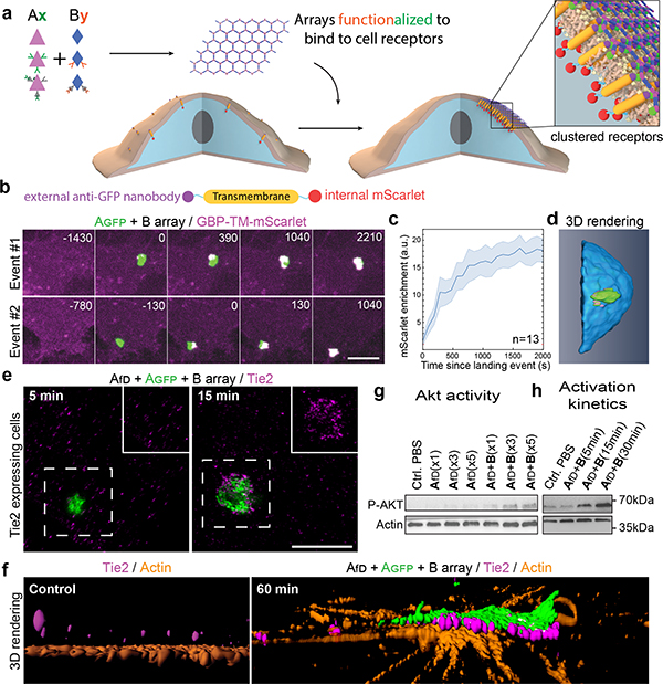
Figure 3. Dynamics of array-induced receptor clustering and biological activation.
(a) Array functionalization by genetic or post-translational fusions. (b) 3T3 cells expressing GBP-TM-mScarlet (GBP: anti-GFP nanobody; TM: transmembrane domain) were incubated with preformed Agfp+B arrays, and mScarlet clustering was monitored by confocal microscopy. (c) Quantification of the effects seen in (b). (d) 3D rendering of an array binding event. (e-h) Tie2 receptor clustering induced by preformed Afd+Agfp+B arrays (fD: F domain of angiopoietin). (e) Tie2 receptors were imaged 5 or 15 minutes after binding of arrays to cells. Insets: high-magnification of the dashed area, omitting the array signal. (f) 3D reconstruction in the absence of arrays (left panel) or 60 minutes post array-binding (right panel) showing the alignment between array and clustered Tie2. (g) Effect of array-induced Tie2 clustering on p-AKT activation. fD monomer concentration: x1/x3/x5: 17.8/53.4/89 nM respectively. (h) Dynamics of Tie2 activation. Scale bars: (b) 3 μm; (e) 2.5 μm.

