Abstract
In vitro embryo production systems are limited by their inability to consistently produce embryos with the competency to develop to the blastocyst stage, survive cryopreservation, and establish a pregnancy. Previous work identified a combination of three cytokines [fibroblast growth factor 2 (FGF2), leukemia inhibitory factor (LIF), and insulin-like growth factor 1 (IGF1)], called FLI, that we hypothesize improve preimplantation development of bovine embryos in vitro. To test this hypothesis, FLI was supplemented into oocyte maturation or embryo culture medium. Embryos were produced in vitro using abattoir-derived oocytes and fertilized with sperm from a single bull known to have high fertility. After an 18–20 h fertilization period, putative zygotes were cultured in synthetic oviductal fluid (SOF) for 8 days. The addition of FLI to the oocyte maturation medium increased (P < 0.05) the dissociation of transzonal projections at 12, 18, and 24 h of maturation, as well as, the proportion of oocytes that reached the metaphase II stage of meiosis. Additionally, lipid content was decreased (P < 0.05) in the blastocyst stage embryo. The addition of FLI during the culture period increased development to the blastocyst stage, cytoskeleton integrity, and survival following slow freezing, as well as, decreased post thaw cell apoptosis (P < 0.05). In conclusion, the supplementation of these cytokines in vitro has the potential to alleviate some of the challenges associated with the cryo-survival of in vitro produced bovine embryos through improving embryo development and embryo quality.
Introduction
In vitro embryo production is an assisted reproductive technique used in livestock production to maximize the use of valuable genetics [1]. Of the livestock species, Bos taurus heavily rely on these technologies compared to others. In 2018, a total of 1,499,367 transferrable embryos were produced worldwide, from which 68.7% were in vitro produced (IVP) [2]. Though the industry is trending toward IVP embryos, the majority (73.2%), are transferred fresh; in part due to the reduced ability of IVP embryos to withstand cryopreservation as compared to embryos produced in vivo [3–5]. Therefore, it is important to understand the IVP system to increase viable embryo production in vitro as well as survival after cryopreservation.
Currently, less than 50% of the IVP bovine embryos will develop to the blastocyst stage. One of the main causes of this low yield has been attributed to inadequate oocyte cytoplasmic and nuclear maturation [6, 7]. A second critical period is culture after fertilization. There is evidence that the environment of the embryo can affect the transcript abundance for genes important for preimplantation development [e.g. GJA1, BAX, Interferon-tau (IFNT)], increase lipid content, reduce numbers of mitochondria, and alter the cytoplasmic:nuclear ratio [8, 9]. One well-reported characteristic of IVP embryos is their increased susceptibility to cryopreservation, likely due to the variation in blastocyst quality observed in IVP embryos [9].
Quality parameters that define the competency of an oocyte or embryo to survive cryopreservation and establish a pregnancy are not well defined. In the oocyte, transzonal projections have been posed as indicators of oocyte quality because they serve as bidirectional communication channels between the oocyte and surrounding cumulus cells [10, 11]. Previous work has shown that transzonal projections may facilitate fatty acid binding protein transport from cumulus cells into the oocyte, and abnormal dissociation of the projections could lead to increased lipid accumulation and metabolic dysfunction [12].
In the embryo, cell number and allocation of cells within the blastocyst (inner cell mass and trophectoderm) have been suggested as indicators of embryo developmental potential [13–15]. However, embryos that appeared cleaved and developed to the blastocyst stage quicker than their counterparts did not have an increased number of cells in the inner cell mass [16]. Another indicator of embryo quality or competence is the accumulation of reactive oxygen species, which can be indicators of cellular stress and altered metabolism [17–19]. Furthermore, cellular integrity, determined by cytoskeleton analysis and cell organization, is suggested to be an indicator of embryo quality in other species [20, 21]. It is evident that there is a need to more clearly define the contribution of these parameters to embryo quality, specifically during in vitro production where embryo viability appears to be compromised [22–25].
One approach to increase the efficiency of the IVP system would be the addition of embryokines that are present in the female reproductive tract following ovulation [26]. The addition of molecules such as LIF or FGF2 to maturation or IGF1 or IL6 to culture medium have improved the embryo’s ability to develop to the blastocyst stage [27, 28], increase cell number [29], survive cryopreservation [30–32], and resist heat stress/shock [33]. Furthermore, certain cytokine combinations such as, LIF and IGF-1, IGF-I, IGF-II, bFGF, TGF-β1, GM-CSF, and LIF, or EGF and IGF-1 have acted additively to improve in vitro embryo production through an increase in embryo development and/or quality [34–36]. Three cytokines, FGF2 (40 ng/ml), LIF (20 ng/ml), and IGF1 (20 ng/ml) (when combined termed FLI), were identified for their potential roles as candidates to improve embryonic development in vitro in the pig [37]. In cattle, FGF2 and LIF are involved in pluripotency maintenance, stimulation of IFNT production [38], and cryotolerance [30, 31], while IGF1 is thought to increase cell survival in response to stress [33]. Thus, we hypothesize, that these three cytokines have the potential to increase oocyte maturation and embryonic development to the blastocyst stage as well as oocyte and embryo quality when added to oocyte maturation medium or embryo culture medium.
Materials and methods
In vitro embryo production media
All embryo production media: Oocyte maturation (OMM), fertilization (IVF-TALP), wash (HEPES-TALP), culture (SOF-BE2), and the supplement penicillamine, hypotaurine, and epinephrine (PHE) were prepared in house unless otherwise noted following recipes previously published [39]. Sperm purification gradient Isolate® (Irvine Scientific, Santa Ana, CA, USA) was acquired ready to use. FLI was supplemented at the dosage of FGF2 (40ng/ml), LIF (20ng/ml), and IGF1 (20ng/ml) [37] to the maturation medium or culture medium.
FLI supplementation during oocyte maturation
The experimental design is presented in Fig 1.
Fig 1. Experimental setup of FLI supplemented to oocyte maturation medium.
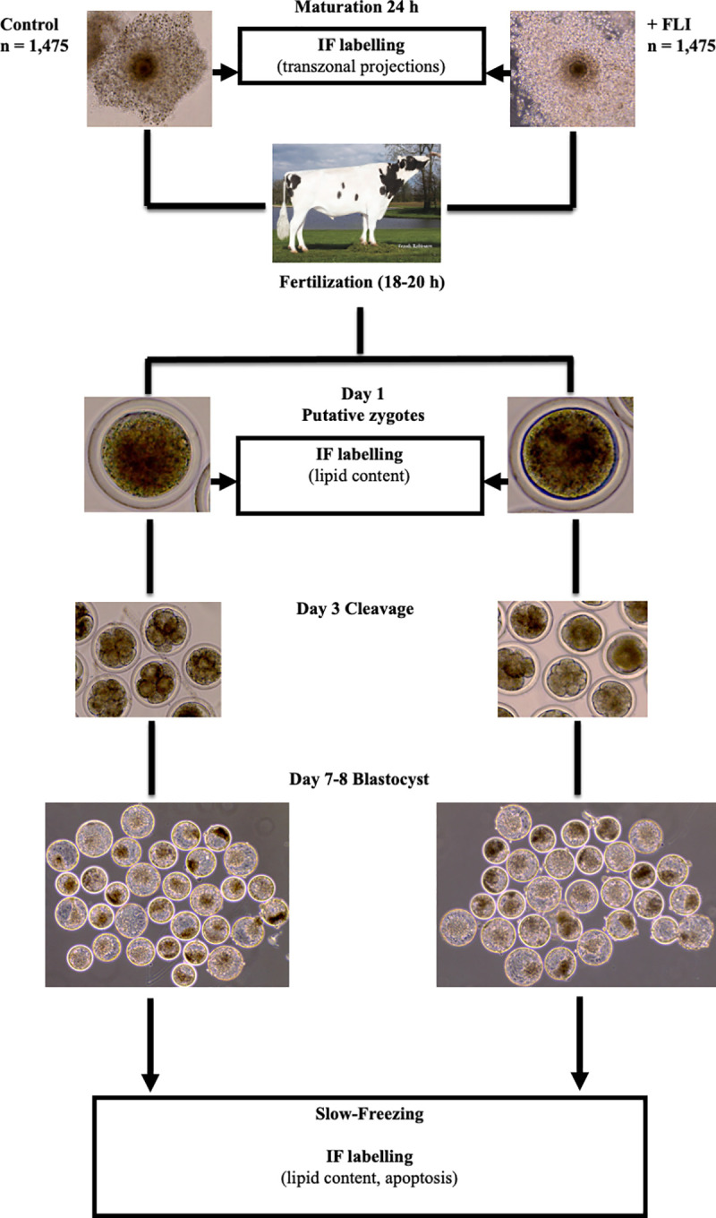
Cumulus oocyte complexes (COCs) were matured in oocyte maturation medium (OMM) with (n = 1475) or without (n = 1475) FLI. FLI was supplemented at the dosage of FGF2 (40ng/ml), LIF (20ng/ml), and IGF1 (20ng/ml). For analysis of transzonal projections, COCs were collected at 6 h, 12 h, 18 h, 24 h after placement into OMM. At 24 h, maturation stage was recorded. Following fertilization, a subset of putative zygotes (day 1) was collected for lipid content quantification. Cleavage (at least one cellular division) was recorded on day 3 and development to the blastocyst stage was recorded on days 7 and 8. On day 7 or 8, blastocyst stage embryos were either collected for quantification of lipid content or slow frozen, thawed, and subjected to a TUNEL assay to analyze apoptosis. IF = immunofluorescent labeling.
Detection of transzonal projections
The number of transzonal projections in cumulus oocyte complexes (COCs) was determined by immunolocalization of F-actin filaments that connect the oocyte and cumulus cells using confocal microscopy. COCs (n = 198), with at least 2 layers of cumulus cells, and dark homogenous ooplasm [40] were matured in OMM [41] supplemented with (n = 98) or without FLI (n = 100), and collected at 6, 12, 18, and 24 h after placement into OMM. At the moment of collection, COCs were washed with phosphate buffered saline (PBS) containing 0.1% polyvinylpyrrolidone (PVP) and fixed for 20 min in droplets of PBS-PVP containing 4% paraformaldehyde. Fixed COCs were permeabilized (0.5%Triton X-100 diluted in PBS) for 30 min at room temperature, washed three times with PBS-PVP, and placed in Rhodamine phalloidin (Cytoskeleton Inc., Denver, CO, USA) diluted at 0.04 μg/ml [37]. Incubation proceeded in the dark for 45 min at room temperature. COCs were washed again three times with PBS-PVP and incubated in Hoechst 33342 (Invitrogen Molecular Probes, Waltham, MA, USA) at 1 μg/ml for 20 min in the dark. Finally, COCs were mounted on slides in Slow fade® Gold antifade reagent (Thermo Fisher Scientific, Waltham, MA, USA) and analyzed by using confocal microscopy (Leica TCS SP8 MP). Excitation/emission was 350/461 for Hoechst 33342 and 540/585 for Rhodamine phalloidin. ImageJ Software 1.46r (National Institutes of Health, Bethesda, MD, USA) was used to count the number of intact transzonal projection filaments present, which was defined by an obvious connection from the cumulus cells to the oocyte through the zona pellucida. Disrupted transzonal projections, identified by obvious breaks in the connection, were not counted.
Nuclear staining
In a second experiment, following maturation, oocytes [n = 304, 148 controls and 156 with FLI, across 3 replicates] were denuded and fixed with 4% paraformaldehyde. Meiotic stage was determined by nuclear labeling as previously described [42, 43]. Briefly, oocytes were permeabilized in 0.1% Triton X-100 for 30 min at room temperature, washed three times with PBS-PVP and incubated in Hoechst 33342 (1μg/ml) for 20 min in the dark. Following staining with Hoechst, oocytes were mounted on slides in Slow fade® Gold antifade reagent (Thermo Fisher Scientific) and analyzed using fluorescence microscopy. Excitation/emission was 350/461 for Hoechst 33342. The proportion of oocytes in each meiotic stage [germinal vesicle (GV), GV breakdown, metaphase 1, anaphase 1, telophase 1, and metaphase II] was recorded.
In vitro embryo production
For all experiments, abattoir derived COCs (n = 2,950, across 12 replicates) were received from a single company that provides abattoir derived COCs. COCs were placed in equilibrated OMM (50 COCs/tube) overlaid with oil and shipped via portable incubator set to 38.5˚C and left in tubes until collection or fertilization. OMM was supplemented with (n = 1475) or without (n = 1475) FLI. Only COCs surrounded by multiple layers of cumulus cells and with homogenous ooplasm were used. For each replicate, up to 300 mature COCs were washed three times in HEPES-TALP and placed in a dish containing 1.7 ml of IVF-TALP. The COCs were inseminated with sperm from a single Holstein bull known to have high fertility in vitro. Sperm were purified from frozen-thawed straws by using isolate gradient, washed two times by centrifugation at 100×g with HEPES-TALP and diluted in IVF-TALP to achieve a final concentration of 1 × 10⁶/ml in the fertilization dish. To improve sperm motility and capacitation, 80 μl PHE was added to the fertilization plate [44].
Fertilization proceeded for 18–20 h in a humidified atmosphere of 5% (v/v) CO2. At the end of fertilization, putative zygotes (oocytes exposed to sperm) were denuded from cumulus cells by vortexing for five min in 200 μl HEPES-TALP. Embryos were then washed three times in HEPES-TALP and up to 50 putative zygotes were placed in a 4-well dish containing 500 μl of SOF-BE2 overlaid with 300 μl mineral oil. Embryos were cultured at 38.5°C in a humidified atmosphere of 5% (v/v) O2 and 5% (v/v) CO2 with the balance N2 until collection. Cleavage of the embryos was recorded on day 3 and development to the blastocyst stage was recorded on days 7 and 8 post insemination.
Cryopreservation of embryos by slow-freezing
On day 7–8 post insemination, blastocysts were collected, grouped according to their stage and quality grade using IETS standards [45]. Blastocyst stage embryos that were quality 1 and stage 6 or 7 were washed three times in holding medium (HEPES-TALP, 10% FBS, 0.05% DTT) [46, 47] equilibrated to 38°C, to remove residual culture medium. Next, embryos were placed in a drop of freezing medium (BioLife Freeze Medium Ethylene Glycol with Sucrose, AgTech Inc, Manhattan, KS, USA), and allowed to equilibrate by sinking to the bottom of the dish. Straws were loaded by adding a column of freezing medium, followed by 2–3 mm of air, another column of freezing medium containing two to five embryos of the same stage and quality grade, 2–3 mm of air, and a final column of freezing medium. Loaded straws were maintained at room temperature for 5 to 15 min, then placed in the freezing machine (Cryogenic L854 Temperature Controller) equilibrated to -6°C. After 2 min, the individual straws were seeded by placing two cotton swabs in liquid nitrogen, then on each side of the top column of freezing medium until ice crystals formed. Loaded straws were held at -6°C for 10 min, and then subjected to a temperature gradient of -0.5°C/min until reaching -32°C and kept at this temperature for 10 min [32, 48]. Immediately after, straws were immersed in liquid nitrogen and stored until thawed for further analysis.
To thaw, straws were removed from liquid nitrogen, held at room temperature for 3 sec, then placed in a cryobath set at 30°C for 45 sec. Embryos were removed from straws and washed in holding medium three times. After washing, embryos were grouped according to their treatment and placed in culture plates containing SOF-BE2, with no more than 10 embryos per well. As previously described, 10% FBS was added to each well [32, 46, 49]. Hatching from the zona pellucida in vitro was used to determine cryo-survival. A total of 148 embryos (n = 73 control, n = 75 +FLI) across 5 replicates were evaluated at 24, 48, and 72 h post thaw for cryo-survival.
TUNEL assay
DNA fragmentation in blastocysts that re-expanded by 72 h post-thawing was determined using a terminal deoxynucleotidyl transferase dUTP nick end labeling (TUNEL) reaction kit assay (In Situ Cell Death Detection Kit with Fluorescein, Millipore Sigma, Burlington, MA, USA). At 72 h post thawing, expanded and hatched blastocysts were collected, washed with PBS-PVP, fixed, and permeabilized as described above. Samples were then separated into three treatments: positive control (DNAse and TUNEL solution), negative control (label solution), and the TUNEL treatment (label solution and enzyme) and subjected to the TUNEL reaction. Briefly, embryos were placed in 25 μl drops of their respective solutions in a four-well plate with water around the wells to allow for a humidified environment. Embryos were placed in a warm incubator for one h, washed in PBS-PVP, then incubated in Hoechst 33342 (1 μg/ml) for 20 min in the dark. Samples were mounted on slides in Slow fade® Gold antifade reagent (Thermo Fisher Scientific) and analyzed by fluorescence microscopy. Excitation/emission was 350/461 for Hoechst 33342 and 520/570 for TUNEL positive cells. ImageJ Software 1.46r (National Institutes of Health) was used to determine the total number of nuclei as well as the number of TUNEL positive (fragmented DNA) nuclei in each embryo (n = 36 controls, n = 38 +FLI, across 5 replicates).
Detection of lipid content
Lipid content in embryos was determined as previously described [50]. Briefly, putative zygotes (day 1) and expanded blastocyst (days 7 and 8) were collected and washed three times in PBS-PVP. At least two embryos were set aside for negative controls, and the remaining embryos were fixed as described before. Negative control embryos were incubated in 100 μl of 100% ethanol for 30 min to dissolve the lipids present. Following fixation, the remaining embryos were placed into a droplet of Nile Red Solution (Invitrogen Molecular Probes) for 30 min (1 μg/ml dilution). After incubation, embryos were washed with PBS-PVP three times and then nuclei were stained with Hoechst 33342 (1 μg/ml dilution) for 20 min in the dark. Samples were mounted on slides in Slow fade® Gold antifade reagent (Thermo Fisher Scientific) and analyzed by using fluorescence microscopy. Excitation/emission was 350/461 for Hoechst 33342 and 552/636 for Nile Red. ImageJ Software 1.46r (National Institutes of Health) was used to quantify mean intensity units per embryo to estimate the amount of lipids. A total of 230 zygotes (109 control, 120 +FLI) and 223 blastocyst stage embryos (116 control, 106 +FLI) were analyzed across 4 replicates in each experiment.
FLI supplementation during embryo culture
The experimental design is presented in Fig 2.
Fig 2. Experimental setup of FLI supplemented to embryo culture medium.
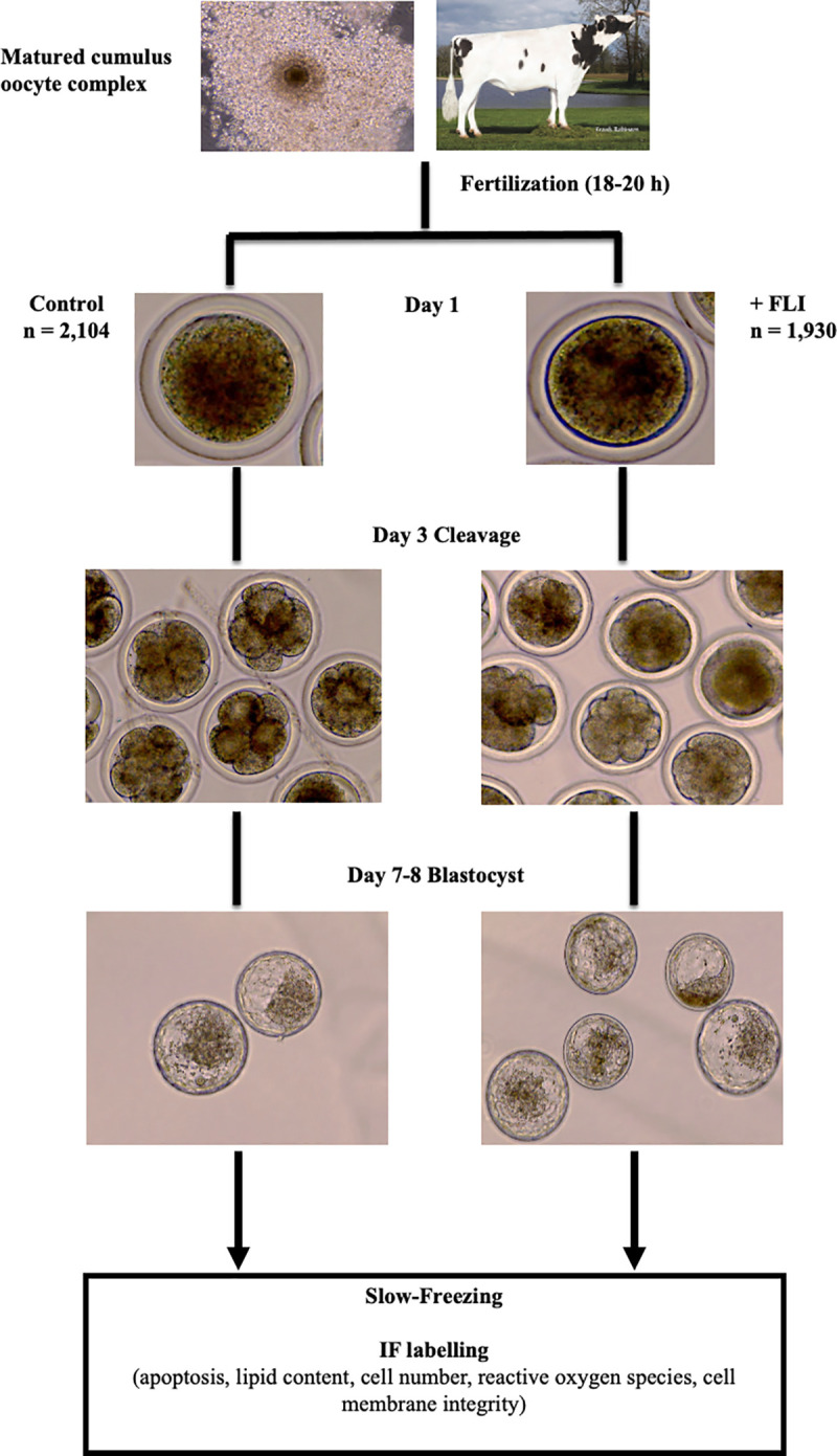
Cumulus oocyte complexes (COCs) were matured in oocyte maturation medium (OMM). Following fertilization, putative zygotes were placed in culture with (n = 1,930) or without (n = 2,104) FLI. FLI was supplemented at the dosage of FGF2 (40ng/ml), LIF (20ng/ml), and IGF1 (20ng/ml). Cleavage (at least one cellular division) was recorded on day 3 and development to the blastocyst stage was recorded on day 7 and 8. Day 7 or 8, blastocyst stage embryos were either collected for cell counting, quantification of lipid content, membrane integrity analysis, quantification of reactive oxygen species, or slow frozen, and subjected to TUNEL assay to analyze apoptosis, membrane integrity analysis, or quantification of reactive oxygen species post-thaw. IF = immunofluorescent labeling.
In vitro embryo production
Abattoir derived COCs (n = 4,400 across 16 replicates) were received in oocyte maturation medium. COCs were received from two different companies that provide abattoir derived COCs. From both sources, COCs were placed in equilibrated OMM (50 COCs/tube) overlaid with oil and shipped via portable incubator set to 38.5˚C and left in tubes until fertilization. For 6 replicates, oocytes were assessed upon receiving and classified as poor quality or good quality (Fig 3A). Poor quality COCs (n = 470) had few expanded cumulus cells, dark and granulated ooplasm, or appeared very pale. Good quality COCs (n = 596) had multiple layers of cumulus cells, and ooplasm that was uniform and dark in color. Given the apparent variation in oocyte quality, the remaining 10 replicates only used COCs from company 2 (consistent cumulus cell expansion and homogenous ooplasm).
Fig 3. FLI Supplementation to culture medium improves embryo development.
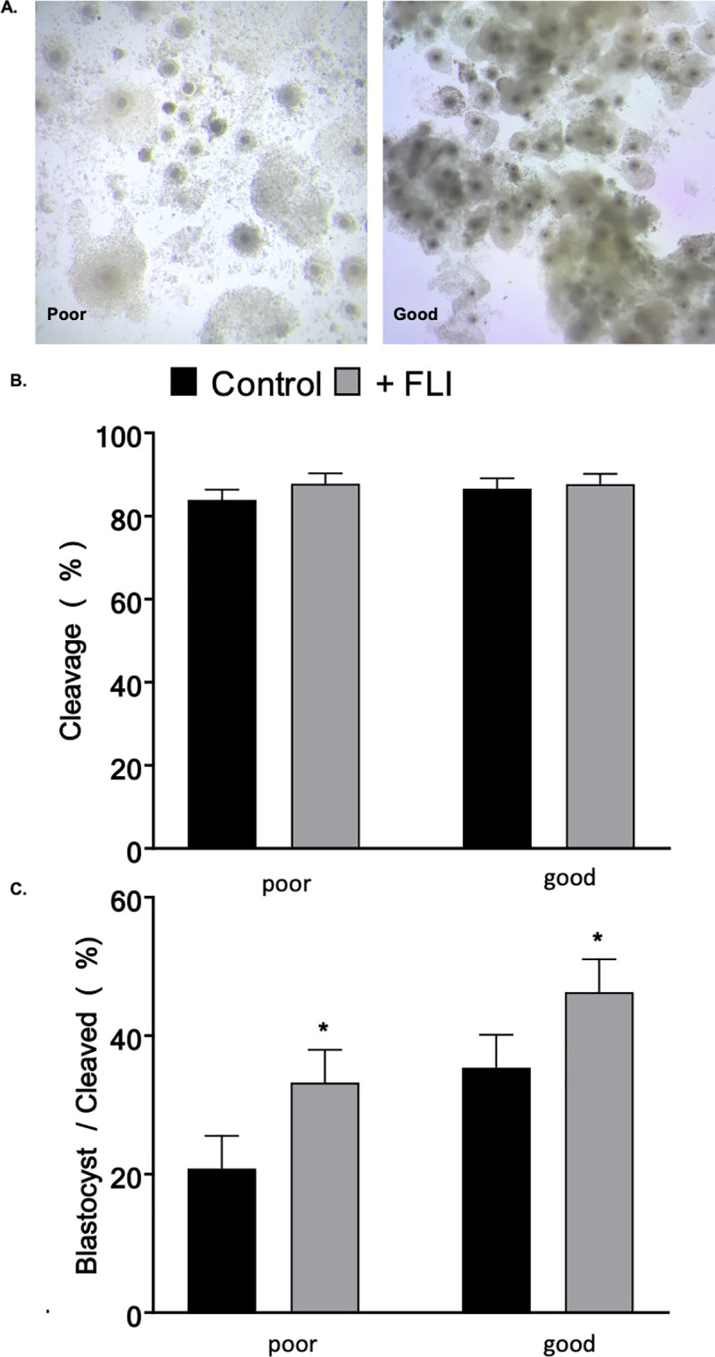
Bovine oocytes (n = 1,066) were of different qualities. (A) Example of oocytes classified as poor quality (470) or good quality (596) (B) Proportion of zygotes that underwent at least one cellular division. (C) The extent of the effect of FLI supplementation in culture medium on development to blastocyst stage varies based on oocyte quality/source. Asterisk (*) indicates statistical differences (P < 0.05). Values are LSMEANS ± SEM.
Fertilization and embryo culture were performed as described for previous experiments. For culture, 50 putative zygotes were placed in a 4-well dish containing 500 μl of SOF-BE2 topped with 300 μl mineral oil. Half of the putative zygotes were placed in culture medium supplemented with FLI. Embryos were cultured in the same environment as previously described. Embryos cultured with or without FLI were subjected to cryopreservation (n = 117; 51 control, 66 +FLI, across 5 replicates), thawed, and those that re-expanded were subjected to TUNEL (n = 79; 31 control, 48 +FLI) or determination of lipid content (n = 162; 72 control, 90 +FLI) as described above.
Cell number
The number of nuclei in the trophectoderm and inner cell mass was determined after immunolocalization of the trophectoderm marker CDX2 and a nuclear stain (Hoescht) in blastocyst (6–1) stage embryos. Day 7 or day 8 embryos were collected, washed with PBS-PVP and fixed for 20 min in 4% paraformaldehyde. Fixed embryos were permeabilized as previously described, washed, and incubated in blocking buffer [5% BSA (w/v)] for one h at room temperature. Embryos were incubated in the primary antibody [anti-CDX2 (mouse monoclonal antibody) BioGenex Fremont, CA, USA] in the dark at 4˚C overnight. Embryos were washed and placed into the secondary antibody (mouse anti-goat, Alexa 488) for one h at room temperature. Embryos were washed with PBS-PVP three times and nuclei were stained with Hoechst 33342 (1 μg/ml dilution) for 20 min in the dark. Each replicate contained a secondary antibody control embryo that was subjected to the above method minus incubation in the primary antibody (S1 Fig). Samples were mounted on slides in Slow fade® Gold antifade reagent (Thermo Fisher Scientific) and analyzed by fluorescent microscopy. Excitation/emission was 350/461 for Hoechst 33342 and 485/535 for Alexa 488. The number of trophectoderm nuclei was subtracted from the total number of nuclei to determine the inner cell mass number. Embryos (n = 160; 72 control and 88 +FLI) were analyzed across three replicates.
Classification of cytoskeleton integrity
Blastocyst stage embryos were collected fresh at day 7 or at 24 h post thaw. A 24 h thawing period allowed for most of the embryos to re-expand but not to the point of hatching from the zona pellucida. Blastocysts were washed in PBS-PVP and fixed for 20 min in 4% paraformaldehyde. After permeabilization, they were washed three times with PBS-PVP, and placed in rhodamine phalloidin (Cytoskeleton Inc.) at 0.04 μg/ml. Incubation proceeded in the dark for 45 min at room temperature. Blastocysts were washed three times with PBS-PVP and incubated in Hoechst 33342 staining (1 μg/ml) for 20 min in the dark. Finally, blastocysts were mounted on slides in Slow fade® Gold antifade reagent (Thermo Fisher Scientific) and analyzed by using confocal microscopy (Leica TCS SP8 MP). Excitation/emission was 350/461 for Hoechst 33342 and 540/585 for rhodamine phalloidin. ImageJ Software 1.46r (National Institutes of Health) was used to determine the number of disruptions (breaks) in the cytoskeleton of the cells and embryos were graded as previously described [20, 21] (Fig 4). Fresh (non-cryopreserved embryos: n = 101, 52 control and 49 +FLI) and thawed (n = 52, 22 control and 30 +FLI) embryos were analyzed across three replicates.
Fig 4. Representative images of bovine blastocysts showing the actin filament staining.

(A) Grade 1 blastocyst stage embryos displayed sharp staining of actin filaments and cells are clearly organized as inner cell mass or trophectoderm. (B) Grade 2 shows less distinct outlining of the cells and some breaks in the cytoskeleton structure. (C) Grade 3 is characterized by large gaps in actin staining and some actin within the cytoplasm. Actin filaments were visualized in 101 fresh (n = 52 control, n = 49 +FLI) and 52 (n = 22 control, 30 +FLI) thawed embryos.
Detection of reactive oxygen species (ROS)
CellROX®Green (Thermo Fisher Scientific) was used to measure oxidative stress [41]. Briefly, day 7 fresh or 24 h post-thaw blastocysts were transferred to fresh SOF-BE2 containing CellROX®Green (10 mM) and cultured at 38.5°C in a humidified atmosphere of 5% (v/v) O2 and 5% (v/v) CO2 with the balance N2 for 30 min. Embryos were collected, washed with PBS-PVP, and fixed for 20 min in 4% paraformaldehyde. Nuclei were stained with Hoechst 33342 (1 μg/ml dilution) for 20 min in the dark. Samples were mounted on slides in Slow fade® Gold antifade reagent (Thermo Fisher Scientific) and analyzed by using fluorescence microscopy. Excitation/emission was 350/461 for Hoechst 33342 and 485/520 for CellROX®Green. ImageJ Software 1.46r (National Institutes of Health) was used to quantify the mean intensity units per embryo to estimate ROS levels. A total of 99 fresh embryos and 70 cryopreserved embryos were analyzed across 3 replicates.
Statistical analysis
All analyses were conducted using SAS (V 9.4, SAS Institute, Cary, NC). Differences in transzonal projection dissociation were analyzed by analysis of variance using PROC GLM. The model included treatment, hour and the interaction of treatment and hour. Maturation rate was analyzed using PROC GLIMMIX. The model included treatment as a fixed factor and replicate as a random factor. A binomial regression model was used. Development, lipid content, and apoptosis (in both FLI added to OMM and FLI added to culture medium) were analyzed by analysis of variance using PROC GLM. The model included replicate, treatment, and the interaction of replicate and treatment. In the analysis for development for FLI added to culture medium, company was included as a covariate. Differences in cell number and cell ratio were analyzed by analysis of variance using PROC GLM. The model included replicate, treatment, and the interaction of replicate and treatment. Embryo hatching following cryopreservation and reactive oxygen species were analyzed by analysis of variance using PROC GLM. The model included replicate and treatment. Differences in cell membrane integrity (pre and post thaw) were analyzed using PROC GLIMMIX. The model included treatment and replicate as well as the interaction of the two. A binomial regression model was included. In all analyses significance was determined as P < 0.05 and results are shown as least square means ± standard error of the mean.
Results
Effects of FLI supplementation in oocyte maturation medium
Transzonal projection dissociation, oocyte maturation, and embryo development
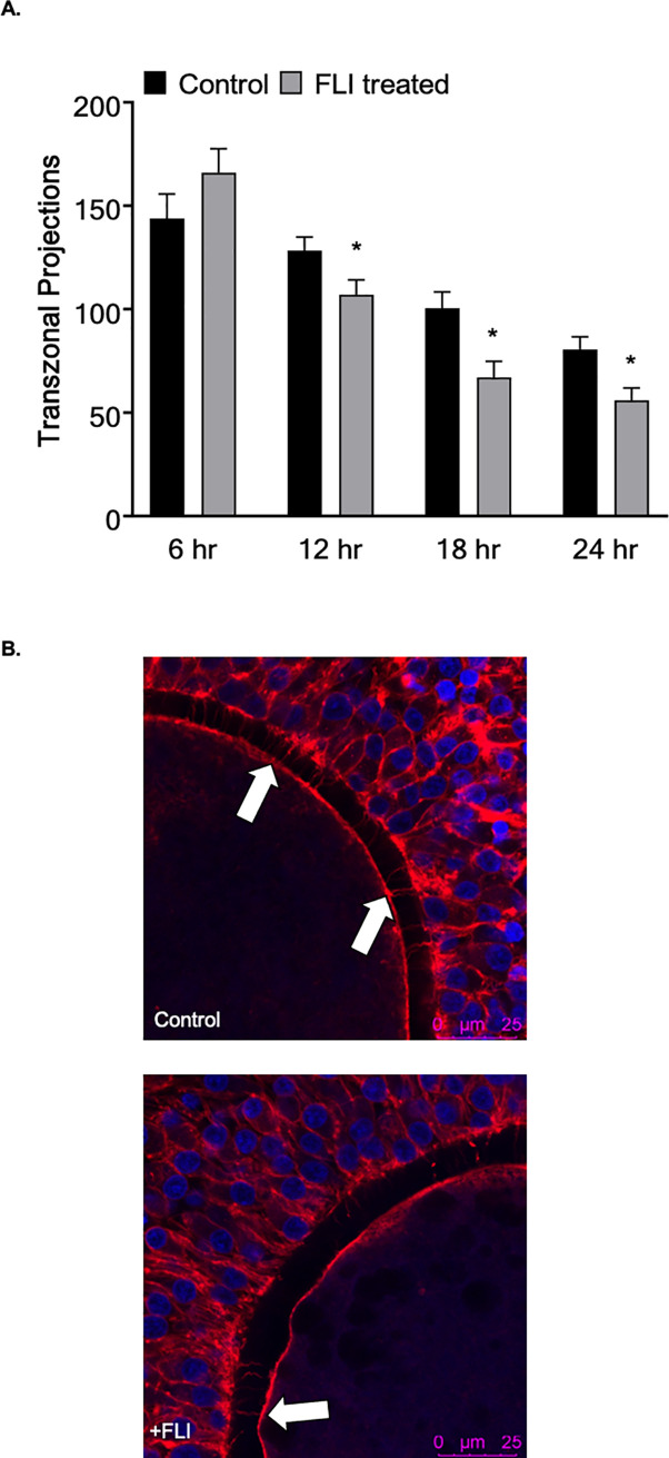
Supplementation of FLI to our current OMM was investigated to understand the effects on oocyte maturation and preimplantation embryo development in vitro. The addition of FLI to OMM had no effect (P > 0.05) on the number of transzonal projections at 6 h between FLI treated and control groups, (166.3 ± 8.6 vs 143.9 ± 8.8), respectively. However, there was a significant treatment by time interaction as FLI-treated COCs had fewer (P < 0.05) transzonal projections at 12 h, 18 h, and 24 h compared to the controls (Fig 5A). Additionally, supplementing FLI to OMM resulted in an increase (P < 0.05) in the percentage of oocytes that reached metaphase II (Table 1). However, there was no difference in cleavage or blastocyst percent between treatments (Table 1).
Fig 5. FLI supplementation advances transzonal projection dissociation.
(A) The effect of the addition of FLI to oocyte maturation medium on the number of TZP at 6, 12, 18, and 24hr. (n = 198; 100 control and 98 +FLI). (B) The images are representative of TZP dissociation at 24hr of maturation. Arrows indicate intact TZP. Asterisk (*) indicates statistical differences (P < 0.05).
Table 1. In vitro development of bovine embryos supplemented with and without FLI during maturation.
| Group1 | n | MII oocytes (%)2 | Cleavage (%) | Blastocyst (%) | Blastocyst/Cleaved (%) |
|---|---|---|---|---|---|
| Control | 1495 | 62.6 ± 0.40a | 80.0 ± 0.73 | 34.2 ± 1.07 | 42.9 ± 1.35 |
| FLI | 1495 | 75.6 ± 0.34b | 80.3 ± 0.73 | 35.4 ± 1.07 | 44.0 ± 1.35 |
a,bNumbers with different superscripts differ (P<0.05).
1All results are reported as least square means ± standard error of the mean.
2An analysis from a subset of oocytes (n = 304) collected from the total.
Lipid content and cryo-survival
Nile Red staining was used to quantify intracellular lipid content in zygotes and blastocyst stage embryos. The addition of FLI into the maturation medium had no effect (P > 0.05) on the lipid content at the zygote stage between the two treatments (Fig 6A). Interestingly, the embryos from the FLI supplemented group had decreased lipid content at the blastocyst stage compared to the controls (P < 0.001, Fig 6A). Supplementation of maturation medium with FLI, did not improve the ability of the embryo to withstand cryopreservation between the treatments (P > 0.05, Table 2) when survivability is measured by hatching rate in vitro. Likewise, there was no difference in DNA fragmentation between the two treatments (P > 0.05).
Fig 6. FLI supplementation during oocyte maturation decreases lipid content at the blastocyst stage.
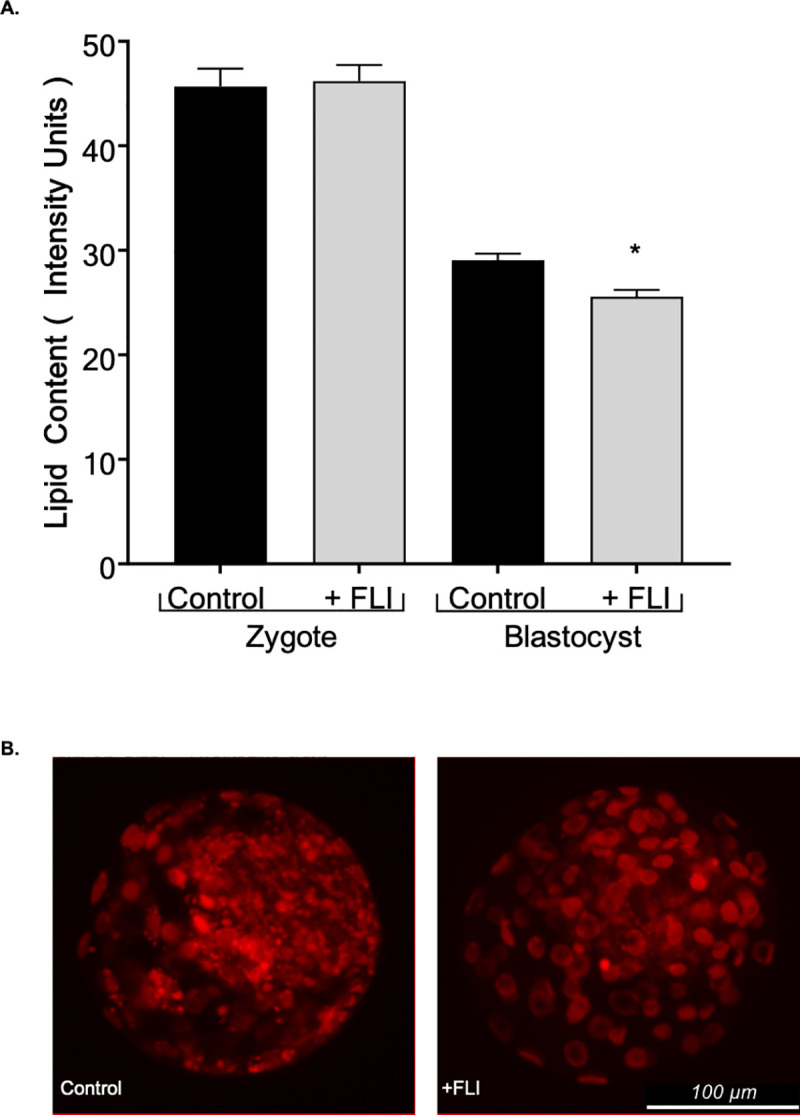
(A) Lipid content was compared at the zygote (n = 239; 109 controls and 120 +FLI) and blastocyst stage (n = 222; 116 controls and 106 +FLI). (B) Blastocyst stage bovine embryos illustrating Nile red staining by fluorescence microscopy used to determine lipid content. Asterisk (*) indicates statistical differences (P < 0.05).
Table 2. Hatching rates post-thaw of bovine embryos supplemented with and without FLI during maturation following cryopreservation by slow-freezing.
| Hatching (%)1 | ||||
|---|---|---|---|---|
| Group | n | 24h | 48h | 72h |
| Control | 51 | 36.2 ± 0.05 | 58.8 ± 0.07 | 67.0 ± 0.07 |
| FLI | 66 | 27.1 ± 0.05 | 60.6 ± 0.07 | 63.3 ± 0.07 |
1Results reported as least square means ± standard error.
Effects of FLI supplementation in embryo culture medium
Embryo development
The aim of this experiment was to analyze the effect of FLI on embryo culture. Supplementation of SOF-BE2 with FLI had no effect (P > 0.05) on cleavage percentage between the control and FLI treated groups (Table 3). However, the addition of FLI to the culture medium resulted in an increased (P < 0.0001) proportion of embryos that developed to the blastocyst stage (Table 3). Oocytes of variable quality were used for IVP during this study. To understand the effect of FLI supplementation on oocytes of different qualities, oocytes were assessed upon receiving and classified as poor quality or good quality (Fig 3A). The extent of the effect of FLI supplementation in culture medium on development to the blastocyst stage varied based on oocyte quality/source (Fig 3C). In both cases those treated with FLI had an increase (P < 0.05) in development to the blastocyst stage.
Table 3. In vitro development and number of nuclei in bovine embryos supplemented with and without FLI in culture.
| Cell Number3 | |||||||
|---|---|---|---|---|---|---|---|
| Group1 | n | Cleavage (%)2 | Blastocyst (%) | Blastocyst / Cleaved (%) | TE4 | ICM5 | Total |
| Control | 2104 | 82.4 ± 0.6 | 27.2 ± 1.1a | 32.9 ± 1.3a | 73.0 ± 2.7 | 66.3 ± 2.4 | 140.2 ± 4.3 |
| FLI | 1930 | 83.7 ± 0.6 | 38.6 ± 1.1b | 46.2 ± 1.3b | 68.0 ± 2.7 | 64.9 ± 2.4 | 133.2 ± 4.3 |
a,bNumbers with different superscripts differ (P<0.05).
1Values are presented as least square means ± standard error.
2Embryos that underwent at least one cellular division.
3A subset (160) of embryos was used for cell number analysis.
4Trophectoderm cells.
5Inner cell mass (equal to total nuclei minus trophectoderm).
Cell number, lipid content, oxidative stress, cytoskeleton integrity
The objective of this experiment was to identify quality parameters affected when zygotes are cultured in the presence of FLI. Embryo cell number, lipid content, oxidative stress, and cytoskeleton integrity were assessed at the blastocyst stage. The supplementation of FLI to culture medium had no effect (P > 0.05) on the number of trophectoderm, inner cell mass, or total cells (Table 3) compared to the control group. Likewise, there was no effect (P > 0.05) on the ratio of trophectoderm to inner cell mass nuclear number (1.35 ± 0.06 control vs 1.24 ± 0.05 +FLI). Lipid content in the control group (42.8 ± 1.5 mean intensity units) was not different (P > 0.05) compared to the FLI treatment (44.5 ± 1.6 mean intensity units). In addition, no difference (P > 0.05) was found in the presence of reactive oxygen species, measured by CellROX®Green, between the control and FLI treated groups (56.98 ± 4.93 vs 56.09 ± 4.13 mean intensity units), respectively.
To determine the integrity of the cytoskeleton, blastocyst stage embryos were stained with rhodamine phalloidin, imaged via confocal microscopy, and graded (Fig 4). A greater (P < 0.05) proportion of FLI-treated embryos were quality grade 1 (15.9% ± 4.07 vs 39.2% ± 4.07), indicating that when cultured in the presence of FLI, embryos have a higher quality cytoskeleton structure and cell organization.
Cryo-survival
Blastocyst stage embryos [quality grade 1 and stage 6 or 7 [45] were slow frozen and hatching from the zona pellucida post-thaw was recorded as an indicator of survivability. Embryos cultured in the presence of FLI had an increased (P < 0.05) ability to withstand cryopreservation by slow-freezing. At 24 h post thawing the percentage of FLI treated embryos (40.4% ± 0.04) that had hatched from the zona pellucida was double that of the control group (19.8% ± 0.04). This difference was maintained at 48 and 72 h (Fig 7A). A TUNEL assay indicated that embryos supplemented with FLI have a decreased (P < 0.05) proportion (6.70% ± 1.32 vs 18.06% ± 1.62) of apoptotic cells compared to control embryos (Fig 7B). For the control and the FLI treated groups, the average total cell numbers were 149.89 ± 22.20 and 199.03 ± 18.10, respectively. The average number of apoptotic cells was 23.04 ± 4.78 for the control group and 9.52 ± 3.90 for the FLI treated group. Reactive oxygen species and cytoskeleton integrity were also determined following cryopreservation. The cytoskeleton integrity of embryos supplemented with FLI was not different (P > 0.05) compared to the control embryos (42.2% ± 4.77 +FLI vs 20.2% ± 4.77 controls). Likewise, there was no difference (P > 0.05) in reactive oxygen species between the FLI (40.9 mean intensity units ± 2.59) and control (33.8 mean intensity units ± 3.16) groups.
Fig 7. FLI supplementation during embryo culture improves cryopreservation.
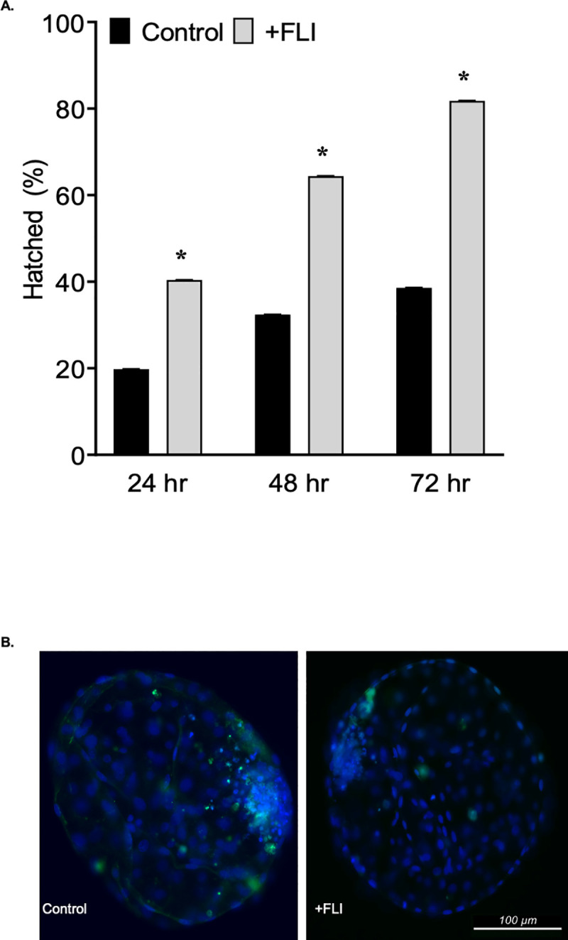
(A) The effect of the addition of FLI to culture medium and post-thaw hatching rate (n = 117; 51 control and 66 +FLI). (B) Images of bovine embryos post-thaw subjected to the TUNEL assay (green) and fluorescence imaging. Asterisk (*) indicates statistical differences (P < 0.05).
Discussion
Supplementation of the cytokine combination FGF2, LIF, and IGF1 (FLI) has been shown to increase the efficiency of in vitro porcine embryo production in [37]. These cytokines have been used individually in in vitro bovine embryo production systems and yielded positive results [27, 35, 51–53], so it is possible that combining these cytokines has the potential to increase bovine embryo development in vitro. Thus, the objective of this study was to investigate the role of FLI in the in vitro bovine embryo production system and evaluate the phenotypic effects on the oocyte and embryo.
The supplementation of FLI into maturation medium resulted in an earlier dissociation of the transzonal projections from the cumulus cell to the oocyte. Transzonal projections have a role in lipid accumulation within the embryo [12]. Our results support this finding as blastocyst stage embryos that developed from the oocytes supplemented with FLI had reduced lipid accumulation compared to those that did not receive FLI supplementation. It is possible that the transzonal projections act as an entryway for lipids or lipid precursors to enter the oocyte. Therefore, the earlier dissociation of the entry mechanism may prevent these molecules from traversing into the oocyte. Furthermore, it has been shown that lipid content decreases around the time of fertilization and the first cleavage division, then increases following the maternal to zygotic transition [54]. Suboptimal maturation conditions may result in excessive lipid uptake that is accumulated instead of used for energy. It is possible that FLI supplementation provides the maturation medium with a necessary component for regulation of lipid synthesis and accumulation. Growth factors such as LIF, IGF1, and FGF2 activate the mitogen‐activated protein kinase (MAPK) signaling cascade [51, 55, 56], which has a role in cellular metabolism and lipolysis [57–59].
One explanation is that the cytokines that compose FLI are interacting with lipid modulators that have a role in lipid droplet lipolysis and mobilization of energy stores. Interestingly, the decreased lipid content at the blastocyst stage did not result in an increased survivability or reduced DNA fragmentation following cryopreservation. This differs from previous studies showing that decreased lipid content resulted in increased survival after cryopreservation [60, 61]. Nile red staining was used to quantify the intracellular lipid content within the embryo. This method tags neutral lipids, specifically, triglycerides, the most abundant lipid in oocytes and embryos [54]. Triglycerides have been detected in higher amounts in in vitro derived embryos compared to in vivo [62]. Our results indicate that lipid content, or neutral lipids rather, may not be the factor that determines survival through cryopreservation, and further research is needed to understand the quality parameters that are important for survival of embryos to slow-freezing.
An increase in lipid content was observed at the blastocyst stage in the control group compared to the FLI supplemented but not in the zygote stage embryos. It is possible that the addition of FLI during maturation results in changes in lipid metabolism regulation that carry through to the blastocyst stage. There is evidence that embryos accumulate lipids from their culture environment [63]. At the zygote stage, the embryo has had minor exposure to the culture environment, but by the blastocyst stage the embryo has been exposed to the medium for 7 days. During this time, embryos with dysregulated lipid metabolism could accumulate excess lipids. We have shown that the addition of FLI to the maturation medium affects the regulation of lipid metabolism in blastocyst stage embryos as evidenced by a decrease in lipid content at this stage.
The addition of FLI during maturation did increase the proportion of oocytes that reached the metaphase II stage of meiosis. After removal from the ovary, timing of resumption and progression of meiosis depend largely on the maturation conditions [64]. Specifically, leukemia inhibitory factor (LIF), has been shown to increase both cytoplasmic and nuclear maturation when supplemented to in vitro oocyte maturation medium [11]. Oocyte maturation has a key role in determining the developmental competence of the oocyte to reach the blastocyst stage [6]. Interestingly, in this study, the increase in oocytes reaching the metaphase II stage did not result in an increase in the proportion of putative zygotes that underwent at least one cleavage division or reached the blastocyst stage. This suggests a role of FLI in driving the progression to metaphase II during nuclear maturation, however, the role of FLI during cytoplasmic maturation remains to be elucidated. In both groups, the percentage of oocytes that reached metaphase II by twenty-four hours of maturation was lower than the cleavage rate. One explanation would be that oocytes with competence to cleave had not reached metaphase 2 by twenty-four hours. Aspiration of the COC from the ovary did not occur in house so the size and maturity of the follicle is unknown. Some of the COCs may have finished maturation to metaphase 2 during fertilization and therefore had the capacity to cleave. In addition, some of them could be parthenotes, as is common in IVP. It should be noted that the current oocyte maturation medium used in this system contains serum with undefined growth factors. We recognize that the undefined molecules present in the serum may be masking the effect of FLI and that this combination of cytokines may be increasingly beneficial in a serum free system.
The addition of FLI at the beginning of culture increased embryonic development to the blastocyst stage. When compared to the US average (25.0%) [45] this is an increase of over ten percentage points. This is consistent with other work that has shown that these cytokines individually, or combinations of these cytokines can increase embryo development in vitro [27, 35, 51–53], indicating that these molecules may be stimulating key regulatory pathways that are important for embryo development.
In this study we observed a difference in cytoskeleton integrity between embryos treated with and without FLI. Fewer disruptions in the cell cytoskeleton and formation of a well-defined inner cell mass and blastocoel within the embryo would indicate that these embryos may be more prepared to undergo stressful processes such as embryo transfer or cryopreservation. While little is known about these cytokines and cell allocation, it is possible that FLI is stimulating pathways involved in cell-to-cell junction formation, cell organization, and compaction. FGFs acting through FGFR have been shown to have a role in cell differentiation, migration, and regeneration [65]. In mice, IGF-1 receptor activation is linked with trophectoderm cell survival and E-Cadherins, molecules involved in cell clustering [66]. Both of these cytokines work through activation of both the MAPK signaling cascade and PI3K pathways which are known to be involved in cell division and self-renewal.
Cryopreserving bovine embryos is an important component when it comes to the adoption and use of IVP. Supplementing FLI at beginning of culture increases the survivability of embryos following slow freezing, and decreased DNA fragmentation post-thawing. Previous studies have indicated that LIF supplementation prior to cryopreservation resulted in an increase in the post thaw survivability of embryos [30]. Additionally, it has been shown that IGF-1 alone or when combined with LIF increases the number of total cells and trophectoderm cells within the embryo following freezing [34]. In this study, lipid content was not a determining factor in post-thaw embryo survival. We have shown a role of FLI in improving post-thaw embryo survival in vitro but further studies are needed to understand if these results translate into improved pregnancy outcomes for IVP and frozen embryos.
The effects of FLI were not expressed through a difference in nuclear number, lipid content, or oxidative stress, indicating that the effects of FLI on quality are not reflected in the blastocyst stage embryo, but rather FLI is affecting the embryo earlier in development, which remains to be elucidated. Recent research has shown that Bovine Serum Albumin (BSA) interferes with the fibroblast growth factor signaling cascade [67]. With BSA present in the current culture medium, it is possible that it is blocking exogenous FGF2 from exerting its effect. The pathway associated with lineage commitment has been altered in mouse blastocyst stage embryos when they were cultured in the presence of BSA [67], thus, the same could be true for bovine.
In summary, the supplementation of FLI during the in vitro production of bovine embryos proves to be beneficial by improving preimplantation embryonic development and embryo quality. The positive influence of FLI is evident in oocyte maturation and embryo lipid content when added to the maturation medium, as well as embryo development and survivability when added to the culture medium. Further research will help to elucidate the mechanisms through which FLI modulates embryo development, quality, and survivability.
Supporting information
Secondary antibody control for localization of CDX2. Embryos were prepared as described above for determination of cell number with the primary antibody step omitted.
(TIF)
Acknowledgments
The authors would like to thank Kelsey Clark and Jason Rizo for their assistance with in vitro embryo production. The authors would also like acknowledge Dr. Bo Harstine from Select Sires, Inc. (Plain City, Ohio) for the bull semen used for this project and Angelica Giraldo from DeSoto BioSciences, Inc. (Seymour, TN) for help with the oocyte maturation experiment. The authors would like to thank Dr. Frank Baker and Dr. Alexander Jurkevich from the University of Missouri Molecular Cytology Core for their training and assistance with the confocal microscope, as well as, Dr. Ye Yuan for his help with transzonal projection analysis and Raissa Cecil for her help with the TUNEL assays.
Data Availability
All relevant data are within the manuscript and its Supporting Information files.
Funding Statement
This research is supported by the Clifton Murphy Scholarship Fund, and USDA National Needs Fellowship funded by USDA NIFA Grant 2019-38420-28972.The funders had no role in study design, data collection and analysis, decision to publish, or preparation of the manuscript.
References
- 1.Hansen PJ, Block J. Towards an embryocentric world: the current and potential uses of embryo technologies in dairy production. Reprod Fertil Dev 2004:15 10.10371/RD03073 [DOI] [PubMed] [Google Scholar]
- 2.Grothmann H. From Stem Cells to Neonates—The Embryo at the Center of All 2019:37 10.1016/j.theriogenology.2019.03.028 [DOI] [PubMed] [Google Scholar]
- 3.Fair T, Lonergan P, Dinnyes A, Cottell DC, Hyttel P, Ward FA, et al. Ultrastructure of bovine blastocysts following cryopreservation: Effect of method of blastocyst production. Mol Reprod Dev 2001; 58:186–195. [DOI] [PubMed] [Google Scholar]
- 4.Leibo SP, Loskutoff NM. Cryobiology of in vitro-derived bovine embryos. Theriogenology 1993; 39:81–94. [Google Scholar]
- 5.Říha J, Machatková M, Pavlok A. Viability of fresh and frozen transferred IVP bovine embryos. Czech J Anim Sci 2002; 47:261–267. [Google Scholar]
- 6.Sirard M-A, Richard F, Blondin P, Robert C. Contribution of the oocyte to embryo quality. Theriogenology 2006; 65:126–136. 10.1016/j.theriogenology.2005.09.020 [DOI] [PubMed] [Google Scholar]
- 7.Sugimura S, Richani D, Gilchrist RB. Follicular guidance for oocyte developmental competence. Anim Reprod 2018; 15:721–726. [DOI] [PMC free article] [PubMed] [Google Scholar]
- 8.Crosier AE, Farin PW, Dykstra MJ, Alexander JE, Farin CE. Ultrastructural Morphometry of Bovine Compact Morulae Produced In Vivo or In Vitro. Biol Reprod 2000; 62:1459–1465. 10.1095/biolreprod62.5.1459 [DOI] [PubMed] [Google Scholar]
- 9.Lonergan P, Rizos D, Gutierrez-Adán A, Moreira PM, Pintado B, de la Fuente J, et al. Temporal divergence in the pattern of messenger RNA expression in bovine embryos cultured from the zygote to blastocyst stage in vitro or in vivo. Biol Reprod 2003; 69:1424–1431. 10.1095/biolreprod.103.018168 [DOI] [PubMed] [Google Scholar]
- 10.Macaulay AD, Gilbert I, Caballero J, Barreto R, Fournier E, Tossou P, et al. The Gametic Synapse: RNA Transfer to the Bovine Oocyte. Biol Reprod 2014; 91 10.1095/biolreprod.114.119867 [DOI] [PubMed] [Google Scholar]
- 11.Makita M, Miyano T. Steroid hormones promote bovine oocyte growth and connection with granulosa cells. Theriogenology 2014; 82:605–612. 10.1016/j.theriogenology.2014.05.020 [DOI] [PubMed] [Google Scholar]
- 12.Collado M del, Silveira JC da, Sangalli JR, Andrade GM, Sousa LR da S, Silva LA, et al. Fatty Acid Binding Protein 3 And Transzonal Projections Are Involved In Lipid Accumulation During In Vitro Maturation Of Bovine Oocytes. Sci Rep 2017; 7:2645 10.1038/s41598-017-02467-9 [DOI] [PMC free article] [PubMed] [Google Scholar]
- 13.Iwasaki S, Yoshiba N, Ushijima H, Watanabe S, Nakahara T. Morphology and proportion of inner cell mass of bovine blastocysts fertilized in vitro and in vivo. Reproduction 1990; 90:279–284. 10.1530/jrf.0.0900279 [DOI] [PubMed] [Google Scholar]
- 14.Mori M, Otoi T, Suzuki T. Correlation between the Cell Number and Diameter in Bovine Embryos Produced In Vitro. Reprod Domest Anim 2002; 37:181–184. 10.1046/j.1439-0531.2002.00354.x [DOI] [PubMed] [Google Scholar]
- 15.Papaioannou VE, Ebert KM. The preimplantation pig embryo: cell number and allocation to trophectoderm and inner cell mass of the blastocyst in vivo and in vitro. Development 1988; 102:793–803. [DOI] [PubMed] [Google Scholar]
- 16.Soom AV, Boerjan M, Ysebaert M-T, Kruif A de. Cell allocation to the inner cell mass and the trophectoderm in bovine embryos cultured in two different media. Mol Reprod Dev 1996; 45:171–182. [DOI] [PubMed] [Google Scholar]
- 17.Cagnone GLM, Sirard M-A. Transcriptomic signature to oxidative stress exposure at the time of embryonic genome activation in bovine blastocysts. Mol Reprod Dev 2013; 80:297–314. 10.1002/mrd.22162 [DOI] [PubMed] [Google Scholar]
- 18.de Assis PM, Castro LS, Siqueira AFP, Delgado J de C, Hamilton TR dos S, Goissis MD, et al. System for evaluation of oxidative stress on in-vitro-produced bovine embryos. Reprod Biomed Online 2015; 31:577–580. 10.1016/j.rbmo.2015.06.014 [DOI] [PubMed] [Google Scholar]
- 19.Sudano MJ, Paschoal DM, da Silva Rascado T, Magalhães LCO, Crocomo LF, de Lima-Neto JF, et al. Lipid content and apoptosis of in vitro-produced bovine embryos as determinants of susceptibility to vitrification. Theriogenology 2011; 75:1211–1220. 10.1016/j.theriogenology.2010.11.033 [DOI] [PubMed] [Google Scholar]
- 20.Tharasanit T, Colenbrander B, Stout T a. E. Effect of cryopreservation on the cellular integrity of equine embryos. Reproduction 2005; 129:789–798. 10.1530/rep.1.00622 [DOI] [PubMed] [Google Scholar]
- 21.Zijlstra C, Kidson A, Schoevers EJ, Daemen AJJM, Tharasanit T, Kuijk EW, et al. Blastocyst morphology, actin cytoskeleton quality and chromosome content are correlated with embryo quality in the pig. Theriogenology 2008; 70:923–935. 10.1016/j.theriogenology.2008.05.055 [DOI] [PubMed] [Google Scholar]
- 22.Enright BP, Lonergan P, Dinnyes A, Fair T, Ward FA, Yang X, et al. Culture of in vitro produced bovine zygotes in vitro vs in vivo: implications for early embryo development and quality. Theriogenology 2000; 54:659–673. 10.1016/S0093-691X(00)00381-2 [DOI] [PubMed] [Google Scholar]
- 23.Lonergan P, Rizos D, Kanka J, Nemcova L, Mbaye AM, Kingston M, et al. Temporal sensitivity of bovine embryos to culture environment after fertilization and the implications for blastocyst quality 2003:10. [DOI] [PubMed] [Google Scholar]
- 24.Luciano AM, Franciosi F, Barros RG, Dieci C, Lodde V. The variable success of in vitro maturation: can we do better? Anim Reprod 2018; 15:727–736. [DOI] [PMC free article] [PubMed] [Google Scholar]
- 25.Salhab M, Dhorne‐Pollet S, Auclair S, Guyader‐Joly C, Brisard D, Dalbies‐Tran R, et al. In vitro maturation of oocytes alters gene expression and signaling pathways in bovine cumulus cells. Mol Reprod Dev 2013; 80:166–182. 10.1002/mrd.22148 [DOI] [PubMed] [Google Scholar]
- 26.Tríbulo P, Siqueira LGB, Oliveira LJ, Scheffler T, Hansen PJ. Identification of potential embryokines in the bovine reproductive tract. J Dairy Sci 2018; 101:690–704. 10.3168/jds.2017-13221 [DOI] [PubMed] [Google Scholar]
- 27.Fields SD, Hansen PJ, Ealy AD. Fibroblast growth factor requirements for in vitro development of bovine embryos. Theriogenology 2011; 75:1466–1475. 10.1016/j.theriogenology.2010.12.007 [DOI] [PubMed] [Google Scholar]
- 28.Mo X, Wu G, Yuan D, Jia B, Liu C, Zhu S, et al. Leukemia inhibitory factor enhances bovine oocyte maturation and early embryo development. Mol Reprod Dev 2014; 81:608–618. 10.1002/mrd.22327 [DOI] [PubMed] [Google Scholar]
- 29.Wooldridge LK, Ealy AD. Interleukin-6 increases inner cell mass numbers in bovine embryos. BMC Dev Biol 2019; 19. [DOI] [PMC free article] [PubMed] [Google Scholar]
- 30.Han YM, Lee ES, Mogoe T, Lee KK, Fukui Y. Effect of human leukemia inhibitory factor on in vitro development of IVF-derived bovine morulae and blastocysts. Theriogenology 1995; 44:507–516. 10.1016/0093-691x(95)00222-t [DOI] [PubMed] [Google Scholar]
- 31.Yamanaka M, Kudo T, Kimura N, Amano T, Itagaki Y. Effect of Bovine Leukemia Inhibitory Factor on Hatching and Numbers of Inner Cell Mass and Trophectoderm of Bovine Intact and Biopsied Blastocysts. J Anim Sci 1999; 70:444–450. [Google Scholar]
- 32.Zolini AM, Carrascal-Triana E, Ruiz de King A, Hansen PJ, Alves Torres CA, Block J. Effect of addition of l-carnitine to media for oocyte maturation and embryo culture on development and cryotolerance of bovine embryos produced in vitro. Theriogenology 2019; 133:135–143. 10.1016/j.theriogenology.2019.05.005 [DOI] [PubMed] [Google Scholar]
- 33.Jousan FD, Hansen PJ. Insulin-like Growth Factor-I as a Survival Factor for the Bovine Preimplantation Embryo Exposed to Heat Shock. Biol Reprod 2004; 71:1665–1670. 10.1095/biolreprod.104.032102 [DOI] [PubMed] [Google Scholar]
- 34.Kocyigit A, Cevik M. Effects of leukemia inhibitory factor and insulin-like growth factor-I on the cell allocation and cryotolerance of bovine blastocysts. Cryobiology 2015; 71:64–69. 10.1016/j.cryobiol.2015.05.068 [DOI] [PubMed] [Google Scholar]
- 35.Neira JA, Tainturier D, Peña MA, Martal J. Effect of the association of IGF-I, IGF-II, bFGF, TGF-β1, GM-CSF, and LIF on the development of bovine embryos produced in vitro. Theriogenology 2010; 73:595–604. 10.1016/j.theriogenology.2009.10.015 [DOI] [PubMed] [Google Scholar]
- 36.Sirisathien S, Brackett BG. TUNEL analyses of bovine blastocysts after culture with EGF and IGF-I. Mol Reprod Dev 2003; 65:51–56. 10.1002/mrd.10263 [DOI] [PubMed] [Google Scholar]
- 37.Yuan Y, Spate LD, Redel BK, Tian Y, Zhou J, Prather RS, et al. Quadrupling efficiency in production of genetically modified pigs through improved oocyte maturation. Proc Natl Acad Sci 2017; 114:E5796 10.1073/pnas.1703998114 [DOI] [PMC free article] [PubMed] [Google Scholar]
- 38.Rodina TM, Cooke FNT, Hansen PJ, Ealy AD. Oxygen tension and medium type actions on blastocyst development and interferon-tau secretion in cattle. Anim Reprod Sci 2009; 111:173–188. 10.1016/j.anireprosci.2008.02.014 [DOI] [PubMed] [Google Scholar]
- 39.Tríbulo P, Rivera RM, Ortega Obando MS, Jannaman EA, Hansen PJ. Production and Culture of the Bovine Embryo In: Herrick JR (ed.), Comparative Embryo Culture, vol. 2006 New York, NY: Springer New York; 2019:115–129. [DOI] [PubMed] [Google Scholar]
- 40.Nagano M. Acquisition of developmental competence and in vitro growth culture of bovine oocytes. J Reprod Dev 2019:2019–022. 10.1262/jrd.2019-022 [DOI] [PMC free article] [PubMed] [Google Scholar]
- 41.Ortega MS, Rocha-Frigoni NAS, Mingoti GZ, Roth Z, Hansen PJ. Modification of embryonic resistance to heat shock in cattle by melatonin and genetic variation in HSPA1L. J Dairy Sci 2016; 99:9152–9164. 10.3168/jds.2016-11501 [DOI] [PubMed] [Google Scholar]
- 42.Meiyu Q, Liu D, Roth Z. IGF-I slightly improves nuclear maturation and cleavage rate of bovine oocytes exposed to acute heat shock in vitro. Zygote Camb Engl 2015; 23:514–524. 10.1017/S096719941400015X [DOI] [PubMed] [Google Scholar]
- 43.Kalo D, Roth Z. Involvement of the sphingolipid ceramide in heat-shock-induced apoptosis of bovine oocytes. Reprod Fertil Dev 2011; 23:876–888. 10.1071/RD10330 [DOI] [PubMed] [Google Scholar]
- 44.Ortega MS, Moraes JGN, Patterson DJ, Smith MF, Behura SK, Poock S, et al. Influences of sire conception rate on pregnancy establishment in dairy cattle. Biol Reprod 2018; 99:1244–1254. 10.1093/biolre/ioy141 [DOI] [PMC free article] [PubMed] [Google Scholar]
- 45.International Embryo Technology Society (IETS). https://www.iets.org/pub_manual.asp. Accessed 27 March 2020.
- 46.Sosa F, Block J, Xiao Y, Hansen PJ. Determinants of survival of the bovine blastocyst to cryopreservation stress: treatment with colony stimulating factor 2 during the morula-to-blastocyst transition and embryo sex. CABI Agric Biosci 2020; 1:12. [DOI] [PMC free article] [PubMed] [Google Scholar]
- 47.de Sousa RV, da Silva Cardoso CR, Butzke G, Dode MAN, Rumpf R, Franco MM. Biopsy of bovine embryos produced in vivo and in vitro does not affect pregnancy rates. Theriogenology 2017; 90:25–31. 10.1016/j.theriogenology.2016.11.003 [DOI] [PubMed] [Google Scholar]
- 48.Block J, Hansen PJ, Loureiro B, Bonilla L. Improving post-transfer survival of bovine embryos produced in vitro: Actions of insulin-like growth factor-1, colony stimulating factor-2 and hyaluronan. Theriogenology 2011; 76:1602–1609. 10.1016/j.theriogenology.2011.07.025 [DOI] [PubMed] [Google Scholar]
- 49.Yang QE, Fields SD, Zhang K, Ozawa M, Johnson SE, Ealy AD. Fibroblast Growth Factor 2 Promotes Primitive Endoderm Development in Bovine Blastocyst Outgrowths. Biol Reprod 2011; 85:946–953. 10.1095/biolreprod.111.093203 [DOI] [PubMed] [Google Scholar]
- 50.Acosta DAV, Denicol AC, Tribulo P, Rivelli MI, Skenandore C, Zhou Z, et al. Effects of rumen-protected methionine and choline supplementation on the preimplantation embryo in Holstein cows. Theriogenology 2016; 85:1669–1679. 10.1016/j.theriogenology.2016.01.024 [DOI] [PubMed] [Google Scholar]
- 51.Bonilla AQS, Ozawa M, Hansen PJ. Timing and dependence upon mitogen-activated protein kinase signaling for pro-developmental actions of insulin-like growth factor 1 on the preimplantation bovine embryo. Growth Horm IGF Res 2011; 21:107–111. 10.1016/j.ghir.2011.03.003 [DOI] [PubMed] [Google Scholar]
- 52.Larson RC, Ignotz GG, Currie WB. Transforming growth factor β and basic fibroblast growth factor synergistically promote early bovine embryo development during the fourth cell cycle. Mol Reprod Dev 1992; 33:432–435. 10.1002/mrd.1080330409 [DOI] [PubMed] [Google Scholar]
- 53.Xie M, McCoski S, Johnson S, Rhoads M, Ealy A. Combinatorial effects of epidermal growth factor, fibroblast growth factor 2 and insulin-like growth factor 1 on trophoblast cell proliferation and embryogenesis in cattle. Reprod Fertil Dev 2015; 29. [DOI] [PubMed] [Google Scholar]
- 54.Ferguson EM, Leese HJ. Triglyceride content of bovine oocytes and early embryos. Reproduction 1999; 116:373–378. 10.1530/jrf.0.1160373 [DOI] [PubMed] [Google Scholar]
- 55.Hirai H, Karian P, Kikyo N. Regulation of embryonic stem cell self-renewal and pluripotency by leukaemia inhibitory factor. Biochem J 2011; 438:11–23. 10.1042/BJ20102152 [DOI] [PMC free article] [PubMed] [Google Scholar]
- 56.Ornitz DM, Itoh N. The Fibroblast Growth Factor signaling pathway. Wiley Interdiscip Rev Dev Biol 2015; 4:215–266. 10.1002/wdev.176 [DOI] [PMC free article] [PubMed] [Google Scholar]
- 57.Papa S, Choy PM, Bubici C. The ERK and JNK pathways in the regulation of metabolic reprogramming. Oncogene 2019; 38:2223–2240. 10.1038/s41388-018-0582-8 [DOI] [PMC free article] [PubMed] [Google Scholar]
- 58.Prates E, Nunes J, Pereira R. A Role of Lipid Metabolism during Cumulus-Oocyte Complex Maturation: Impact of Lipid Modulators to Improve Embryo Production. Mediators Inflamm 2014; 2014:692067 10.1155/2014/692067 [DOI] [PMC free article] [PubMed] [Google Scholar]
- 59.Sozen B, Ozturk S, Yaba A, Demir N. The p38 MAPK signalling pathway is required for glucose metabolism, lineage specification and embryo survival during mouse preimplantation development. Mech Dev 2015; 138 Pt 3:375–398. 10.1016/j.mod.2015.05.002 [DOI] [PubMed] [Google Scholar]
- 60.Barceló-Fimbres M, Seidel GE Jr. Effects of either glucose or fructose and metabolic regulators on bovine embryo development and lipid accumulation in vitro. Mol Reprod Dev 2007; 74:1406–1418. 10.1002/mrd.20700 [DOI] [PubMed] [Google Scholar]
- 61.Mucci N, Aller J, Kaiser GG, Hozbor F, Cabodevila J, Alberio RH. Effect of estrous cow serum during bovine embryo culture on blastocyst development and cryotolerance after slow freezing or vitrification. Theriogenology 2006; 65:1551–1562. 10.1016/j.theriogenology.2005.08.020 [DOI] [PubMed] [Google Scholar]
- 62.I.M A, G C, S K, Le Guienne B, Mermillod P, Guyader-Joly C, et al. Difference in lipid composition between in vivo-and in vitro-produced bovine embryos. Theriogenology 2000; 53:346. [Google Scholar]
- 63.McEvoy TG, Robinson JJ, Sinclair KD. Developmental consequences of embryo and cell manipulation in mice and farm animals. Reprod Camb Engl 2001; 122:507–518. [PubMed] [Google Scholar]
- 64.Dominko T, First NL. Timing of meiotic progression in bovine oocytes and its effect on early embryo development. Mol Reprod Dev 1997; 47:456–467. [DOI] [PubMed] [Google Scholar]
- 65.Yun Y-R, Won JE, Jeon E, Lee S, Kang W, Jo H, et al. Fibroblast Growth Factors: Biology, Function, and Application for Tissue Regeneration. J Tissue Eng 2010; 2010. [DOI] [PMC free article] [PubMed] [Google Scholar]
- 66.Bedzhov I, Liszewska E, Kanzler B, Stemmler MP. Igf1r Signaling Is Indispensable for Preimplantation Development and Is Activated via a Novel Function of E-cadherin. PLOS Genet 2012; 8. [DOI] [PMC free article] [PubMed] [Google Scholar]
- 67.Frum T, Ralston A. Culture conditions antagonize lineage-promoting signaling in the mouse blastocyst. Reproduction 2020; 160:V5–V7. 10.1530/REP-20-0107 [DOI] [PMC free article] [PubMed] [Google Scholar]
Associated Data
This section collects any data citations, data availability statements, or supplementary materials included in this article.
Supplementary Materials
Secondary antibody control for localization of CDX2. Embryos were prepared as described above for determination of cell number with the primary antibody step omitted.
(TIF)
Data Availability Statement
All relevant data are within the manuscript and its Supporting Information files.


