Abstract
Citrus canker caused by Xanthomonas citri subsp. citri (Xcc) is one of the most devastating diseases in citrus industry worldwide. Most citrus cultivars such as sweet orange are susceptible to canker disease. Here, we utilized wild citrus to identify canker-resistant germplasms, and found that Atalantia buxifolia, a primitive (distant-wild) citrus, exhibited remarkable resistance to canker disease. Although the susceptibility gene LATERAL ORGAN BOUNDARIES 1 (LOB1) could also be induced in Atalantia after canker infection, the induction extent was far lower than that in sweet orange. In addition, three of amino acids encoded by transcription factor TFIIAγ in Atalantia (AbTFIIAγ) exhibited difference from those in sweet orange (CsTFIIAγ) which could stabilize the interaction between effector PthA4 and effector binding element (EBE) of LOB1 promoter. The mutation of AbTFIIAγ did not change its interaction with transcription factor binding motifs (TFBs). However, the AbTFIIAγ could hardly support the LOB1 expression induced by the PthA4. In addition, the activity of AbLOB1 promoter was significantly lower than that of CsLOB1 under the induction by PthA4. Our results demonstrate that natural variations of AbTFIIAγ and effector binding element (EBE) in the AbLOB1 promoter are crucial for the canker disease resistance of Atalantia. The natural mutations of AbTFIIAγ gene and AbLOB1 promoter in Atalantia provide candidate targets for improving the resistance to citrus canker disease.
Author summary
It has been well documented that most citrus cultivars are susceptible to canker disease, while little is known about the resistance or susceptibility of primitive or wild citrus to canker disease. This study reveals that primitive citrus (Atalantia buxifolia) is highly resistant to citrus canker. Transcriptome data demonstrated that Atalantia had an active resistance response to the infection of Xcc, compared with susceptible sweet orange. Our results indicated that natural variations of AbTFIIAγ gene and AbLOB1 promoter contributed to the resistance. Hence, we propose that the natural mutations of AbTFIIAγ gene and AbLOB1 promoter could provide candidate targets for breeding canker resistant citrus.
Introduction
Citrus canker is one of the most devastating bacterial diseases in citrus industry worldwide. It causes severe necrosis symptoms on the leaves and fruits, accelerating the dropping of citrus fruit and leaves [1]. This disease causes considerable yield losses, increases in bactericide cost, ecosystem risks, and even negative impacts on socio-economy [2]. One of economic and environment-friendly approaches is to improve the resistance of the host plants. The pathogenic bacteria of Xanthomonas citri strains harbor different host range. For example, XccAW and XccA* strains have a narrow host range including Mexican lime, whereas XccA can cause disease in most commercial citrus varieties [3–5].
Since almost all citrus cultivars are susceptible to canker disease [6], there have been extensive studies of its pathogenesis. Previous studies have shown that Xanthomonas bacteria contain the type III secretion system (T3SS), and that different types of Xanthomonas strains have different numbers of type III secretion (T3S) effectors. Transcription activator-like effectors (TALEs) belong to the AvrBs3/PthA family of T3SS effectors, and they are vital for canker bacteria to form pustule on citrus [5,7]. A typical TALE processes an N-terminal translocation signal, a central repeat domain consisting of 34 amino acids, 3 nuclear localization signals, transcription factor binding motifs (TFBs), and C-terminal amino acid activation domain [8,9]. PthA4 of Xcc can activate LATERAL ORGAN BOUNDARIES 1 (LOB1) gene expression [10], and the expression of susceptibility genes can be suppressed either by variation of the TFIIAγ or by mutation of the effector binding element (EBE) [11]. LOB1 is a transcription factor belonging to the LATERAL ORGAN BOUNDARIES (LOB) gene family, which not only controls the expansion and growth of plant cells, but also promotes bacterial growth and pustule formation in citrus [12,13].
Transcription factor IIA (TFIIA) is vital for the transcriptional regulation in eukaryotes. In humans, TFIIA contains three subunits, including α, β and γ subunits, among which α and β subunits are encoded by the same gene, while in yeast, TFIIA only contains the TOA1 and TOA2 subunits with TOA2 homologous to human TFIIAγ subunit [14,15]. Several studies have illustrated that TFIIA is required for RNA polymerase II-dependent transcription, and it can stabilize the TATA box-binding protein (TBP)-TFIID complex in the TATA box region of the promoters [16–18]. In plants, TFIIAγ is expressed in most tissues, especially in the young and active tissues [19]. In addition, TFIIAγ is associated with the disease resistance of rice [20]. In rice, it has been reported that TFBs are found to be vital for the infection of Xanthomonas bacteria, that TFBs can interact with the host basal transcription factor IIA gamma subunit (OsTFIIAγ5) to promote the expression of susceptibility genes, and that silencing OsTFIIAγ5 results in the increased resistance to Xanthomonas oryzae pv. oryzae (Xoo) and Xanthomonas oryzae pv. oryzicola (Xoc) [20]. However, its allele xa5, which carries a V to E mutation at the 39th amino acid position, could not interact with the TFBs of Xoo and Xoc [21,22]. The mutation at the 39th amino acid could lead to the interaction of xa5 with an acidic activation domain (AAD) of avirulence protein Avrxa5, thus impeding host cell transcription, eventually resulting in subsequent cell death as well as the resistance reaction [23,24]. In sweet orange, TALE TFBs could also interact with basal transcription factor (CsTFIIAγ) to promote Xcc infection [20].
In rice, SWEET genes belong to the sucrose transporter family, and are required for the susceptibility to rice blight. Natural mutations in the EBEs of SWEET genes have been reported in rice blight resistant varieties [11,25]. Editing the EBE regions of TALE-induced SWEET genes impedes the expression of corresponding susceptibility genes [26]. To the best of our knowledge, the polymorphism of the EBEs in the susceptibility gene promoter has not been reported in citrus canker resistant varieties so far.
Citrus has a broad spectrum of resources ranging from primitive, wild, and cultivated varieties. Currently, little is known about the resistance or susceptibility of primitive or wild citrus to canker disease. Atalantia buxifolia (Chinese box orange), as a primitive citrus, shows tolerance to diverse abiotic and biotic stresses and is sometimes used as rootstock for the grafting of commercial citrus [27,28]. It becomes possible to identify resistant genes to citrus canker disease with the availability of the genomes of Atalantia and other wild germplasms [29,30]. In this study, we found that Atalantia is resistant to citrus canker. The susceptibility gene LOB1 was induced in Atalantia after inoculation with Xcc, but its induction degree was much lower in Atalantia than in sweet orange. In contrast to CsTFIIAγ of sweet orange, AbTFIIAγ of Atalantia could hardly support the expression of the CsLOB1 gene induced by PthA4, and the activity of the AbLOB1 promoter was significantly lower than that of CsLOB1 induced by PthA4. Our results indicate that the natural mutations of AbTFIIAγ gene and AbLOB1 promoter are vital for the canker resistance of Atalantia.
Results
Resistance of Atalantia to citrus canker
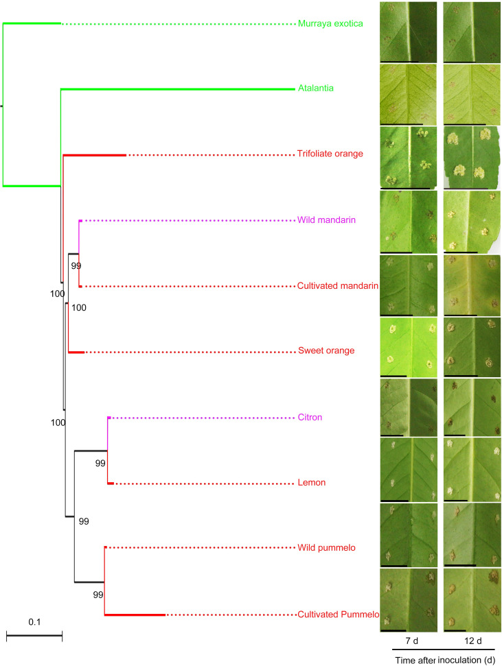
Great research efforts have been made to evaluate the canker disease resistance of various commercial citrus cultivars and the relatives, and almost all cultivars have been found to be susceptible to the disease [2,7]. To explore the canker resistant germplasms, we expanded the research scope to investigate wild citrus in tribe Citrinae belonging to the subfamily Aurantioideae. Ten citrus varieties were chosen for canker disease evaluation. The disease lesion area was determined at 12 d after the inoculation with Xcc. Based on the results, the citrus varieties were classified into three categories: susceptible (lesion area = 2–4 mm2), tolerant (lesion area = 1–2 mm2), and resistant (lesion area = 0–1 mm2) (S1 Fig). Three wild citrus varieties including wild mandarin, wild pummelo, and citron showed different degrees of susceptibility to citrus canker. Wild mandarin and citron showed tolerance to citrus canker in the early stages, but exhibited small lesions at 12 d after inoculation. The wild pummelo of purple pummelo were susceptible to citrus canker. However, remarkable resistance was observed in Atalantia buxifolia, a distant wild relative of citrus (Fig 1).
Fig 1. Atalantia buxifolia resistance to citrus canker.
The phylogenetic tree was constructed by using the maximum likelihood tree method and the substitution model GTRGAMMA in the RAxML software. Murraya koenigii was used as the outgroup. Fully expanded leaves were treated with Xcc (108 CFU/mL) to evaluate citrus canker resistance. The symptoms were observed at 7 and 12 d after inoculation. Red indicates canker susceptible citrus (Wild pummelo: purple pummelo; Cultivated pummelo: guanxi pummelo; Cultivated mandarin: Ponkan; Lemon: Eureka lemon). Purple indicates canker tolerant citrus (Wild mandarin: mangshan mandarin). Green indicates canker-resistant citrus. Bootstrap values greater than 60 are labelled at the node in the tree. Scale bars, 1 cm.
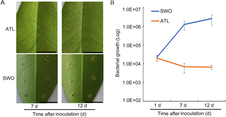
A low bacterial titer of Xcc was used to further evaluate the canker disease development in Atalantia. Sweet orange, a variety known to be susceptible to canker disease, was used as the control. Canker development was assessed at 7 d and 12 d after Xcc inoculation (Fig 2). At 7 d, canker lesion was not observed on the leaves of Atalantia, but obvious canker symptoms appeared on the leaves of sweet orange (Fig 2A). Bacterial growth evaluation results showed that the population of Xcc decreased at 7 and 12 d in Atalantia, compared with that in sweet orange, indicating the inhibition of bacterial growth (Fig 2B).
Fig 2. Inability to grow of Xcc in Atalantia at low bacterial titer.
(A) Fully expanded mature leaves of Atalantia (ATL) and sweet orange (SWO) were treated with Xcc (106 CFU/mL). Photographs were taken at 7 and 12 d after inoculation. (B) Bacterial growth in ATL and SWO at 1, 7, and 12 d after Xcc inoculation. Error bars indicate standard deviation of three independent tests. Scale bars, 1 cm.
Differentially expressed genes of Atalantia in response to canker infection
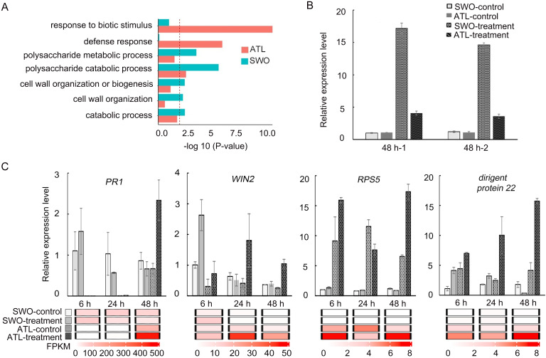
To reveal the molecular basis for the resistance of Atalantia to Xcc, samples were collected from leaves inoculated with Xcc (treatment) and leaves inoculated with sterile water (control) at different time points (hour 6, 24 and 48). A total of approximately 772 million paired-end reads (32 million reads for each library on average) were obtained by Illumina sequencing technology (S1 Table). Gene ontology (GO) analysis results indicated that the up-regulated genes in Atalantia were enriched in pathways such as defense response and biotic stimulus response, while those in sweet orange were enriched in polysaccharide metabolic and catabolic pathways at 48 h after the inoculation (Fig 3A). The cross-comparison Venn diagram of differentially expressed genes (DEGs) showed that eight DEGs were overlapped between Atalantia and sweet orange, one of which was citrus canker susceptibility gene LOB1 (S2 Table) [10]. However, the transcriptome data showed that the relative expression level of AbLOB1 was much lower than that of CsLOB1. Quantitative real-time PCR data confirmed that the expression level of CsLOB1 was three folds as high as that of AbLOB1 (Fig 3B). To confirm expression of the DEGs (S3 Table), quantitative RT-PCR was conducted for 10 candidate genes chosen from the transcriptional profile. These unigenes such as pathogenesis-related 1 (PR1), HopW1-1-interacting 2 (WIN2), dirigent-like protein 22, and pathogenesis-related 4 (PR4) were up-regulated in Atalantia from 6 h to 48 h after Xcc inoculation, while these genes were down-regulated in sweet orange. Gene RPS5 (Resistant to pseudomonas syringae 5) and quinone oxidoreductase gene were only significantly induced in Atalantia at 6 h, 24 h, 48 h after Xcc inoculation. Other genes such as repressor GA3 (RGA3), cytochrome P450 (CYP76C4) were up-regulated in Atalantia and down-regulated in sweet orange at 48 h after Xcc inoculation (Figs 3C and S2). Correlations between the ten genes expression and RNA-seq data were shown in S2C Fig. Overall, the expression profile of most genes selected for qRT-PCR validation was consistent with the RNA-Seq data (S2C Fig).
Fig 3. Differentially expressed genes in Atalantia and sweet orange after Xcc (108 CFU/ml) inoculation.
(A) GO enrichment analysis of the up-regulated genes in Atalantia (ATL) and sweet orange (SWO) at 48 h after inoculation. (B) qRT-PCR validation of LOB1 expression in Atalantia and sweet orange at 48 h after inoculation with Xcc and sterile water. (C) Expression levels of four DEGs: pathogenesis-related gene 1 (PR1), Resistance to Pseudomonas syringae 5 (RPS5), HopW1-1-Interacting 2 (WIN2), and dirigent-like protein 22 genes, and their corresponding expression patterns in RNA-Seq data in Atalantia and sweet orange. The treatment group was inoculated with Xcc and the control group was inoculated with sterile water at 6 h, 24 h, and 48 h. All the gene expressions were normalized according to the gene expression of the sweet orange control group at 6 h post inoculation. The relative expression level was calculated by 2-△△Ct method with EF1a as the reference gene. Error bars indicate standard deviation of three independent repetitions.
Genetic variations of TFIIAγ in resistant and susceptible citrus and their interaction with TAL effectors
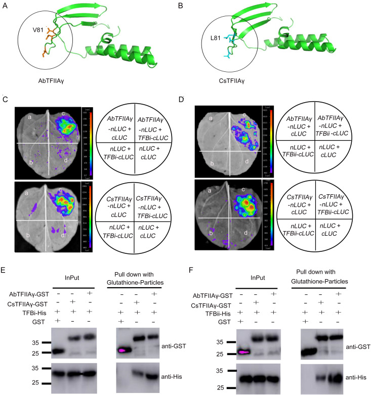
Previous studies have shown that TFIIAγ could stabilize TAL effectors in the TATA box region and induce LOB1 expression and susceptibility [20]. We further investigated the sequence variations of the TFIIAγ gene in the resistant and susceptible germplasms. Full-length nucleotide sequences of TFIIAγ were cloned from primitive, wild, and cultivated varieties. Amino acid alignment indicated that the TFIIAγ was highly conserved in the investigated varieties except the resistant citrus Murraya and Atalantia from primitive varieties, and that Murraya and Atalantia showed slight differences in amino acid sequences from susceptible and tolerant citrus (S3 Fig). Furthermore, the three-dimensional structures of AbTFIIAγ and CsTFIIAγ protein were modeled. Homology modeling of AbTFIIAγ and CsTFIIAγ proteins (Fig 4A and 4B) and their mutant proteins (S4 Fig) revealed that the 81st residue of AbTFIIAγ and CsTFIIAγ was exposed on the surface of a β-sheet, and that the side chain of leucine (L81) in CsTFIIAγ was longer than that of valine (V81) in AbTFIIAγ.
Fig 4. Interactions of AbTFIIAγ or CsTFIIAγ with TALE TFBs.
(A) Homology modeling of AbTFIIAγ protein. (B) Homology modeling of CsTFIIAγ protein. The structures of TFIIAγ proteins were homology modeled using SWISS-MODEL online service with default parameters (Fig 4A and 4B). (C) Interactions between AbTFIIAγ-nLuc or CsTFIIAγ-nLuc and TFBi-cLuc. (D) Interactions between AbTFIIAγ-nLuc or CsTFIIAγ-nLuc and TFBii-cLuc. Note: a, b, and d indicate the negative controls of tobacco leaves; c denotes the interactions between TFBi-cLuc or TFBii-cLuc and AbTFIIAγ-nLuc or CsTFIIAγ-nLuc in tobacco leaves in luciferase complementation assay (Fig 4C and 4D). (E) The interaction of AbTFIIAγ or CsTFIIAγ with TFBi in pull-down assays. (F) The interaction of AbTFIIAγ or CsTFIIAγ with TFBii in Pull-down assays. Note: GST-tagged AbTFIIAγ or GST-tagged CsTFIIAγ and His-tagged TFBi or His-tagged TFBii were incubated with immobilized glutathione S-transferase (GST) (Fig 4E and 4F). The experiments were repeated three times independently.
To determine whether AbTFIIAγ can interact with TALE TFBs, luciferase complementation and pull-down assay were carried out. Luciferase activity was detected when AbTFIIAγ-nLuc or CsTFIIAγ-nLuc was co-expressed with TFBi-cLuc/TFBii-cLuc (Fig 4C and 4D), indicating both AbTFIIAγ and CsTFIIAγ could interact with TFBs. GST pull-down assay in vitro was performed using purified AbTFIIAγ-GST/CsTFIIAγ-GST and TFBi-His/TFBii-His proteins to further verify their interaction. Pull-down assay results indicated that His-tagged TFBi or TFBii interacted with the GST-tagged AbTFIIAγ and CsTFIIAγ proteins, but not with GST alone (Fig 4E and 4F).
Enhancement of resistance to Xcc by silencing of TFIIAγ in sweet orange
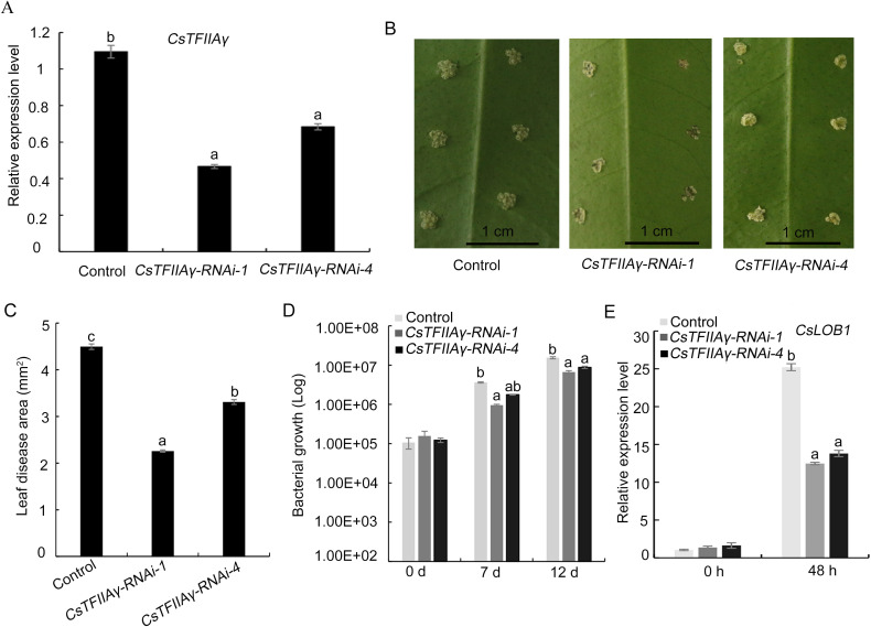
A previous report has revealed that transient interference of CsTFIIAγ in sweet orange enhanced its resistance to Xcc [20]. Here, we suppressed the CsTFIIAγ gene expression in sweet orange leaves by RNA interference (RNAi) for the functional complementation of AbTFIIAγ because of the unavailability of the gene transformation system for Atalantia. The results showed that the reduction of CsTFIIAγ gene expression resulted in the decrease in the disease lesion area and the inhibition of the bacterial growth and the CsLOB1 expression, compared with the control (Fig 5).
Fig 5. Enhancement of sweet orange resistance to Xcc through CsTFIIAγ RNA interference.
(A) Relative expression levels of CsTFIIAγ in CsTFIIAγ-RNAi group and control group. (B) Symptoms of CsTFIIAγ-silenced lines (RNAi-1 and RNAi-4) and control leaves after inoculation with Xcc (108 CFU/mL). The photos were taken at 12 d after inoculation. (C) Disease lesion area on CsTFIIAγ-silenced lines and control leaves at 12 d after inoculation. The disease lesion area was calculated by ImageJ 2.0. (D) Bacterial growth in CsTFIIAγ-RNAi plants and control plants at 0, 7, and 12 d after inoculation. (E) Relative expression level of CsLOB1 at 48 h after inoculation. Data from three independent replicates were expressed as mean ± SD. Different letters above the bars represent significant differences (P < 0.05) in Duncan’s multiple range test.
Complementation assay of AbTFIIAγ and CsTFIIAγ
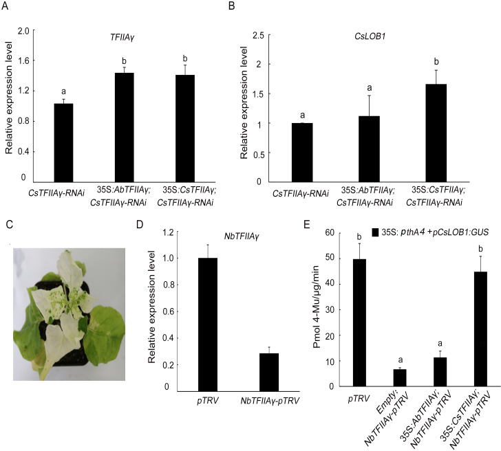
AbTFIIAγ or CsTFIIAγ was transformed into a vector containing the promoter CaMV 35S and terminator NOS, respectively. Subsequently, the resultant two vectors were respectively transformed into CsTFIIAγ-silenced citrus line (RNAi-1). Complementary CsTFIIAγ in RNAi-1 line treated with Xcc promoted the CsLOB1 gene expression, whereas the complementary AbTFIIAγ hardly supported this gene expression (Fig 6A and 6B). In order to further clarify the function of AbTFIIAγ, we performed the complementary assay in Nicotiana benthamiana. First, N. benthamiana TFIIAγ (NbTFIIAγ) was silenced, and the N. benthamiana phytoene dehydrogenase gene was used as the positive control to confirm the effectiveness of the virus-induced gene silencing (VIGS). Obvious albino phenotype was observed at 15 d (Fig 6C). CsLOB1 promoter fused with the β-glucuronidase (GUS) reporter gene vector and 35S promoter-driven pthA4 vector were transformed into NbTFIIAγ-silenced tobacco to detect the function of AbTFIIAγ and CsTFIIAγ. We first confirmed that gene AbTFIIAγ and CsTFIIAγ were expressed in NbTFIIAγ-silenced tobacco (S5 Fig), then we checked the promoter activity. The results showed that the complementary CsTFIIAγ restored the reduced GUS activity of CsLOB1 promoter in NbTFIIAγ-silenced plants, while the complementary AbTFIIAγ hardly restored the reduced activity of CsLOB1 promoter which was similar to that of complementary empty vector (Fig 6D and 6E).
Fig 6. CsLOB1 gene expression in TFIIAγ-silenced sweet orange and tobacco hardly supported by AbTFIIAγ.
(A) Relative expression of TFIIAγ. (B) Relative expression of CsLOB1. Transient expression of AbTFIIAγ, CsTFIIAγ, and empty vector in CsTFIIAγ-RNAi sweet orange line (RNAi-1) at 4 days after Xcc (108 CFU/ml) inoculation (Fig 6A and 6B). (C) Albino phenotype exhibited by NbPDS-pTRV tobacco at 15 d after silencing NbPDS. (D) Relative expression level of NbTFIIAγ in NbTFIIAγ-pTRV and control (pTRV) tobacco. (E) Expression of vector pCsLOB1:GUS induced by 35S:pthA4 in NbTFIIAγ-pTRV plants after complementation of AbTFIIAγ, CsTFIIAγ, or empty vector. Data from three independent replicates were expressed as mean ± SD. Different letters above the bars represent significant differences (P < 0.05) in Duncan’s multiple range test.
Significant lower promoter activity of AbLOB1 than that of CsLOB1 mediated by PthA4
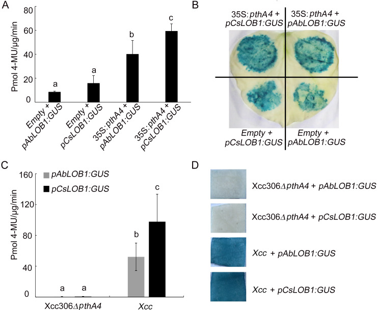
Previous research has shown that the PthA4 of Xcc306 can bind to the promoter of LOB1 to increase its expression [10]. We further compared AbLOB1 and CsLOB1 promoters and coding sequences, and found two single nucleotide polymorphisms (SNPs) in their EBE regions of promoters (S6A Fig). Amino acid alignment revealed that LOB1 was highly conserved (S6B Fig). To further verify the activities of the two promoters, AbLOB1 and CsLOB1 promoters were fused to pKGWFS7 vector containing the β-glucuronidase (GUS) reporter gene [10]. No significant difference in GUS activity was observed between AbLOB1 or CsLOB1 co-infiltrated with empty vector PBI121, but the GUS activity of AbLOB1 was significantly lower than that of CsLOB1 when these two promoters were co-infiltrated with vector 35S:pthA4 (Fig 7A and 7B). We further compared the activities of these two promoters in sweet orange leaves by transient expression mediated by Xcc [10]. The results showed that GUS activity of AbLOB1 was also significantly lower than that of CsLOB1 when these two promoters were co-infiltrated with the Xcc, whereas no GUS activity of two promoters was observed upon the co-infiltration with the mutant strain Xcc306ΔpthA4 (Fig 7C and 7D).
Fig 7. Significantly lower promoter activity of AbLOB1 than CsLOB1 induced by PthA4.
(A) Promoter activity of AbLOB1 and CsLOB1 induced by PthA4 in tobacco. (B) GUS staining assay in tobacco leaves. Agrobacteria containing pAbLOB1:GUS or pCsLOB1:GUS mixed with 35S: pthA4 or empty vector were infiltrated into tobacco leaves. Samples were collected at 2 days (Fig 7A and 7B). (C) Transient GUS activity related to pAbLOB1 and pCsLOB1 promoters after inoculation with Xcc or Xcc306ΔpthA4 in sweet orange. Samples were collected for further analysis at 4 days post inoculation. (D) GUS staining assay of sweet orange leaves upon ectopic expression of pAbLOB1:GUS or pCsLOB1:GUS post inoculation with Xcc or Xcc306ΔpthA4. Error bars indicate standard deviation of three independent replicates. Different letters above the bars represent significant differences (P < 0.05) in Duncan’s multiple range test.
Discussion
It has been reported that cultivated citrus varieties are more susceptible to canker disease than wild citrus, and that wild citrus varieties have a broad spectrum of disease resistance, whereas cultivated citrus varieties have lost certain resistance to different degrees during domestication [2,6]. For example, Meiwa kumquat obtain a durable resistance to Xcc by upregulating a host susceptibility gene to elicit immune responses [31]. Tracing back to wild germplams is an alternative strategy for identifying gene resources for breeding purpose. Chinese box orange (Atalantia buxifolia) is widely spread in Southern China, Philippines, and Japan [32,33]. It is a distant wild relative of Citrus, and it is occasionally used as rootstock due to its tolerance to diverse biotic and abiotic stresses [28]. In this study, we demonstrated that Atalantia, as a wild variety, was resistant to citrus canker.
LOB1 is a susceptibility gene whose expression is crucial for the formation of pustule in different citrus varieties [10,12]. We found that the expression of LOB1 was remarkably lower in Atalantia than in sweet orange. Previous studies have indicated that TFIIAγ can stabilize and increase the expression of the LOB1 gene [20]. Considering this, we examined the genetic variations of TFIIAγ in resistant and susceptible varieties, and found that natural mutations in Atalantia, AbTFIIAγ could hardly support TALE-mediated CsLOB1 gene expression (Fig 6). Homology modeling of AbTFIIAγ and CsTFIIAγ proteins and their mutant proteins revealed that the 81st residues of AbTFIIAγ and CsTFIIAγ were exposed on the surface of a β-sheet, and that the side chain of leucine (L81) was longer than that of valine (V81). We speculated that leucine (L81) in sweet orange might be more conducive to the stabilization of the protein complex (TBP)-TFIID in the TATA box region of LOB1 promoter due to its longer alkyl side chain than valine (V81) in Atalantia, which may partly explain the difference expression of LOB1 in sweet orange and Atalantia. Based on the above results, it could be concluded that Atalantia possessed an unique allele AbTFIIAγ, which contributed to the canker resistance in Atalantia.
The mutation of EBE on the AbLOB1 promoter also contributed to the disease resistance in Atalantia. We found two substitutions at the 7th (C to T) and 13th (T to C) position in the EBE region of promoter AbLOB1 (S6A Fig). A previous study has shown that a single base insertion in the EBE region could offset PthA4-mediated CsLOB1 expression in citrus, and that the substitution at the 9th (C to T) position could compromise PthA4-induced CsLOB1 expression [10]. Consistently, a substitution at the 10th position in the EBE region appeared to compromise AvrXa7-induced OsSWEET14 expression in a study of rice blight [25]. Our results revealed that the natural mutation of EBE on the AbLOB1 promoter also compromised PthA4-induced LOB1 expression (Fig 7). To our knowledge, the expression of LOB1 is crucial for the formation of pustule. A comparison of LOB1 gene expression 48 h after inoculation with Xcc or Xcc306ΔpthA4 in Atalantia and sweet orange revealed that the expression of AbLOB1 inoculated with Xcc was similar to that of CsLOB1 inoculated with Xcc306ΔpthA4 (S7 Fig). It is known that the Xcc306ΔpthA4 strain is not pathogenic in citrus, and that low expression of LOB1 can reduce the formation of pustule [10]. On the other hand, the inability of Xcc to grow on the leaf of Atalantia (Fig 2B) and the up-regulation of the disease defense-related genes (Figs 3C and S2) suggested that an active response occurred in Atalantia after Xcc infection.
Our data demonstrated that both cis- and trans-regulatory elements of AbLOB1 contributed to the canker resistance of Atalantia. The mutations might provide some useful target sites for improving the resistance to citrus canker, a devastating and stubborn disease which is mostly controlled by chemicals at present [34,35]. The current control approaches are costly due to the use of bactericide, and environmentally- and ecologically-unfriendly [36,37]. Breeding canker-resistant cultivars is a promising strategy for solving the above problems. Recently, genome editing technology has been widely applied to breeding disease-resistant cultivars due to its high efficiency and accuracy. In rice, SWEET genes are the members of sucrose transporter gene family, and are required for the susceptibility to rice blight. Gene editing in the EBE regions of SWEET11, SWEET13, and SWEET14 promoters could confer rice with broad-spectrum resistance to bacterial blight [38]. In citrus, editing the susceptibility gene CsLOB1 and its promoter could enhance citrus canker resistance. However, most of the edited citrus might still be infected with canker disease to different degrees [39,40]. Our data indicated the CsTFIIAγ could regulate the expression of gene CsLOB1. It is worthwhile to evaluate the phenotype and agronomic performances of citrus plants whose TFIIAγ gene and EBEs of LOB1 are edited. Homology recombination [41,42] and single base editing [43] are promising approaches to simultaneously modifying TFIIAγ and EBEs of LOB1 in susceptible citrus.
Experimental procedures
Materials and methods
Plant materials and pathogen
All the citrus varieties used in this study were taken from the National Citrus Breeding Center of Huazhong Agricultural University. All the transgenetic and control plants were grown in a greenhouse at 25–30°C. The X. citri strain 3213 (hereafter referred to as Xcc) and the mutant strain Xcc306ΔpthA4 were grown on nutrient agar medium at 28°C as previously reported [44].
Assay of pathogenicity
Fully expanded leaves were inoculated with Xcc (106 and 108 CFU/ml) and Xcc306ΔpthA4 (108 CFU/ml) with an inoculating needle (0.5 mm in diameter). Each inoculation site consisted of six pricks according to previous reports [45] with minor modifications. Bacterial suspension was dropped into each inoculation site. The disease lesion area was measured (36 punctures on average) with ImageJ 2.0. To examine the bacterial growth in citrus plants, leaf disk with 0.5 cm diameter in inoculated area was punched at 0, 1, 7, and 12 d after inoculation. DNA was extracted from 100 mg fresh weight of inoculated leaf disks, and 100 ng of total nucleic acid per inoculated sample was used as the template for quantitative polymerase chain reaction (qPCR). pthA of Xcc was used to calculate the bacterial population using the following formula [46,47].
Construction of phylogenetic tree of Citrinae
SNPs contained in the conserved single-copy genes in citrinae genomes were used for the construction of the phylogenetic tree of 10 varieties, namely, Murraya exotica, Atalantia, Citron, Wild mandarin (mangshan mandarin), Cultivated mandarin (Ponkan), Trifoliate orange, Lemon (Eureka lemon), Wild pummelo (purple pummelo), Cultivated pummelo (guanxi pummelo), Sweet orange. The maximum likelihood tree was constructed by using the substitution model GTRGAMMA in the RAxML software with Murraya koenigii as the outgroup. A total of 1,000 rapid bootstrap inferences were performed. The bootstrap value greater than 60 was labelled at each node in the tree.
Gene expression analysis
Total RNA was extracted using the HiPure HP Plant RNA Mini Kit (Magen, Guangzhou, China) according to the manufacturer’s protocol. RNA samples were reverse transcribed into cDNA using the Maxima H Minus First-Strand cDNA Synthesis Kit (Thermo Scientific, Shanghai, China). The qRT-PCR was performed with SYBR Green PCR Master Mix (Applied Biosystems, American), and amplification was performed on ABI 7900 Fast Real Time System (PE; Applied Biosystems). The elongation factor (EF1α) was used as the endogenous gene of citrus, while Ubiquitin (UBQ) was selected as the endogenous gene of tobacco. The used primers were listed in S4 Table. The relative gene expression was calculated using 2−ΔΔCt method.
Transcriptome data analysis
Total RNA was extracted from sterile water- or Xcc (108 CFU/ml)- inoculated Atalantia and sweet orang leaves at 6 h, 24 and 48 h after inoculation for RNA sequencing. The RNA-seq reads were mapped to the reference genome of sweet orange by HISAT2 [48]. The mapping results were transformed and sorted by samtools [49]. Gene expression values were normalized as Fragments Per Kilobase per Million (FPKM) by Cufflinks package [50]. The genes with P < 0.01 and an absolute value (fold change) log2 ratio ≥ 1 were defined as differentially expressed genes (DEGs). DEGs from the RNA seq data were analyzed and presented in S3 Table. Gene ontology term (GO) enrichment analysis was performed using Agrigo (https://www.Agrigo.com/) with FDR < 0.05. Our RNA-seq data were submitted to NCBI with genbank accession numbers of PRJNA612768 and PRJNA612769.
Molecular cloning and sequence analysis of TFIIAγ and LOB1
The TFIIAγ gene sequences were cloned from the cDNA of the ten citrus varieties (Murraya exotica, Atalantia, Citron, mangshan mandarin, Ponkan, Trifoliate orange, Eureka lemon, purple pummelo, guanxi pummelo, Sweet orange). Gene AbLOB1 and CsLOB1 and their promoter sequences were amplified from the cDNA and DNA of Atalantia and sweet orange. The primers used for amplification were listed in S4 Table. The gene ID of AbTFIIAγ was sb14580, and that of CsTFIIAγ gene was Cs3g16970 in the citrus genome database (http://citrus.hzau.edu.cn/cgi-bin/orange/blast). Sequence alignment of TFIIAγ and LOB1 was conducted by ClustalW2 and GENEDOC software.
Homology modeling of TFIIAγ
The structures of AbTFIIAγ and CsTFIIAγ were homology modeled using SWISS-MODEL online service with default parameters (https://swissmodel.expasy.org/). The crystal structure of transcription initiation factor IIA from human HsTFIIA (PDB ID: 5M4S) was used as the template. AbTFIIAγ and CsTFIIAγ exhibited 53% and 49% sequence identity with the HsTFIIA template, respectively. The structures of AbTFIIAγ and CsTFIIAγ were generated by PyMOL.
Luciferase complementation assay
The full-length coding sequence (CDS) of TFIIAγ without stop codons was cloned into vector JW-like-771-nLuc, and sequence TFBi/TFBii were cloned into vector JW-like-772-cLuc. The primers were listed in S4 Table. All the vectors were transformed into Agrobacterium tumefacien strain GV3101. Then, the Agrobacterium strain carrying the two constructed reporter vectors (JW-like-771-nLuc and JW-like-772-cLuc at a ratio of 1:1) was infiltrated into tobacco leaves at OD 600 nm = 0.8. The images of luciferase signal were taken at 2 d via a charge coupled device camera (LB985 NightSHADE) from the Key Laboratory of Horticultural Plant Biology, Ministry of Education.
Pull-down assay
The full-length TFIIAγ and TFBs were fused into GST-tagged PGEX-6p and HIS-tagged PET-32a, respectively. The proteins with GST-tag or His-tag were expressed in E.coli BL21 strain in vitro and purified with a GST purification kit (Sangon Biotech, Shanghai, China) or Ni-NTA purification kit (Sangon Biotech, Shanghai, China). Both types of proteins were purified following the manufacturer’s protocol. The GST-tagged proteins were incubated with Mag-Beads GST fusion protein purification kit (Sangon Biotech, Shanghai, China) for two hours, and washed four times. After one-hour incubation of His-tagged protein, the Mag-Beads were eluted and target protein was separated. The eluted proteins were tested with anti-GST (Smart-lifesciences, Changzhou, China) or anti-His (Smart-lifesciences, Changzhou, China) by using western blotting.
CsTFIIAγ silencing assay
CsTFIIAγ gene sequence (174 bp from the ATG start codon) was cloned from sweet orange cDNA and fused into vector pk7GWIWG2D (provided by professor Chunying Kang of Key Laboratory of Horticultural Plant Biology). The self-complementary RNAi vector pk7GWIWG2D had the selection tag eGFP controlled by 35S promoter. The vector CsTFIIAγ-pk7GWIWG2D was transformed into Agrobacterium tumefaciens strain EHA105. The resultant Agrobacterium was further transformed into sweet orange epicotyl explants, as previously reported [51,52].
Vector construction
NbTFIIAγ (174 bp from ATG start codon) and NbPDS (369 bp of the coding sequence) were amplified from the Nicotiana benthamiana genome and fused into the tobacco pTRV2 vector. The complete CDS of AbTFIIAγ and CsTFIIAγ was amplified and fused into the overexpression vector PK7WG2D. Promoter CsLOB1 and AbLOB1 were amplified (296 bp upstream from LOB1 coding sequence) and fused into uidA (β-glucuronidase (GUS) reporter gene vector pKGWFS7. The pthA4 was amplified from X. citri strain 3213 genome and transferred into the PBI121 vector. Primer information was presented in S4 Table. All the constructs were transferred into GV3101 and suspended in the solution containing 10 mM MgCl2, 10 mM Mes (pH 5.6) and 100 μM acetosyringone.
Virus-induced gene silencing (VIGS) and AbTFIIAγ functional analysis
To investigate the putative function of AbTFIIAγ, VIGS assay was performed according to previous reports [53]. Phytoene desaturase (PDS) gene was used as the positive control. The mixed Agrobacterium GV3101 strains containing pTRV1and pTRV2 or pTRV1 and NbTFIIAγ-pTRV vectors were infiltrated into three-week-old tobacco, respectively. The inoculated tobacco was then transferred to a culture room and incubated at 22°C in the dark for two days, and then exposed to the cycle of 16-hour light and 8-hour darkness for one week. Subsequently, the plants were cultured at 25°C in the cycle of 16-hour light and 8-hour darkness for one week. When an obvious albino phenotype was observed, Agrobacterium GV3101 strains containing the 35S:pthA4 and pCsLOB1:GUS mixed with 35S:CsTFIIAγ or 35S:AbTFIIAγ, or empty vector were co-infiltrated into NbTFIIAγ-silenced tobacco. Samples were collected 2 days after co-infiltration for gene expression and GUS activity analysis.
For a further analysis of AbTFIIAγ gene function, we complemented AbTFIIAγ into CsTFIIAγ-silenced citrus line (RNAi-1). In brief, the overexpression vector 35S:AbTFIIAγ, 35S:CsTFIIAγ, and empty vector PK7WG2D were separately transferred into CsTFIIAγ-silenced sweet orang leaves. Five hours later, Xcc was infiltrated at the same area at an OD 600nm = 0.3, as previously described [10]. Gene expression was analyzed 4 days after Xcc inoculation.
GUS activity assay
Firstly, GUS activity assay was performed in tobacco, vector 35S:pthA4 and empty vector mixed with pAbLOB1:GUS or pCsLOB1:GUS were co-injected into tobacco leaves, respectively. The treated leaves were collected for GUS staining and GUS activity assay at day 2 after co-injection. We further analyzed the GUS activity in sweet orang leaves mediated by Xcc. Vector pAbLOB1:GUS or pCsLOB1:GUS was injected into sweet orange leaves, as previously reported [10]. Samples were collected at day 4 after vector injection. GUS staining assay was performed with GUS staining kit (MREDA, Shanghai, China) according to the manufacturer’s protocol with minor modifications. GUS activity assay was performed with the GUS activity assay kit (FCNCS, Nanjing, China) according to the manufacturer’s protocol.
Supporting information
(A) Bacterial growth of 10 citrus varieties at 1 and 7 d after Xcc (108 CFU/ml) inoculation. (B) Disease lesion area of 10 citrus varieties at 12 d after inoculation. Disease lesion area was calculated by ImageJ 2.0. Note: Wild mandarin: mangshan mandarin; Cultivated mandarin: Ponkan; Lemon: Eureka lemon; Wild pummelo: purple pummelo; Cultivated pummelo: guanxi pummelo. Error bars indicate standard deviation of three independent replicates.
(TIF)
(A) Three DEGs: pathogenesis-related gene 4 (PR4), quinone oxidoreductase, and repressor GA3 (RGA3). (B) Three DEGs: cytochrome P450 (CYP76C4), RED elongated 1 (RED1), and gibberellin 2-oxidase (GA2OX1). The treatment group was inoculated with Xcc, and the control was inoculated with sterile water at 6 h, 24 h, and 48 h. All the gene expressions were normalized according to the gene expression of the sweet orange control group at 6 h post inoculation. The relative expression level was calculated by 2-△△Ct method with EF1a as the reference gene. Error bars indicate standard deviation of three independent repetitions. (C) Correlations between qRT-PCR gene expression and RNA-seq data. A linear regression line (green). Correlation coefficients and y = x line (black, dotted) are also shown in each panel. The x and y axes represent qRT-PCR Log2 (fold change) and RNA-seq Log2 (fold change), respectively. For RNA-seq data, fold-changes of gene expression level (fragments per kilobase of transcript per million mapped reads, FPKMs) were normalized to the FPKM of the control (inoculation with sterile water at 6 h, 24 h, and 48 h). For qRT-PCR data gene expression levels were calculated by 2-△△Ct method with EF1a as the reference gene.
(TIF)
These 10 varieties include Wild mandarin: mangshan mandarin; Cultivated mandarin: Ponkan; Lemon: Eureka lemon; Wild pummelo: purple pummelo; Cultivated pummelo: guanxi pummelo, and other varieties. The red box represents the 81th, 87th, and 90th amino acid, respectively. The sequence alignment was conducted by ClustalW2 and GENEDOC software.
(TIF)
(A) Homology modeling of AbTFIIAγ protein. (B) Homology modeling of mutated AbTFIIAγV81L proteins. (C) Comparison of AbTFIIAγ and mutated AbTFIIAγV81L proteins. (D) Homology modeling of CsTFIIAγ protein. (E) Homology modeling of mutated CsTFIIAγL81V proteins. (F) Comparison of CsTFIIAγ and mutated CsTFIIAγL81V proteins. The structures of TFIIAγ proteins were homology modeled using SWISS-MODEL online service with default parameters.
(TIF)
Gene expression was detected after 2 days of transient expression. Data from three independent replicates were expressed as mean ± SD. Different letters above the bars represent significant differences (P < 0.05) in Duncan’s multiple range test.
(TIF)
(A) Sequence alignment of AbLOB1 and CsLOB1 promoter. The promoter pAbLOB1 and pCsLOB1 represent AbLOB1 and CsLOB1 promoter (296 bp upstream from LOB1 coding sequence), respectively. The red box represents effector binding element (EBE). (B) Alignment of the predicted amino acid sequence of AbLOB1 and CsLOB1. Sequence alignment was performed using ClustalW2 and GENEDOC software.
(TIF)
Atalantia and Sweet orange leaves were respectively treated with Xcc (108 CFU/mL), Xcc306ΔpthA4 (108 CFU/mL), sterile water, and a control group was subjected to no treatment. RNA was extracted 48 h after inoculation. Relative expression level was calculated by the method of 2-△△Ct with EF1a as the reference gene. Error bars indicate standard deviation of three independent tests.
(TIF)
Note: ATL and SWO represent Atalantia and sweet orange, respectively. The treatment group and control group were inoculated with Xcc and sterile water at 6 h, 24 h, and 48 h.
(DOCX)
Note: fold-changes of gene expression level (fragments per kilobase of transcript per million mapped reads, FPKMs) were normalized to the FPKM of the control (inoculation with sterile water at 48 h). ‘-’ indicates down-regulation.
(DOCX)
ATL and SWO represent Atalantia and sweet orange, respectively. T6, T24, and T48 represent the treatment time points (inoculation with Xcc at 6 h, 24 h and 48 h); C6, C24, C48 represent the control (inoculation with sterile water at 6 h, 24 h and 48 h).
(XLSX)
(DOCX)
Acknowledgments
We are grateful to Prof. Huasong Zou of Plant Protection Institute in Fujian Agriculture University for providing us X. citri strain 3213, to Fang Ding (Huazhong Agricultural University) for helpful discussion. We also thank linguistics professor Zuoxiong Liu and Ping Liu from Foreign Language College, Huazhong Agriculture University, Wuhan, China for their work at English editing and language polishing.
Data Availability
All transcriptome date files are available from the NCBI with accession PRJNA612768 and PRJNA612769.
Funding Statement
This project was supported by National Natural Science Foundation of China (31925034 and 31872052), National Key Research and Development Program of China (2018YFD1000101), the Science and Technology Major Project of Guangxi (Gui Ke AA18118046), and the Fundamental Research Funds for the Central Universities. The funders had no role in study design, data collection and analysis, decision to publish, or preparation of the manuscript.
References
- 1.Stover E, Driggers R, Richardson ML, Hall DG, Duan YP, Lee RF. Incidence and severity of asiatic citrus canker on diverse citrus and citrus-related germplasm in a florida field planting. Hortscience. 2014;49(1):4–9. [Google Scholar]
- 2.Gottwald TR, Graham JH, Schubert TS. Citrus Canker: The Pathogen and Its Impact. Plant Health Progress. 2002;3(1):15. [Google Scholar]
- 3.Sun XA, Stall RE, Jones JB, Cubero J, Gottwald TR, Graham JH, et al. Detection and characterization of a new strain of citrus canker bacteria from Key/Mexican Lime and Alemow in South Florida. Plant Disease. 2004;88(11):1179–1188. 10.1094/PDIS.2004.88.11.1179 [DOI] [PubMed] [Google Scholar]
- 4.Schubert TS, Rizvi SA, Sun X, Gottwald TR, Graham JH, Dixon WN. Meeting the challenge of Eradicating Citrus Canker in Florida—Again. Plant Disease. 2001;85(4):340–356. 10.1094/PDIS.2001.85.4.340 [DOI] [PubMed] [Google Scholar]
- 5.Brunings AM, Gabriel DW. Xanthomonas citri: breaking the surface. Molecular Plant Pathology. 2003;4(3):141–157. 10.1046/j.1364-3703.2003.00163.x [DOI] [PubMed] [Google Scholar]
- 6.Gottwald TR, Graham JH, Civerolo EL, Barrett HC, Hearn CJ. Differential host range reaction of citrus and citrus relatives to citrus canker and citrus bacterial spot determined by leaf mesophyll susceptibility. Plant Disease. 1992;77(10). [Google Scholar]
- 7.Moreira LM, Almeida NF, Potnis N, Digiampietri LA, Adi SS, Bortolossi JC, et al. Novel insights into the genomic basis of citrus canker based on the genome sequences of two strains of Xanthomonas fuscans subsp. aurantifolii. BMC Genomics. 2010;(11):238 10.1186/1471-2164-11-238 [DOI] [PMC free article] [PubMed] [Google Scholar]
- 8.Doyle EL, Stoddard BL, Voytas DF, Bogdanove AJ. TAL effectors: highly adaptable phytobacterial virulence factors and readily engineered DNA targeting proteins. Trends in Cell Biology. 2013;23(8):390–398. 10.1016/j.tcb.2013.04.003 [DOI] [PMC free article] [PubMed] [Google Scholar]
- 9.Sugio A, Yang B, Zhu T, White FF. Two type III effector genes of Xanthomonas oryzae pv. oryzae control the induction of the host genes OsTFIIAγ1 and OsTFX1 during bacterial blight of rice. Proc Natl Acad Sci U S A. 2007;104(25):10720–10725. 10.1073/pnas.0701742104 [DOI] [PMC free article] [PubMed] [Google Scholar]
- 10.Hu Y, Zhang JL, Jia HG, Sosso D, Li T, Frommer WB, et al. Lateral organ boundaries 1 is a disease susceptibility gene for citrus bacterial canker disease. Proc Natl Acad Sci U S A. 2014;111(4):E521–E529. 10.1073/pnas.1313271111 [DOI] [PMC free article] [PubMed] [Google Scholar]
- 11.Bogdanove AJ, Schornack S, Lahaye T. TAL effectors: finding plant genes for disease and defense. Curr Opin Plant Biol. 2010;13(4):394–401. 10.1016/j.pbi.2010.04.010 [DOI] [PubMed] [Google Scholar]
- 12.Xu CZ, Luo F, Hochholdinger F. LOB Domain Proteins: Beyond Lateral Organ Boundaries. Trends in Plant Science. 2016;21(2):159–167. 10.1016/j.tplants.2015.10.010 [DOI] [PubMed] [Google Scholar]
- 13.Zhang JL, Tapia-Huguet JC, Hu Y, Jones JB, Wang N, Liu SZ, et al. Homologues of CsLOB1 in citrus function as disease susceptibility genes in citrus canker. Molecular Plant Pathology. 2017;18(6):798–810. 10.1111/mpp.12441 [DOI] [PMC free article] [PubMed] [Google Scholar]
- 14.Ranish JA, Lane WS, Hahn S. Isolation of two genes that encode subunits of the yeast transcription factor IIA. Science. 1992;255(5048):1127–1129. 10.1126/science.1546313 [DOI] [PubMed] [Google Scholar]
- 15.Ozer J, Moore PA, Bolden AH, Lee A, Rosen CA, Lieberman PM. Molecular cloning of the small (gamma) subunit of human TFIIA reveals functions critical for activated transcription. Genes & Development. 1994;8(19):2324–2335. 10.1101/gad.8.19.2324 [DOI] [PubMed] [Google Scholar]
- 16.Tan S, Hunziker Y, Sargent DF, Richmond TJ. Crystal structure of a yeast TFIIA/TBP/DNA complex. Nature. 1996;381(6578):127–134. 10.1038/381127a0 [DOI] [PubMed] [Google Scholar]
- 17.Dejong J, Bernstein R, Roeder RG. Human general transcription factor TFIIA: characterization of a cDNA encoding the small subunit and requirement for basal and activated transcription. Proc Natl Acad Sci U S A. 1995;92(8):3313–3317. 10.1073/pnas.92.8.3313 [DOI] [PMC free article] [PubMed] [Google Scholar]
- 18.Hieb AR, Halsey WA, Betterton MD, Perkins TT, Kugel JF, Goodrich JA. TFIIA changes the conformation of the DNA in TBP/TATA complexes and increases their kinetic stability. Journal of Molecular Biology. 2007;372(3):619–632. 10.1016/j.jmb.2007.06.061 [DOI] [PubMed] [Google Scholar]
- 19.Gentile A, Cruz PD, Tavares RG, Krugbaldacin MG, Menossi M. Molecular characterization of ScTFIIAγ, encoding the putative TFIIA small subunit from sugarcane. Plant Cell Reports. 2010;29(8):857–864. 10.1007/s00299-010-0871-3 [DOI] [PubMed] [Google Scholar]
- 20.Huang RY, Hui SG, Zhang M, Li P, Xiao JH, Li XH, et al. A conserved basal transcription factor is required for the function of diverse TAL effectors in multiple plant hosts. Frontiers in Plant Science. 2017;(8):1919 10.3389/fpls.2017.01919 [DOI] [PMC free article] [PubMed] [Google Scholar]
- 21.Yuan M, Ke YG, Huang RY, Ma L, Yang ZY, Chu ZH, et al. A host basal transcription factor is a key component for infection of rice by TALE-carrying bacteria. eLife. 2016;(5). 10.7554/eLife.19605 [DOI] [PMC free article] [PubMed] [Google Scholar]
- 22.Huang S, Antony G, Li T, Liu B, Obasa K, Yang B, et al. The broadly effective recessive resistance gene xa5 of rice is a virulence effector-dependent quantitative trait for bacterial blight. Plant J. 2016;86(2):186–194. Epub 2016/03/19. 10.1111/tpj.13164 . [DOI] [PubMed] [Google Scholar]
- 23.Jiang GH, Xia ZH, Zhou Y, Wan J, Li DY, Chen RS, et al. Testifying the rice bacterial blight resistance gene xa5 by genetic complementation and further analyzing xa5 (Xa5) in comparison with its homolog TFIIA. Molecular Genetics and Genomics. 2006;(275):354–366. 10.1007/s00438-005-0091-7. [DOI] [PubMed] [Google Scholar]
- 24.Iyer AS, Mccouch SR. The rice bacterial blight resistance gene xa5 encodes a novel form of disease resistance. Molecular Plant-microbe Interactions. 2004;17(12):1348–1354. 10.1094/MPMI.2004.17.12.1348 [DOI] [PubMed] [Google Scholar]
- 25.Zaka A, Grande G, Coronejo T, Quibod IL, Chen CW, Chang SJ, et al. Natural variations in the promoter of OsSWEET13 and OsSWEET14 expand the range of resistance against Xanthomonas oryzae pv. oryzae. PLoS One. 2018;13(9):e0203711 Epub 2018/09/14. 10.1371/journal.pone.0203711 [DOI] [PMC free article] [PubMed] [Google Scholar]
- 26.Li T, Liu B, Spalding MH, Weeks DP, Yang B. High-efficiency TALEN-based gene editing produces disease-resistant rice. Nat Biotechnol. 2012;30(5):390–392. Epub 2012/05/09. 10.1038/nbt.2199 . [DOI] [PubMed] [Google Scholar]
- 27.Shi MM, Guo XM, Chen YZ, Zhou LX, Zhang DX. Isolation and characterization of 19 polymorphic microsatellite loci for Atalantia buxifolia (Rutaceae), a traditional medicinal plant. Conservation Genetics Resources. 2014;6(4):857–859. [Google Scholar]
- 28.Yang YY, Yang W, Zuo WJ, Zeng YB, Liu SB, Mei WL, et al. Two new acridone alkaloids from the branch of Atalantia buxifolia and their biological activity. Journal of Asian Natural Products Research. 2013;15(8):899–904. 10.1080/10286020.2013.803073 [DOI] [PubMed] [Google Scholar]
- 29.Wang X, Xu YT, Zhang SQ, Cao L, Huang Y, Cheng J, et al. Genomic analyses of primitive, wild and cultivated citrus provide insights into asexual reproduction. Nature Genetics. 2017;49(5):765–772. 10.1038/ng.3839 [DOI] [PubMed] [Google Scholar]
- 30.Wang L, He F, Huang Y, He JX, Yang SZ, Zeng JW, et al. Genome of wild mandarin and domestication history of mandarin. Molecular Plant. 2018;11(8):1024–1037. 10.1016/j.molp.2018.06.001 [DOI] [PubMed] [Google Scholar]
- 31.Teper D, Xu J, Li JY, Wang N. The immunity of Meiwa kumquat against Xanthomonas citri is associated with a known susceptibility gene induced by a transcription activator-like effector. PLoS Pathog. 2020;16(9):e1008886 10.1371/journal.ppat.1008886 ; PMCID: PMC7518600. [DOI] [PMC free article] [PubMed] [Google Scholar]
- 32.Hung TH, Wu ML, Su HJ. Identification of alternative hosts of the fastidious bacterium causing citrus greening disease. Journal of Phytopathology. 2000;148(6):321–326. [Google Scholar]
- 33.Lin SJ Ke YF, Tao CC. Bionomics observation and integrated control of citrus psylla, Diaphorina citri Kuwayama. Journal of Horticultural Society of China. 1973;19(4):234–242. [Google Scholar]
- 34.Hasabi V, Askari H, Alavi SM, Zamanizadeh H. Effect of amino acid application on induced resistance against citrus canker disease in lime plants. Journal of Plant Protection Research. 2014;54(2):144–149. [Google Scholar]
- 35.Riera N, Wang H, Li Y, Li J, Pelzstelinski KS, Wang N. Induced systemic resistance against citrus canker disease by rhizobacteria. Phytopathology. 2018;108(9):1038–1045. 10.1094/PHYTO-07-17-0244-R [DOI] [PubMed] [Google Scholar]
- 36.Vidaver AK. Uses of Antimicrobials in Plant Agriculture. Clinical Infectious Diseases. 2002;(34). 10.1086/340247 [DOI] [PubMed] [Google Scholar]
- 37.Choi J, Park E, Lee S, Hyun J, Baek AK. Selection of small synthetic antimicrobial peptides inhibiting Xanthomonas citri subsp. citri causing citrus canker. Plant Pathology Journal. 2017;33(1):87–94. 10.5423/PPJ.NT.09.2015.0188 [DOI] [PMC free article] [PubMed] [Google Scholar]
- 38.Oliva R, Ji C, Atienzagrande G, Huguettapia JC, Perezquintero AL, Li T, et al. Broad-spectrum resistance to bacterial blight in rice using genome editing. Nature Biotechnology. 2019;37(11):1344–1350. 10.1038/s41587-019-0267-z [DOI] [PMC free article] [PubMed] [Google Scholar]
- 39.Peng AH, Chen SS, Lei TG, Xu LZ, He YR, Wu L, et al. Engineering canker-resistant plants through CRISPR/Cas9-targeted editing of the susceptibility gene CsLOB1 promoter in citrus. Plant Biotechnology Journal. 2017;15(12): 1509–1519. 10.1111/pbi.12733 [DOI] [PMC free article] [PubMed] [Google Scholar]
- 40.Jia HG, Zhang YZ, Orbović V, Xu J, White FF, Jones JB, et al. Genome editing of the disease susceptibility gene CsLOB1 in citrus confers resistance to citrus canker. Plant Biotechnology Journal. 2017;15(7): 817–823. 10.1111/pbi.12677 [DOI] [PMC free article] [PubMed] [Google Scholar]
- 41.Li SY, Li JJ, Zhang JH, Du WM, Fu JD, Sutar S, et al. Synthesis-dependent repair of Cpf1-induced double strand DNA breaks enables targeted gene replacement in rice. Journal of Experimental Botany. 2018;69(20): 4715–4721. 10.1093/jxb/ery245 [DOI] [PMC free article] [PubMed] [Google Scholar]
- 42.Čermák T, Baltes NJ, Čegan R, Zhang Y, Voytas DF. High-frequency, precise modification of the tomato genome. Genome Biology. 2015;16(1): 232 10.1186/s13059-015-0796-9 [DOI] [PMC free article] [PubMed] [Google Scholar]
- 43.Komor AC, Kim YB, Packer MS, Zuris JA, Liu DR. Programmable editing of a target base in genomic DNA without double-stranded DNA cleavage. Nature. 2016;533(7603):420–424. 10.1038/nature17946 [DOI] [PMC free article] [PubMed] [Google Scholar]
- 44.Yan Q, Wang N. High-throughput screening and analysis of genes of Xanthomonas citri subsp. citri involved in citrus canker symptom development. Mol Plant Microbe Interact. 2012;25(1):69–84. 10.1094/MPMI-05-11-0121 [DOI] [PubMed] [Google Scholar]
- 45.Fu XZ, Gong XQ, Zhang YX, Wang Y, Liu JH. Different Transcriptional response to Xanthomonas citri subsp. citri between Kumquat and Sweet Orange with contrasting canker tolerance. Plos One. 2012;7(7). 10.1371/journal.pone.0041790 [DOI] [PMC free article] [PubMed] [Google Scholar]
- 46.McCollum G, Stange R, Albrecht U, Bowman K, Niedz R, Stover E. Development of a qPCR technique to screen for resistance to Asiatic citrus canker. Acta Horticulturae. 2011;(892):173–181. [Google Scholar]
- 47.Stover EW, Mccollum TG. Levels of Candidatus Liberibacter asiaticus and Xanthomonas citri in diverse citrus genotypes and relevance to potential transmission from pollinations. Hortscience. 2011;46(6):854–857. [Google Scholar]
- 48.Kim D, Langmead B, Salzberg SL. HISAT: a fast spliced aligner with low memory requirements. Nature Methods. 2015; 12(4):357–360. 10.1038/nmeth.3317 [DOI] [PMC free article] [PubMed] [Google Scholar]
- 49.Li H, Handsaker B, Wysoker A, Fennell T, Ruan J, Homer N, et al. The Sequence Alignment/Map format and SAMtools. Bioinformatics. 2009; 25(16):2078–2079. 10.1093/bioinformatics/btp352 [DOI] [PMC free article] [PubMed] [Google Scholar]
- 50.Trapnell C, Roberts A, Goff LA, Pertea G, Kim D, Kelley DR, et al. Differential gene and transcript expression analysis of RNA-seq experiments with TopHat and Cufflinks. Nature Protocols. 2012;7(3):562–578. 10.1038/nprot.2012.016 [DOI] [PMC free article] [PubMed] [Google Scholar]
- 51.Peng AH, Xu LZ, He YY, Lei TG, Yao LX, Zou SC, et al. Efficient production of marker-free transgenic ‘Tarocco’ blood orange (Citrus sinensis Osbeck) with enhanced resistance to citrus canker using a Cre/lox P site-recombination system. Plant Cell Tissue & Organ Culture. 2015;123(1):1–13. [Google Scholar]
- 52.Pena L, Perez RM, Cervera M, Juarez J, Navarro L. Early events in Agrobacterium-mediated genetic transformation of citrus explants. Annals of Botany. 2004;94(1):67–74. 10.1093/aob/mch117 [DOI] [PMC free article] [PubMed] [Google Scholar]
- 53.Padmanabhan M, Dinesh-Kumar SP. Virus-induced gene silencing as a tool for delivery of dsRNA into plants. Cold Spring Harbor Protocols. 2009;2009(2). 10.1101/pdb.prot5139 [DOI] [PubMed] [Google Scholar]
Associated Data
This section collects any data citations, data availability statements, or supplementary materials included in this article.
Supplementary Materials
(A) Bacterial growth of 10 citrus varieties at 1 and 7 d after Xcc (108 CFU/ml) inoculation. (B) Disease lesion area of 10 citrus varieties at 12 d after inoculation. Disease lesion area was calculated by ImageJ 2.0. Note: Wild mandarin: mangshan mandarin; Cultivated mandarin: Ponkan; Lemon: Eureka lemon; Wild pummelo: purple pummelo; Cultivated pummelo: guanxi pummelo. Error bars indicate standard deviation of three independent replicates.
(TIF)
(A) Three DEGs: pathogenesis-related gene 4 (PR4), quinone oxidoreductase, and repressor GA3 (RGA3). (B) Three DEGs: cytochrome P450 (CYP76C4), RED elongated 1 (RED1), and gibberellin 2-oxidase (GA2OX1). The treatment group was inoculated with Xcc, and the control was inoculated with sterile water at 6 h, 24 h, and 48 h. All the gene expressions were normalized according to the gene expression of the sweet orange control group at 6 h post inoculation. The relative expression level was calculated by 2-△△Ct method with EF1a as the reference gene. Error bars indicate standard deviation of three independent repetitions. (C) Correlations between qRT-PCR gene expression and RNA-seq data. A linear regression line (green). Correlation coefficients and y = x line (black, dotted) are also shown in each panel. The x and y axes represent qRT-PCR Log2 (fold change) and RNA-seq Log2 (fold change), respectively. For RNA-seq data, fold-changes of gene expression level (fragments per kilobase of transcript per million mapped reads, FPKMs) were normalized to the FPKM of the control (inoculation with sterile water at 6 h, 24 h, and 48 h). For qRT-PCR data gene expression levels were calculated by 2-△△Ct method with EF1a as the reference gene.
(TIF)
These 10 varieties include Wild mandarin: mangshan mandarin; Cultivated mandarin: Ponkan; Lemon: Eureka lemon; Wild pummelo: purple pummelo; Cultivated pummelo: guanxi pummelo, and other varieties. The red box represents the 81th, 87th, and 90th amino acid, respectively. The sequence alignment was conducted by ClustalW2 and GENEDOC software.
(TIF)
(A) Homology modeling of AbTFIIAγ protein. (B) Homology modeling of mutated AbTFIIAγV81L proteins. (C) Comparison of AbTFIIAγ and mutated AbTFIIAγV81L proteins. (D) Homology modeling of CsTFIIAγ protein. (E) Homology modeling of mutated CsTFIIAγL81V proteins. (F) Comparison of CsTFIIAγ and mutated CsTFIIAγL81V proteins. The structures of TFIIAγ proteins were homology modeled using SWISS-MODEL online service with default parameters.
(TIF)
Gene expression was detected after 2 days of transient expression. Data from three independent replicates were expressed as mean ± SD. Different letters above the bars represent significant differences (P < 0.05) in Duncan’s multiple range test.
(TIF)
(A) Sequence alignment of AbLOB1 and CsLOB1 promoter. The promoter pAbLOB1 and pCsLOB1 represent AbLOB1 and CsLOB1 promoter (296 bp upstream from LOB1 coding sequence), respectively. The red box represents effector binding element (EBE). (B) Alignment of the predicted amino acid sequence of AbLOB1 and CsLOB1. Sequence alignment was performed using ClustalW2 and GENEDOC software.
(TIF)
Atalantia and Sweet orange leaves were respectively treated with Xcc (108 CFU/mL), Xcc306ΔpthA4 (108 CFU/mL), sterile water, and a control group was subjected to no treatment. RNA was extracted 48 h after inoculation. Relative expression level was calculated by the method of 2-△△Ct with EF1a as the reference gene. Error bars indicate standard deviation of three independent tests.
(TIF)
Note: ATL and SWO represent Atalantia and sweet orange, respectively. The treatment group and control group were inoculated with Xcc and sterile water at 6 h, 24 h, and 48 h.
(DOCX)
Note: fold-changes of gene expression level (fragments per kilobase of transcript per million mapped reads, FPKMs) were normalized to the FPKM of the control (inoculation with sterile water at 48 h). ‘-’ indicates down-regulation.
(DOCX)
ATL and SWO represent Atalantia and sweet orange, respectively. T6, T24, and T48 represent the treatment time points (inoculation with Xcc at 6 h, 24 h and 48 h); C6, C24, C48 represent the control (inoculation with sterile water at 6 h, 24 h and 48 h).
(XLSX)
(DOCX)
Data Availability Statement
All transcriptome date files are available from the NCBI with accession PRJNA612768 and PRJNA612769.


