PLOS ONE logoLink to PLOS ONE
. 2021 Feb 4;16(2):e0246630. doi: 10.1371/journal.pone.0246630

Prenylated quinolinecarboxylic acid compound-18 prevents sensory nerve fiber outgrowth through inhibition of the interleukin-31 pathway

Masato Ogura 1,*, Kumiko Endo 1, Toshiyuki Suzuki 1, Yoshimi Homma 1
Editor: Ed Manser2
PMCID: PMC7861556  PMID: 33539470

Abstract

Interleukin-31 (IL-31) is involved in excessive development of cutaneous sensory nerves in atopic dermatitis (AD), leading to severe pruritus. We previously reported that PQA-18, a prenylated quinolinecarboxylic acid (PQA) derivative, is an immunosuppressant with inhibition of p21-activated kinase 2 (PAK2) and improves skin lesions in Nc/Nga mice as an AD model. In the present study, we investigate the effect of PQA-18 on sensory nerves in lesional skin. PQA-18 alleviates cutaneous nerve fiber density in the skin of Nc/Nga mice. PQA-18 also inhibits IL-31-induced sensory nerve fiber outgrowth in dorsal root ganglion cultures. Signaling analysis reveals that PQA-18 suppresses phosphorylation of PAK2, Janus kinase 2, and signal transducer and activator of transcription 3 (STAT3), activated by IL-31 receptor (IL-31R), resulting in inhibition of neurite outgrowth in Neuro2A cells. Gene silencing analysis for PAK2 confirms the requirement for STAT3 phosphorylation and neurite outgrowth elicited by IL-31R activation. LC/MS/MS analysis reveals that PQA-18 prevents the formation of PAK2 activation complexes induced by IL-31R activation. These results suggest that PQA-18 inhibits the IL-31 pathway through suppressing PAK2 activity, which suppresses sensory nerve outgrowth. PQA-18 may be a valuable lead for the development of a novel drug for pruritus of AD.

Introduction

Atopic dermatitis (AD) is known as chronic inflammatory dermatitis with severe pruritus [13]. Pruritus often causes scratching behavior, leading to disturbance of sleep and exacerbation of AD [4,5]. Since pruritus reduces the quality of life of AD patients, and the intensity of pruritus negatively correlates with psychosocial well-being, suppression of pruritus plays an important role in the improvement of AD [5,6]. However, it has been reported that most pruritus in AD is not suppressed by antihistamines [5,7], and the pruritus leads to the prolongation of AD treatment and the occurrence of side effects due to long-term administration [5,7,8]. Although the underlying mechanism of pruritus development in AD is not fully understood, recent studies have demonstrated that excessive development of cutaneous sensory nerves and production of sensory nerve stimulants, such as interleukin-31 (IL-31), are involved in the development of pruritus [911]. Therefore, there is a demand for the development of new therapeutic agents for AD that target the cutaneous sensory nerves and IL-31.

IL-31 is a cytokine mainly produced by activated helper type 2 T cells, and the level of IL-31 is increased in lesional skin of patients with AD and Nc/Nga mice as an AD model [1214]. IL-31 activates the Janus kinase (JAK)/signal transducer and activator of the transcription (STAT) pathway through the IL-31 receptor (IL-31R), which is composed the IL-31Rα subunit and oncostain M receptor [5]. IL-31R is reported to be highly expressed in sensory neurons, and activates sensory nerves, promotes the development of sensory nerve fibers, and lowers the pruritus threshold [9,15]. Indeed, in addition to Nc/Nga mice, IL-31 transgenic mice have been shown to develop increased scratching behavior, enhanced sensory nervous system and atopic-like dermatitis [15]. Moreover, intradermal administration of IL-31 also causes severe pruritus in normal mice [9,16]. Thus, suppressing the development of the sensory nerve density in AD and inhibiting the IL-31 pathway related to development could lead to attenuation of pruritus in AD.

Cellular slime molds are soil microorganisms that produce many pharmacologically active molecules and are an important source of lead compounds for medical research [1722]. We have previously identified prenylated quinolinecarboxylic acid (PQA)-18, as a novel immunosuppressant with p21-activated kinase 2 (PAK2) inhibitory activity, from a group of slime mold-derived PQA derivative [21]. We reported that the application of PQA-18 ointment to lesional skin improved dermatitis and scratching behavior in Nc/Nga mice [21]. In this study, we examined the effect of PQA-18 on the cutaneous sensory nervous system in order to elucidate the mechanism of PQA-18 to improve dermatitis. PQA-18 suppressed the excessive development of sensory nerves in the lesional skin of Nc/Nga mice. Furthermore, PQA-18 suppressed IL-31-induced neurite outgrowth in dorsal root ganglion (DRG) neurons. Analysis of the effect of PQA-18 on the IL-31 pathway revealed that PQA-18 suppresses activation of PAK2, JAK2 and STAT3 induced by IL-31R stimulation. IL-31-induced neurite outgrowth and STAT3 activation were inhibited by suppressing PAK2 expression. These results suggest that PQA-18 suppresses excessive development of cutaneous sensory nerves through inhibition of the IL-31 pathway. PQA-18 may be a promising compound for improving pruritus of AD.

Materials and methods

Antibodies and chemicals

The PQA-18 in this study was synthesized and purified as previously described [20,21], and the structure and purity were confirmed by 1H and 13C NMR spectroscopy and high-resolution mass spectroscopy. The purity of PQA-18 was greater than 98%. Mouse anti-β-actin (A5316) monoclonal antibody (mAb), mouse anti-βIII-tubulin (05–559) mAb, and dimethyl sulfoxide (DMSO) were purchased from Sigma-Aldrich (St. Louis, MO); rabbit anti-phospho-STAT3 (Tyr705) (#9145) mAb, rabbit anti-STAT3 (#12640) mAb, rabbit anti-phospho-JAK2 (Tyr1008) (#8082) mAb, rabbit anti-JAK2 (#3230) mAb, rabbit anti-PAK2 (#2608) polyclonal antibody, rabbit anti-phospho-PAK2 (Ser141) (#2606) polyclonal antibody were obtained from Cell Signaling Technology (Beverly, MA); Rabbit anti-PGP9.5 (NE1013) polyclonal antibody was obtained from Merck Millipore (Billerica, MA); Rabbit anti-IL-31Rα (ab113498) polyclonal antibody and rabbit anti-PAK-interacting exchange factor alpha (α-PIX) mAb (ab184569) were obtained from Abcam (Cambridge, MA); Recombinant mouse IL-31 (rIL-31) protein (#210–31) was obtained from PeproTech (Rocky Hill, NJ); FRAX597 was obtained from Cayman Chemical (Ann Arbor, MI). All other chemicals and reagents were of the highest grade commercially available.

Animal study

All the experiments were conducted in accordance with the guidelines of the National Institutes of Health, as well as the Ministry of Education, Culture, Sports, Science and Technology of Japan, and were approved by the Fukushima Medical University Animal Studies Committee. All efforts were made to minimize animal suffering, to reduce the number of animals used, and to utilize alternatives to in vivo techniques. Male Nc/Nga mice (12 weeks old; Japan SLC, Shizuoka, Japan) with spontaneous dermatitis were used as previously described [13,14,21]. The mice were housed at 21°C with a 12:12-h light/dark cycle with free access to water and a commercial diet. After preliminary breeding for one week, the mice were divided into four groups and received no ointment treatment or an application of 100 mg of one of the following vaseline ointments to the skin of the ear, face, neck, and rostral back three times a week: vehicle (0.1% DMSO), 0.05% PQA-18, or 0.1% FK506 [21]. After four weeks of the ointment treatment, the animals were euthanized by cervical dislocation to collect skin samples, followed by fixation with 10% formalin. To collect normal skin and ganglion samples, male C57BL/6J mice (12 weeks old; CLEA Japan, Tokyo, Japan) were used. To prepare sensory neuron cultures from dissociated DRG, C57Bl/6J mice were euthanized by cervical dislocation, and DRGs from the lumbar, thoracic, and cervical regions were removed [15]. DRGs were trimmed of connective tissue and nerve roots and then treated with 3 mg/ml collagenase for 2 h and then 0.25 mg/ml trypsin for 30 min. DRGs were triturated for dissociation to prepare a single-cell suspension. The DRG cells were plated onto culture dishes coated with poly-L-lysine (BD Biosciences) and laminin (BD Biosciences). The cells were cultivated in Neurobasal medium supplemented with 2% B27-supplement, 1% penicillin/streptomycin, 500 μM glutamine. DRG cells were stimulated with 100 ng/ml rIL-31 for 3 days in the absence or presence of PQA-18 at 100 nM.

Immunohistochemistry

Skins were embedded in Tissue Tek (Sakura Finetek, Torrance, CA), frozen on dry ice, and stored at -80°C. Cryostat sections (20 μm in thickness) were prepared, air-dried, and fixed in 10% neutral formaldehyde solution for 10 min [22]. Sections were blocked with 5% swine serum (Vector Laboratories, Burlingame, CA) and stained with rabbit anti-PGP9.5 antibody and rabbit anti-IL-31Rα antibody. Rabbit antibodies were detected with anti-rabbit IgG conjugated with Alexa Fluor 488 (Thermo Fisher Scientific). The sections were further mounted with VECTASHIELD (Vector Laboratories) and analyzed with an FV1000-D confocal microscope (Olympus, Tokyo, Japan). PGP9.5-positive or IL-31Rα-positive areas were measured in 10 different visual fields per section, which were randomly chosen in a blinded fashion. PGP9.5-positive or IL-31Rα-positive nerve fibers were quantified with a computer-assisted imaging program (ImageJ, 1.47V, US National Institutes of Health).

Cell cultures

Mouse neuroblastoma Neuro2A cells (passage numbers 6–10, CCL-131: American Type Culture Collection, Manassas, VA) were cultivated in growth medium consisting in RPMI1640 supplemented with 10% (v/v) heat-inactivated fetal bovine serum (FBS, Sigma-Aldrich) in a humidified atmosphere of 5% CO2 and 95% air at 37°C. Human neuroblastoma SH-SY5Y cells (passage numbers 4–10, CRL-2266: American Type Culture Collection) were cultivated in growth medium consisting in a 1:1 mixture of MEM/Ham’s F-12 supplemented with 10% (v/v) heat-inactivated FBS [22]. For IL-31 pathway analysis, Neuro2A cells were treated with PQA-18 and FRAX597 at different concentrations from 1 to 1000 nM for 30 min in the absence or presence of anti-IL-31Rα antibody at 50 ng/ml.

Immunoblotting

Neuro2A cells and SH-SY5Y cells were solubilized in lysis buffer (PBS, pH 7.4, 1% n-dodecyl-β-D-maltoside [DDM], 1 mM Na3VO4) containing aprotinin (10 μg/ml), leupeptin (10 μg/ml), and phenylmethylsulfonyl fluoride (1 mM) [22]. After incubating on ice for 15 min, the lysates were clarified by centrifugation at 12,000 g for 15 min. After protein determination by a Bio-Rad protein assay reagent (Bio-Rad Laboratories, Hercules, CA), the supernatants (20 μg) were subjected to SDS-PAGE and the proteins were transferred to PVDF filter membranes (Millipore, Billerica, MA). The membranes were blocked with 5% non-fat dry milk in Tris-buffered saline containing 0.05% Tween 20 and incubated with primary antibodies. Blots were probed with goat anti-mouse IgG antibody or anti-rabbit IgG antibody coupled to HRP (Bio-Rad Laboratories), and the positive signals were visualized by ECL (PerkinElmer, Waltham, MA). Band intensities were quantified using Image J software.

Immunocytochemistry

Cultured DRG cells and Neuro2A cells growing on glass coverslips were fixed with 10% neutral formaldehyde solution for 15 min at room temperature [22]. The cells were permeabilized with 0.1% Triton X-100 in PBS containing 5% swine serum for 1 h at room temperature and incubated with the primary antibody overnight at 4°C. The cells were then reacted with anti-rabbit IgG antibody conjugated with Alexa Fluor 488 (Thermo Fisher Scientific) and anti-mouse IgG antibody conjugated with Alexa Fluor 546 (Thermo Fisher Scientific) for 1 h at room temperature, and observed under a confocal laser-scanning microscope system, FV-1000D (Olympus). Nerve fiber length of IL-31Rα-expressed DRG neurons was quantified with a computer-assisted imaging program (ImageJ) in 10 different visual fields per well, which were randomly chosen in a blinded fashion.

RNA interference

The silencer select pre-designed short interfering RNA (siRNA) for mouse PAK2 (s104751) was obtained from Thermo Fisher Scientific. The scramble sequence for the control (5′-AGGUAGUGUAAUCGCCUUGdTdT-3′) was designed as previously described [21]. To achieve gene silencing, Neuro2A cells were transfected with the 80 nM siRNA for 24 h using the Neon Transfection System (Thermo Fisher Scientific) according to the manufacturer’s recommended protocol. In our previous study, we confirmed over 90% transfection efficiency for siRNA [21].

Neurite outgrowth assay

Neuro2A cells were plated at a density of 5 x 10⁴ cells per well in a six-well plate and grown for 6 h in RPMI1640 with 10% FBS and then incubated with RPMI1640 with 0.1% FBS for 16 h prior to any treatment. The cells were treated with rIL-31 at 100 ng/ml, anti-IL-31Rα antibody at 50 ng/ml, and PQA-18 or FRAX597 at different concentrations from 1 to 1000 nM for 48 h. The cells were fixed with 10% formalin solution for 15 min and observed with a microscope. Pictures of 10 random fields/condition were taken per well. The cells with projections of a length at least two times greater than the cell diameter were scored as positive for neurite outgrowth [23]. Neurite outgrowth was quantified as the percentage of cells with neurites of total cells in the fields of corresponding condition.

LC/MS/MS

The lysates of Neuro2A cells treated without or with PQA-18 at 100 nM for 30 min in the absence or presence of anti-IL-31Rα antibody at 50 ng/ml were clarified by centrifugation at 100,000 g for 15 min. To immunoprecipitate PAK2, the supernatants were incubated with PAK2 antibody-conjugated beads for 2 h, and washed with washing buffer (PBS, pH 7.4, 0.05% DDM) [24]. The precipitated proteins were reduced with dithiothreitol (8.3 mM) at 65°C for 10 min, alkylated with iodoacetamide (14.5 mM) at room temperature for 30 min in the dark, and then digested with trypsin (1:40) at 30°C for 12 h. The resulting peptides were purified on a C18 spin column (Pierce), and then dried almost completely in a vacuum centrifuge, and resuspended in 20 μL of 0.1% formic acid in water for LC/MS/MS. Liquid chromatography was performed on an Easy nanoLC II system (Thermo Fisher Scientific) coupled to an Orbitrap Elite mass spectrometer (Thermo Fishier Scientific). The Proteome DiscovererTM 1.4 software (Thermo Fishier Scientific) was used to generate the peak lists of all acquired MS/MS spectra, and these were then were automatically searched against the human SWISSPROT protein sequence database using the SEQUEST searching program (Thermo Fishier Scientific). To confirm the MS results, the precipitated proteins were also blotted with anti-α-PIX antibody and anti-PAK2 antibody.

Data analysis

The statistical significance of differences was determined using the one-way analysis of variance with Turkey-Kramer post-hoc comparisons. Data are expressed as means and SD (**, p < 0.01; *, p < 0.05, as compared with control: ##, p < 0.01; #, p < 0.05, $ $, p < 0.01, as compared with rIL-31 or anti-IL-31R antibody-treated group).

Results

Alleviation of excessive cutaneous nerve fiber density by PQA-18 ointment

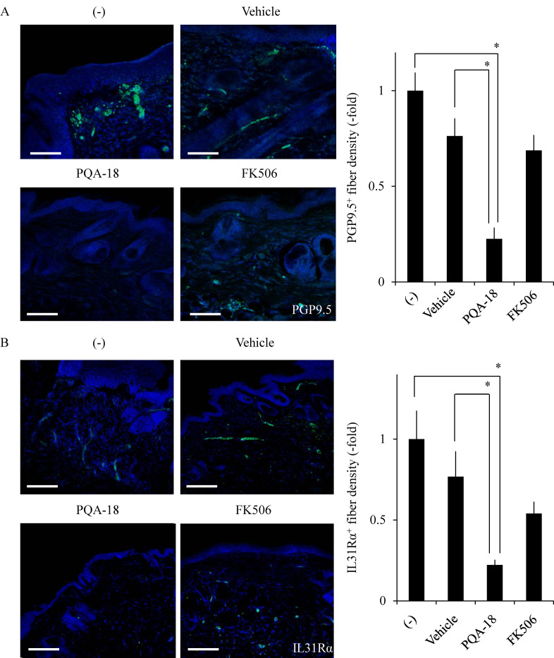
To investigate the effect of PQA-18 on the pruritus in AD, we examined the sensory nerve fiber in lesional skin and treated skin from Nc/Nga mice. Nc/Nga mice (13 weeks of age) were divided into four groups (nine animals in each group) then treated with or without the ointment containing vehicle (DMSO), PQA-18, or FK506. FK506 was used as positive control for improvement of dermatitis [21]. Nerve fiber density was analyzed using immunohistochemistry to visualize protein gene product 9.5 (PGP9.5)-positive nerve fibers. PGP9.5 is known as a peripheral sensory nerve marker [15]. As shown in Fig 1A, treatment with PQA-18 ointment showed a significant decrease in the PGP9.5-positive nerve fiber density in treated skin as compared with that in lesional- or vehicle-treated skin in Nc/Nga mice. FK506 ointment did not significantly affect nerve fiber density. A recent study has demonstrated that the IL-31 pathway is associated with AD and develops a skin sensory neuron network to induce pruritus [15]. Thus, we further examined the effect of PQA-18 on IL-31Rα-positive nerve fibers in lesional skin and treated skin from Nc/Nga mice. Treatment with PQA-18 ointment also showed a significant decrease in the IL-31Rα-positive nerve fiber density in treated skin compared with that in lesional- or vehicle-treated skin in Nc/Nga mice (Fig 1B). FK506 ointment did not significantly affect IL-31Rα-positive fiber density. These results suggest that PQA-18 ointment, but not FK506 ointment, alleviates excessive cutaneous nerve fiber density in the skin of Nc/Nga mice.

Fig 1. Suppression of cutaneous nerve fiber density by PQA-18.

Fig 1

Sections were prepared from skin samples of Nc/Nga mice treated with or without vaseline ointment containing either vehicle, PQA-18 or FK506, and stained by anti-PGP9.5 antibody (green) (A) or the anti-IL-31Rα antibody (green) (B) and Hoechst33342 (blue; Nuclei). Scale bar: 100 μm. The representative images are shown on the left, and quantitative data of the number of nerve fiber are shown on the right. Data are pooled from three independent experiments with nine mice per group and shown as mean and SD. *p < 0.05, as compared with lesioned and vehicle (one-way ANOVA/Tukey-Kramer post-hoc comparisons).

Inhibition of IL-31-induced sensory nerve development by PQA-18

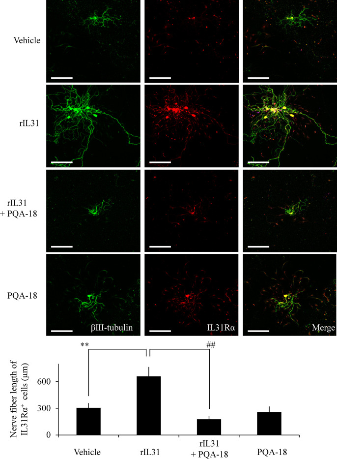
To investigate the effect of PQA-18 on sensory nerve outgrowth, we examined the morphology of DRG neurons prepared from C57BL6J mice. Sensory nerve fiber was analyzed using immunocytochemistry to visualize βIII-tubulin- and IL-31Rα-positive nerve fibers. As shown in Fig 2, treatment with rIL-31 significantly enhanced βIII-tubulin-positive nerve fiber development as compared with vehicle treatment in IL-31Rα-expressed DRG neurons. PQA-18 significantly suppressed the IL-31-induced development, while PQA-18 alone did not affect normal sensory nerve development. These results suggest that PQA-18 inhibits IL-31-induced sensory nerve outgrowth.

Fig 2. Inhibition of IL-31-induced sensory nerve fiver outgrowth by PQA-18.

Fig 2

DRG cells were treated with or without rIL-31 in the absence or presence of PQA-18, and the cell morphology were examined by immunocytochemistry with anti-βIII-tubulin (green) and IL-31Rα (red) antibodies. Scale bar: 300 μm. The representative micrographs are shown (upper), and the quantitative data of the neurite outgrowth are shown (lower). Data are pooled from three independent experiments and shown as mean and SD. **p < 0.01 as compared with control; ##p < 0.01 as compared with the rIL-31-treated group (one-way ANOVA/Tukey-Kramer post-hoc comparisons).

Inhibition of the IL-31 pathway by PQA-18

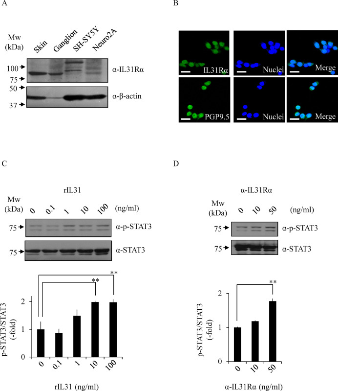
The above results led us to examine the effect of PQA-18 on the IL-31 pathway, using in vitro models of neurons; mouse Neuro2A and human SH-SY5Y neuroblastoma cells. We examined the expression of IL-31Rα in Neuro2A cells and SH-SY5Y cells by immunoblotting analysis with anti-IL-31Rα antibody. Mouse skin and ganglion samples were used as a positive control. We detected IL-31Rα in the lysate of Neuro2A cells, but not in SH-SY5Y cells (Fig 3A). Furthermore, immunocytochemistry analysis showed that Neuro2A cells express IL-31Rα and PGP9.5 (Fig 3B). In order to investigate its receptor function in Neuro2A cells, we examined the effect of rIL-31 and anti-IL-31Rα antibody on phosphorylation of STAT3. As shown in Fig 3C, the phosphorylation of STAT3 was enhanced by treatment with rIL-31 in a dose-dependent manner. Similar results were obtained using anti-IL-31Rα antibody (Fig 3D), indicating that the anti-IL-31Rα antibody also serves as a specific activator for IL-31R. These results suggest that Neuro2A cells functionally express IL-31R.

Fig 3. Functional expression of IL-31R in Neuro2A cells.

Fig 3

The expression of IL-31Rα was determined by immunoblotting with anti-IL-31Rα antibody and anti-β-actin antibody (loading control) (A). The expression of IL-31Rα was analyzed by immunocytochemistry with anti-IL-31Rα antibody (green) or anti-PGP9.5 antibody (green) and Hoechst33342 (blue; Nuclei) (B). The representative micrographs are shown. Scale bar, 100 μm. Phosphorylated STAT3 at Tyr705 was analyzed by immunoblotting. The cells were treated with rIL-31 (C) or anti-IL-31Rα antibody (D) at indicated concentrations, and the cell lysates were examined by immunoblotting with indicated antibodies. The representative images are shown (upper) and the quantitative data of the ratios of phosphorylated STAT3 versus STAT3 are shown (lower). Data are pooled from three independent experiments and shown as mean and SD. **p < 0.01, as compared with control (one-way ANOVA/Tukey-Kramer post-hoc comparisons).

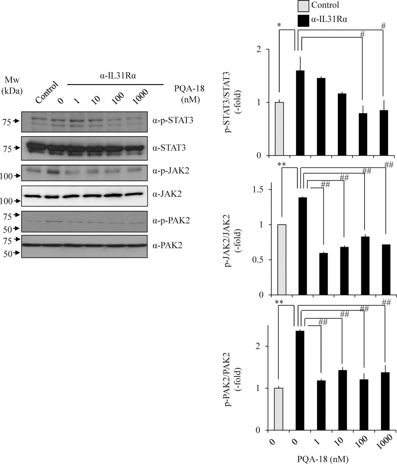
We examined the effect of PQA-18 on anti-IL-31Rα antibody-induced phosphorylation of STAT3 and upstream kinase JAK2 in Neuro2A cells. These phosphorylations were significantly enhanced by anti-IL-31Rα antibody. The enhancements significantly inhibited by treatment with PQA-18 in a dose-dependent manner (Fig 4), indicating that PQA-18 inhibits IL-31 pathway. Since PAKs is involved in cytokine signaling pathway [25] and PQA-18 is a PAK2 inhibitor [21], we further examined the effect of PQA-18 on activation of PAK2 in Neuro2A cells. PQA-18 also significantly inhibited anti-IL-31Rα antibody-induced phosphorylation of PAK2 (Fig 4). These results suggest that PQA-18 inhibits the IL-31 pathway through attenuation of PAK2 activation.

Fig 4. Inhibition of IL-31 pathway by PQA-18.

Fig 4

Phosphorylated STAT3 at Tyr705, JAK2 at Tyr1008, and PAK2 at Ser141 were analyzed by immunoblotting. Neuro2A cells were treated with anti-IL-31Rα antibody in the absence or presence of PQA-18 at indicated concentrations, and the cell lysates were examined by immunoblotting with indicated antibodies. The representative images are shown on the left, and the quantitative data of the ratios of phosphorylated STAT3 versus STAT3, phosphorylated JAK2 versus JAK2, and phosphorylated PAK2 versus PAK2 are shown on the right. Data are pooled from three independent experiments and shown as mean and SD. **p < 0.01, *p < 0.05 as compared with control; ##p < 0.01, #p < 0.05 as compared with anti-IL-31Rα antibody-treated group (one-way ANOVA/Tukey-Kramer post-hoc comparisons).

Inhibition of IL-31-induced neurite outgrowth by PQA-18

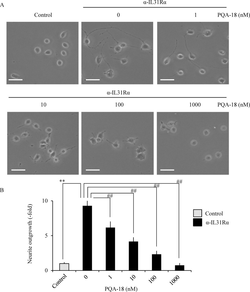
We examined the effect of PQA-18 on IL-31-stimulating neurite outgrowth in Neuro2A cells. As shown in Fig 5, treatment with anti-IL-31Rα antibody significantly increased the number of neurite-positive cells in Neuro2A cells, while that increase was significantly inhibited by PQA-18 in a dose-dependent manner, suggesting that PQA-18 inhibits IL-31-induced neurite outgrowth in Neuro2A cells.

Fig 5. Inhibition of IL-31-induced neurite outgrowth by PQA-18.

Fig 5

Neuro2A cells were treated with anti-IL-31Rα antibody in the absence or presence of PQA-18 at indicated concentrations, and the cell morphology was examined by microscopy. Neurites were defined as a process with lengths equivalent to one diameter of a cell body. The percentage of neurite-bearing cells was calculated from the total number of counted cells. The representative micrographs are shown (A), and the quantitative data of the neurite outgrowth are shown (B). Scale bar: 20 μm. Data are pooled from three independent experiments and shown as mean and SD. **p < 0.01 as compared with control; ##p < 0.01 as compared with anti-IL-31Rα antibody-treated group (one-way ANOVA/Tukey-Kramer post-hoc comparisons).

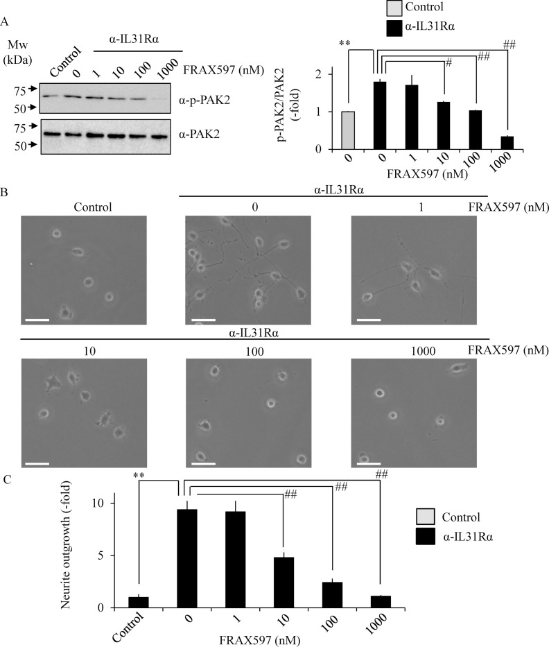
Based on the above results, selective group I PAK inhibitor FRAX597 was used to understand the role of PAK2 in neurite outgrowth [26,27]. Treatment with FRAX597 significantly inhibited anti-IL-31Rα antibody-induced phosphorylation of PAK2 (Fig 6A) and neurite outgrowth (Fig 6B and 6C) in a dose-dependent manner in Neuro2A cells, suggesting the involvement of PAK2 in IL-31-induced neurite outgrowth.

Fig 6. Inhibition of IL-31-induced neurite outgrowth by FRAX597.

Fig 6

Neuro2A cells were treated with anti-IL-31Rα antibody in the absence or presence of FRAX597 at indicated concentrations. Phosphorylated PAK2 at Ser141 were analyzed by immunoblotting (A). The representative images are shown on the left, and the quantitative data of the ratios of phosphorylated PAK2 versus PAK2 are shown on the right. Neurites were defined as a process with lengths equivalent to one diameter of a cell body. The percentage of neurite-bearing cells was calculated from the total number of counted cells. The representative micrographs are shown (B), and the quantitative data of the neurite outgrowth are shown (C).Scale bar: 20 μm. Data are pooled from three independent experiments and shown as mean and SD. **p < 0.01 as compared with control; ##p < 0.01, #p < 0.05 as compared with anti-IL-31Rα antibody-treated group (one-way ANOVA/Tukey-Kramer post-hoc comparisons).

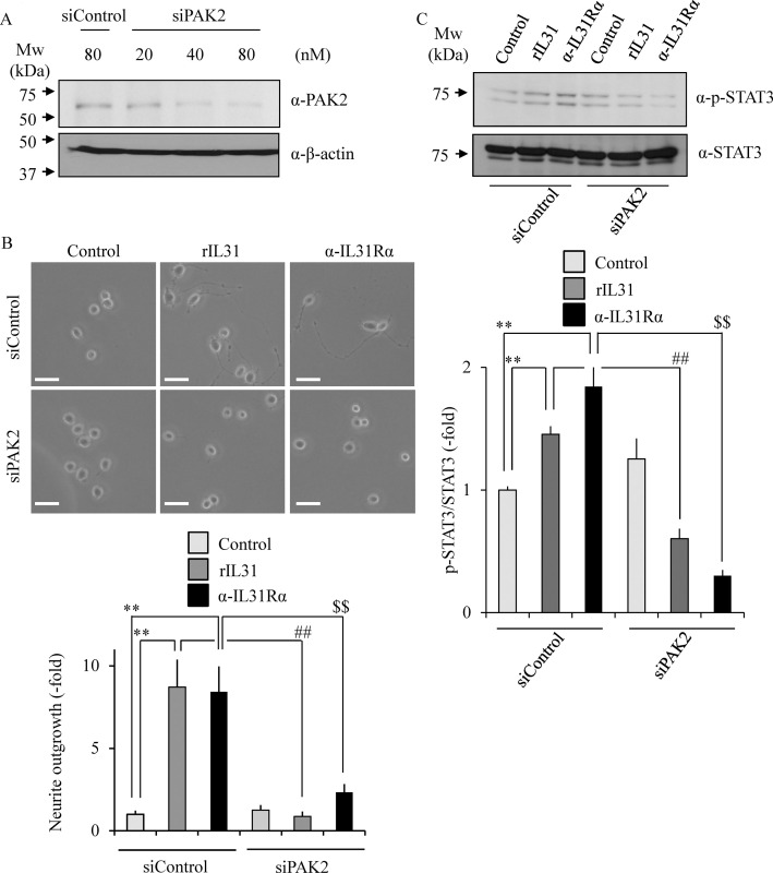
The effect of PAK2 siRNA on the neurite outgrowth was tested to confirm the PAK2 requirement for the IL-31-stimulating nerve fiber outgrowth. We confirmed that the PAK2 expression level is significantly suppressed by treatment with PAK2 siRNA in Neuro2A cells in a dose-dependent manner as compared with the control siRNA (Fig 7A). When the cells were introduced with PAK2 siRNA and then treated with either rIL-31 or anti-IL-31Rα antibody, the number of neurite-positive cells was significantly reduced as compared with control cells (Fig 7B). Furthermore, rIL-31- or anti-IL-31Rα antibody-induced phosphorylation of STAT3 was also significantly reduced by treatment with PAK2 siRNA (Fig 7C). These results suggest an indispensable role for PAK2 in IL-31-induced neurite outgrowth and STAT3 phosphorylation.

Fig 7. Requirement of PAK2 for IL-31-induced neurite outgrowth and STAT3 phosphorylation.

Fig 7

Neuro2A cells were transfected with siRNA for PAK2 or control siRNA. PAK2 expression was determined by immunoblotting (A). Representative images of Neuro2A cells after transfection with siRNA for PAK2 or control siRNA. Neurites were defined as a process with lengths equivalent to one diameter of a cell body (B). The percentage of neurite-bearing cells was calculated from the total number of counted cells. Scale bar: 20 μm. Phosphorylated STAT3 at Tyr705 was analyzed by immunoblotting (C). Neuro2A cells were treated with rIL-31 or anti-IL-31Rα antibody in the absence or presence of siRNA for PAK2, and the cell lysates were examined by immunoblotting with indicated antibodies. The representative images are shown (upper), and the quantitative data of the ratios of phosphorylated STAT3 versus STAT3 are shown (lower). Data are pooled from three independent experiments and shown as mean and SD. **p < 0.01 as compared with control; ##p < 0.01 as compared with rIL-31-treated group; $ $p < 0.01 as compared with anti-IL-31Rα antibody-treated group (one-way ANOVA/Tukey-Kramer post-hoc comparisons).

Prevention of IL-31-induced formation of PAK2 activation complex by PQA-18

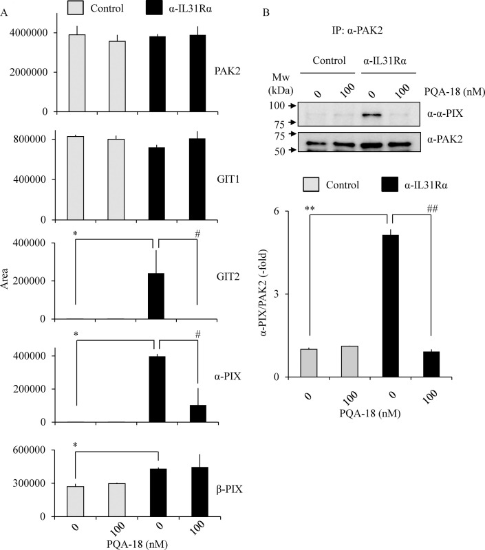
Several scaffold proteins including G protein-coupled receptor kinase interactor (GIT) and PIX are involved in PAK2 activation mechanism [2830]. To understand the molecular mechanisms underlying inhibition of PAK2 by PQA-18, we examined PAK2 complex components by LC/MS/MS. PAK2 complexes were purified from cell lysates prepared from Neuro2A cells treated with PQA-18 in the absence or presence of anti-IL-31Rα antibody using anti-PAK2 antibody conjugated beads. When the purified PAK2 complex was digested with trypsin and the resulting peptides were analyzed by LC/MS/MS, 12 proteins could be identified in each sample (Table 1). Unique peptides for GIT1, GIT2, α-PIX and β-PIX were identified as components of the PAK2 activation complex. On the other hand, these peptides were not detected in normal rabbit IgG-beads as a negative control. As shown in Fig 8A, the peak areas for GIT2, α-PIX, and β-PIX were significantly increased when Neuro2A cells were treated with anti-IL-31Rα antibody. The increase of the peak areas for GIT2 and α-PIX was significantly inhibited by treatment with PQA-18. To confirm the above results, we further examined PAK2 interaction with α-PIX by immunoprecipitation assay. Treatment with anti-IL-31Rα antibody significantly increased PAK2 interaction with α-PIX, while that increase was significantly inhibited by treatment with PQA-18 (Fig 8B). These results suggest that PQA-18 inhibits IL-31-induced PAK2 activation through preventing formation of the PAK2 activation complex with GIT2 and α-PIX.

Table 1. PAK2 interacting proteins as identified by LC/MS/MS.

Description ∑Coverage Number of Unique Peptides
Control PQA-18 α-IL-31R α-IL-31R+PQA-18
Serine/threonine-protein kinase PAK2 55.15 93 80 103 86
Rho guanine nucleotide exchange factor 7 β-PIX 18.68 13 16 50 7
Peripherin 18.11 0 0 31 8
H1 histone family, member X 17.02 0 5 9 8
Tubulin beta-5 chain 15.09 0 3 0 9
ARF GTPase-activating protein GIT1 14.16 12 10 19 15
Rho guanine nucleotide exchange factor 6 α-PIX 9.73 0 1 5 1
ARF GTPase-activating protein GIT2 4.38 0 0 10 0
Casein kinase II subunit alpha-interacting protein 3.61 0 0 1 0
Protein transport protein sec16 3.35 0 2 0 3
Alpha-1,4-N-acetylglucosaminyltransferase 2.93 0 0 1 0
Alpha-actinin-1 2.47 0 0 6 6

Fig 8. Prevention of formation of PAK2 activation complex by PQA-18.

Fig 8

PAK2 interacting proteins as identified by LC/MS/MS (A). LC/MS/MS analysis was performed using tryptic peptides of purified PAK2 prepared from Neuro2A cells treated without or with PQA-18 in the absence or presence of anti- IL-31Rα antibody using anti-PAK2 antibody conjugated beads. The quantitative data of the peak area of the indicated proteins are shown. Immunoprecipitates were obtained from Neuro2A cells treated without or with PQA-18 in the absence or presence of anti- IL-31Rα antibody using anti-PAK2 antibody conjugated beads, and immunoblotted with anti-α-PIX antibody or anti-PAK2 antibody (B). The representative images are shown (upper), and the quantitative data of the ratios of α-PIX versus PAK2 are shown (lower). Data are pooled from three independent experiments and shown as mean and SD. **p < 0.01, *p < 0.05 as compared with control; ##p < 0.01, #p < 0.05 as compared with anti-IL-31Rα antibody-treated group (one-way ANOVA/Tukey-Kramer post-hoc comparisons).

Discussion

In this study, we demonstrated that PQA-18 suppresses sensory nerve outgrowth through inhibition of the IL-31 pathway. IL-31 promotes the development of sensory nerve fibers by activating IL-31R [15]. We observed that neurite outgrowth is significantly promoted by treatment with rIL-31 and anti-IL-31Rα antibody in vitro experiments using DRG neurons (Fig 2) and neuronal model Neuro2A cells (Figs 3 and 5), which express IL-31R. Moreover, PQA-18 treatment suppressed the neurite outgrowth in the two independent cells (Figs 2 and 5). The IL-31 pathway is known to activate the intracellular JAK/STAT pathway [9,15]. JAK has four known molecules, JAK1, JAK2, JAK3, and tyrosine kinase 2, and interacts with various cytokine receptors [31]. After autophosphorylation by cytokine receptor activation, JAKs phosphorylate STATs, which promote dimer formation and nuclear import, and regulate downstream target gene transcription [32]. IL-31 is thought to mainly promote phosphorylation of STAT3 though JAK1 or JAK2 activation [5]. A recent study revealed that phosphorylation of STAT3 promotes the expression of genes involved in neurite outgrowth in the sensory nervous system [15]. It has also been reported that neurite outgrowth is suppressed by pharmacological inhibition of STAT3 and a dominant negative form of STAT3 [15,33]. We observed that stimulation of IL-31R induces phosphorylation of JAK2 and STAT3 in Neuro2A cells (Fig 4). PQA-18 suppressed the IL-31R-induced phosphorylation of JAK2 and STAT3 (Fig 4). These results suggest that PQA-18 inhibits IL-31/JAK2/STAT3 pathway. Increased IL-31 levels and excessive cutaneous sensory innervation are observed in AD patients and AD models including Nc/Nga mice, leading to lowered the pruritus threshold and causing severe pruritus and dermatitis [1214]. These symptoms are improved by application of anti-IL-31 antibody and anti-IL-31R-neutralizing antibody [5]. Hence, IL-31 pathway plays a crucial role in development of cutaneous sensory innervation in vivo. Indeed, we found that administration of the PQA-18 ointment reduces cutaneous sensory nerve density in Nc/Nga mice (Fig 1). Taken together, these results suggest that PQA-18 suppresses sensory nerve outgrowth through inhibition of the IL-31 pathway both in vitro and in vivo. We speculate that PQA-18 could improve the pruritus of AD by suppression of the excessive sensory innervation in dermatitis.

Given that our previous finding of PQA-18 being a PAK2 inhibitor [21], it is conceivable that PAK2 also plays an important role in neurite outgrowth. PAKs, effector molecules of Rac and Cdc42, are serine/threonine kinases that phosphorylate multiple substrates, including those that are involved in cytoskeletal reorganization, cell proliferation, and survival [34]. The PAK family is divided into two groups (group I and group II) based on sequence and structural homology. PAK2, group I PAK, is expressed in neuronal cells and has been reported to be implicated in crucial neuronal functions including synapse formation [35]. We observed that IL-31R stimulation enhances phosphorylation of PAK2 and STAT3, and neurite outgrowth in Neuro2A cells. These enhancements were significantly inhibited by PQA-18 treatment (Figs 4 and 5). In addition, FRAX597 treatment also significantly inhibited IL-31R-induced phosphorylation of PAK2 and neurite outgrowth (Fig 6). These results suggest the involvement of PAK2 in neurite outgrowth system. Although the role of PAK2 in neurite outgrowth is not well understood, the group I PAK inhibitor IPA3 has been reported to inhibit STAT3 phosphorylation and neurite outgrowth [25,36]. We observed inhibition of IL-31-induced STAT3 phosphorylation and neurite outgrowth from analysis using Neuro2A cells that suppress PAK2 expression (Fig 7). These results indicate that PAK2 is required for IL-31-induced activation of STAT3 and neurite outgrowth. Thus, PAK2 may play a key role in regulation of sensory nerve outgrowth. Further analysis of the role of PAK2 in the development of cutaneous sensory nerves is needed to clarify the mechanism of pruritus development in AD.

Although the mechanism of PAK2 activation is not yet fully understood, understanding of the mechanism by which IL-31 activates PAK2 is important in understanding the IL-31 pathway. Group I PAKs exists in an inactive form by dimerization, but dissociates due to the interaction of activated Rac and cdc42, interacts with the PIX/GIT complex known as a scaffold protein, which promotes autophosphorylation due to high local concentrations of PAKs [28]. We have previously reported that autophosphorylation of PAK2 at Ser141 plays an important role in kinase activity [21,34]. From the analysis using the anti-phospho-Ser141 PAK2 antibody, we observed that the phosphorylated PAK2 at Ser141 is increased by the IL-31R activation (Fig 4). PQA-18 inhibited the phosphorylation of PAK2 at Ser141 (Fig 4), indicating that PQA-18 suppresses autophosphorylation of PAK2 induced by IL-31R. Furthermore, when the activated PAK2 complex was analyzed using LC/MS/MS, 12 types of unique peptides including α-PIX, β-PIX, GIT1 and GIT2 were detected (Table 1). Quantitative analysis of these peptides revealed the significant increased the interaction of GIT2, α-PIX and β-PIX with PAK2 by IL-31R stimulation (Fig 8). Among them, the amount of interaction of GIT2 and α-PIX with PAK2 was significantly decreased by PQA-18 treatment (Fig 8). We further confirmed that PQA-18 treatment significantly inhibits IL-31R-induced the interaction of PAK2 with α-PIX by immunoprecipitation assay (Fig 8). These results indicate that the interaction of PAK2 with the PIX/GIT complex plays an important role in the mechanism of PAK2 activation by the IL-31 pathway. PQA-18 might prevent the formation of PAK2 activation complexes by inhibiting GIT2 and α-PIX interactions, supporting previous our observation that PQA-18 inhibits PAK2 activity in non-competitive manner [21]. In future, functional analysis of PAK2 interacting components and the structural analysis of PAK2 activation complexes are necessary to clarify the IL-31 pathway.

Our observations demonstrated that PQA-18 at low concentrations (1–10 nM) strongly inhibits IL-31R-induced phosphorylation of PAK2 (Fig 4) but has a limited effect on neurite outgrowth (Fig 5). On the other hands, FRAX597 had a similar inhibitory dose response between PAK2 phosphorylation and neurite outgrowth (Fig 6). The difference between the two compounds is that PQA-18 inhibits the PAK2 activation complex as described above (Fig 8), whereas FRAX597 inhibits PAK2 activity by the competitive inhibition of ATP [26,27]. Recent study has revealed that PAK-PIX interactions as well as STAT3 activation regulate neurite growth [37]. Interestingly, complete inhibition of PAK-PIX interaction suppressed neurite outgrowth, whereas partial inhibition promoted neurite outgrowth [37]. Therefore, it is possible that partial inhibition of PAK-PIX interaction may be involved in the attenuation of the inhibitory effect of neurite outgrowth by PQA-18 at low concentration. Further analysis of the physiological role of PAK-PIX interaction is required for understanding the mechanism underlying the regulation of neurite outgrowth by PQA-18.

The scratching behavior mechanically damages the skin, lowers the barrier function, enhances the inflammatory reaction by foreign antigens that have penetrated through the epidermis, and aggravates dermatitis and further enhances pruritus. Such vicious cycle of scratching, exacerbation of inflammation, and enhancement of the itch is called the itch-scratch-cycle, and is known to contribute to chronic AD [5,38]. Therefore, the development of an AD therapeutic drug that suppresses not only inflammation but also pruritus is highly desired. Currently, glucorticoids and calcineurin inhibitors are used as therapeutic agents for AD, and mainly target skin inflammation [39]. However, these drugs have serious side effects including adrenal failure, skin atrophy, neurotoxicity, nephrotoxicity, skin cancer, tumor growth, due to long-term administration [39]. Although immunosuppressant FK506 is also used to treat AD, it has been reported to cause not only nephrotoxicity and hepatotoxicity as adverse effects but also itching sensation [40,41]. We observed that application of PQA-18 ointment to Nc/Nga mice significantly improves excessive sensory nerve density and IL-31R expression, but FK506 ointment fail to improve it (Fig 1). Thus, it is conceivable that FK506 did not target the IL-31 pathway, resulting in weak therapeutic effects on pruritus, which is in line with previous study which showed that IL-31-induced scratching behavior is not inhibited by treatment with FK506 [42]. Recently, the development of a novel AD therapeutic drug targeting the IL-31 pathway has advanced. Nemolizumab, an IL-31R-neutralizing humanized antibody, is effective in improving pruritus in AD patients, but clinical studies have shown that exacerbation of dermatitis and peripheral edema are observed in some patients [43,44]. Given our previous findings that PQA-18 suppresses inflammatory cytokine production through inhibition of PAK2 in T cells and does not cause side effects such as renal damage, liver damage or tumor formation even with long-term systemic administration in mice [21], PQA-18 is considered to be a lead compound for the development of a useful therapeutic drug targeting both inflammation and pruritus for AD.

In conclusion, we have demonstrated that PQA-18 is an inhibitor of sensory nerve outgrowth. PQA-18 inhibits the IL-31 pathway by suppressing PAK2 activity, which in turn suppresses STAT3 activation. We have further demonstrated that PQA-18 exhibits an inhibitory effect on excessive cutaneous sensory nerve development in an AD model. These results suggest that PQA-18 may be a feasible lead compound for treatment of pruritus in AD.

Supporting information

S1 Fig. Full blot data of Fig 3A.

(PDF)

S2 Fig. Full blot data of Fig 3C.

(PDF)

S3 Fig. Full blot data of Fig 3D.

(PDF)

S4 Fig. Full blot data of Fig 4A.

(PDF)

S5 Fig. Full blot data of Fig 4B.

(PDF)

S6 Fig. Full blot data of Fig 4C.

(PDF)

S7 Fig. Full blot data of Fig 6A.

(PDF)

S8 Fig. Full blot data of Fig 7A.

(PDF)

S9 Fig. Full blot data of Fig 7C.

(PDF)

S10 Fig. Full blot data of Fig 8.

(PDF)

S1 Table. Minimal underlying data.

(PDF)

Data Availability

All relevant data are within the manuscript and its Supporting Information files.

Funding Statement

This work was funded by the Japan Society for the Promotion of Science (Grant-in-Aid for Scientific Research) [grant number 18K06702 (MO): https://kaken.nii.ac.jp/ja/index/] and Fukushima Medical University (grant for Project Research) [grant number KKI29001-1 (MO)]: https://www.fmu.ac.jp].

References

  • 1.Williams HC. Clinical Practice. Atopic Dermatitis. N Engl J Med. 2005;352: 2314–2324. 10.1056/NEJMcp042803 [DOI] [PubMed] [Google Scholar]
  • 2.Bieber T. Atopic Dermatitis. N Engl J Med. 2008;358: 1483–1494. 10.1056/NEJMra074081 [DOI] [PubMed] [Google Scholar]
  • 3.Boguniewicz M, Leung DYM. Atopic dermatitis: A disease of altered skin barrier and immune dysregulation. Immunol Rev. 2011;242: 233–246. 10.1111/j.1600-065X.2011.01027.x [DOI] [PMC free article] [PubMed] [Google Scholar]
  • 4.Yosipovitch G, Bernhard JD. Clinical Practice. Chronic Pruritus. N Engl J Med. 2013;368: 1625–1634. 10.1056/NEJMcp1208814 [DOI] [PubMed] [Google Scholar]
  • 5.Furue M, Yamamura K, Kido-Nakahara M, Nakahara T, Fukui Y. Emerging Role of interleukin-31 and interleukin-31 Receptor in Pruritus in Atopic Dermatitis. Allergy. 2018;73: 29–36. 10.1111/all.13239 [DOI] [PubMed] [Google Scholar]
  • 6.Gochnauer H, Valdes-Rodriguez R, Cardwell L, Anolik RB. The Psychosocial Impact of Atopic Dermatitis. Adv Exp Med Biol. 2017;1027: 57–69. 10.1007/978-3-319-64804-0_6 [DOI] [PubMed] [Google Scholar]
  • 7.Kabashima K. New concept of the pathogenesis of atopic dermatitis: Interplay among the barrier, allergy, and pruritus as a trinity. J Dermatol Sci. 2013;70: 3–11. 10.1016/j.jdermsci.2013.02.001 [DOI] [PubMed] [Google Scholar]
  • 8.Newsom M, Bashyam AM, Balogh EA, Feldman SR, Strowd LC. New and Emerging Systemic Treatments for Atopic Dermatitis. Drugs. 2020;80: 1041–1052. 10.1007/s40265-020-01335-7 [DOI] [PMC free article] [PubMed] [Google Scholar]
  • 9.Cevikbas F, Wang X, Akiyama T, Kempkes C, Savinko T, Antal A, et al. A sensory neuron-expressed IL-31 receptor mediates T helper cell-dependent itch: Involvement of TRPV1 and TRPA1. J Allergy Clin Immunol. 2014;133: 448–460. 10.1016/j.jaci.2013.10.048 [DOI] [PMC free article] [PubMed] [Google Scholar]
  • 10.Bilsborough J, Leung DYM, Maurer M, Howell M, Boguniewcz M, Yao L, et al. IL-31 is associated with cutaneous lymphocyte antigen-positive skin homing T cells in patients with atopic dermatitis. J Allergy Clin Immunol. 2006;117: 418–425. 10.1016/j.jaci.2005.10.046 [DOI] [PubMed] [Google Scholar]
  • 11.Sonkoly E, Muller A, Lauerma AI, Pivarcsi A, Soto H, Kemeny L, et al. IL-31: A new link between T cells and pruritus in atopic skin inflammation. J Allergy Clin Immunol. 2006;117: 411–417. 10.1016/j.jaci.2005.10.033 [DOI] [PubMed] [Google Scholar]
  • 12.Kim S, Kim HJ, Yang HS, Kim E, Huh IS, Yang JM. IL-31 Serum Protein and Tissue mRNA Levels in Patients With Atopic Dermatitis. Ann Dermatol. 2011;23: 468–473. 10.5021/ad.2011.23.4.468 [DOI] [PMC free article] [PubMed] [Google Scholar]
  • 13.Takaoka A, Arai I, Sugimoto M, Yamaguchi A, Tanaka M, Nakaike S. Expression of IL-31 Gene Transcripts in NC/Nga Mice With Atopic Dermatitis. Eur J Pharmacol. 2005;516: 180–181. 10.1016/j.ejphar.2005.04.040 [DOI] [PubMed] [Google Scholar]
  • 14.Takaoka A, Arai I, Sugimoto M, Honma Y, Futaki N, Nakamura A, et al. Involvement of IL-31 on Scratching Behavior in NC/Nga Mice With Atopic-Like Dermatitis. Exp Dermatol. 2006;15: 161–167. 10.1111/j.1600-0625.2006.00405.x [DOI] [PubMed] [Google Scholar]
  • 15.Feld M, Garcia R, Buddenkotte J, Katayama S, Lewis K, Muirhead G, et al. The Pruritus- And TH2-associated Cytokine IL-31 Promotes Growth of Sensory Nerves. J Allergy Clin Immunol. 2016;138: 500–508 10.1016/j.jaci.2016.02.020 [DOI] [PubMed] [Google Scholar]
  • 16.Arai I, Tsuji M, Miyagawa K, Takeda H, Akiyama N, Saito S. Repeated Administration of IL-31 Upregulates IL-31 Receptor A (IL-31RA) in Dorsal Root Ganglia and Causes Severe Itch-Associated Scratching Behaviour in Mice. Exp Dermatol. 2015;24: 75–78. 10.1111/exd.12587 [DOI] [PubMed] [Google Scholar]
  • 17.Morris HR, Taylor GW, Masento MS, Jermyn KA, Kay RR. Chemical structure of the morphogen differentiation inducing factor from Dictyostelium discoideum. Nature. 1987;328: 811–814. 10.1038/328811a0 [DOI] [PubMed] [Google Scholar]
  • 18.Kubohara Y, Kikuchi H, Matsuo Y, Oshima Y, Homma Y. Mitochondria are the target organelle of differentiation-inducing factor-3, an anti-tumor agent isolated from Dictyostelium Discoideum. PLoS One. 2013;8: e72118 10.1371/journal.pone.0072118 [DOI] [PMC free article] [PubMed] [Google Scholar]
  • 19.Suzuki T, Kikuchi H, Ogura M, Homma MK, Oshima Y, Homma Y. Weight loss by Ppc-1, a novel small-molecule mitochondrial uncoupler derived from slime mold. PLoS One. 2015;10: e0117088 10.1371/journal.pone.0117088 [DOI] [PMC free article] [PubMed] [Google Scholar]
  • 20.Kikuchi H, Suzuki T, Ogura M, Homma MK, Homma Y, Oshima Y. Synthesis of prenylated quinolinecarboxylic acid derivatives and their anti-obesity activities. Bioorg Med Chem. 2015;23: 66–72. 10.1016/j.bmc.2014.11.024 [DOI] [PubMed] [Google Scholar]
  • 21.Ogura M, Kikuchi H, Suzuki T, Yamaki J, Homma MK, Oshima Y, et al. Prenylated quinolinecarboxylic acid derivative suppresses immune response through inhibition of PAK2. Biochem Pharmacol. 2016;105: 55–65. 10.1016/j.bcp.2016.01.020 [DOI] [PubMed] [Google Scholar]
  • 22.Ogura M, Kikuchi H, Shakespear N, Suzuki T, Yamaki J, Homma MK, et al. Prenylated Quinolinecarboxylic Acid Derivative Prevents Neuronal Cell Death Through Inhibition of MKK4. Biochem Pharmacol. 2019;162: 109–122. 10.1016/j.bcp.2018.10.008 [DOI] [PubMed] [Google Scholar]
  • 23.Zorina Y, Iyengar R, Bromberg KD. Cannabinoid 1 Receptor and interleukin-6 Receptor Together Induce Integration of Protein Kinase and Transcription Factor Signaling to Trigger Neurite Outgrowth. J Biol Chem. 2010;285: 1358–1370. 10.1074/jbc.M109.049841 [DOI] [PMC free article] [PubMed] [Google Scholar]
  • 24.Ogura M, Yamaki J, Homma MK, Homma Y. Phosphorylation of flotillin-1 by Mitochondrial c-Src Is Required to Prevent the Production of Reactive Oxygen Species. FEBS Lett. 2014;588: 2837–2843. 10.1016/j.febslet.2014.06.044 [DOI] [PubMed] [Google Scholar]
  • 25.Kurgonaite K, Gandhi H, Kurth T, Pautot S, Schwille P, Weidemann T, et al. Role of Endocytosis for interleukin-4-receptor-mediated JAK/STAT Signalling. J Cell Sci. 2015;128: 3781–3795. 10.1242/jcs.170969 [DOI] [PubMed] [Google Scholar]
  • 26.Licciulli S, Maksimoska J, Zhou C, Troutman S, Kota S, Liu Q, et al. FRAX597, a small molecule inhibitor of the p21-activated kinases, inhibits tumorigenesis of neurofibromatosis type 2 (NF2)-associated Schwannomas. J Biol Chem. 2013;288: 29105–29114. 10.1074/jbc.M113.510933 [DOI] [PMC free article] [PubMed] [Google Scholar]
  • 27.Yeo D, Phillips P, Baldwin GS, He H, Nikfarjam M. Inhibition of group 1 p21-activated kinases suppresses pancreatic stellate cell activation and increases survival of mice with pancreatic cancer. Int J Cancer. 2017;140: 2101–2111. 10.1002/ijc.30615 [DOI] [PubMed] [Google Scholar]
  • 28.Hoefen RJ, Berk BC. The Multifunctional GIT Family of Proteins. J Cell Sci. 2006;119: 1469–1475. 10.1242/jcs.02925 [DOI] [PubMed] [Google Scholar]
  • 29.Daniels RH, Zenke FT, Bokoch GM. alphaPix Stimulates p21-activated Kinase Activity Through Exchange Factor-Dependent and -Independent Mechanisms. J Biol Chem. 1999;274: 6047–6050. 10.1074/jbc.274.10.6047 [DOI] [PubMed] [Google Scholar]
  • 30.Loo TH, Ng YW, Lim L, Manser E. GIT1 Activates p21-activated Kinase Through a Mechanism Independent of p21 Binding. Mol Cell Biol. 2004;24: 3849–3859. 10.1128/mcb.24.9.3849-3859.2004 [DOI] [PMC free article] [PubMed] [Google Scholar]
  • 31.Morris R, Kershaw NJ, Babon JJ. The Molecular Details of Cytokine Signaling via the JAK/STAT Pathway. Protein Sci. 2018;27: 1984–2009. 10.1002/pro.3519 [DOI] [PMC free article] [PubMed] [Google Scholar]
  • 32.Welsch K, Holstein J, Laurence A, Ghoreschi K. Targeting JAK/STAT Signalling in Inflammatory Skin Diseases With Small Molecule Inhibitors. Eur J Immunol. 2017;47:1096–1107. 10.1002/eji.201646680 [DOI] [PubMed] [Google Scholar]
  • 33.Wu YY, Bradshaw RA. Activation of the Stat3 Signaling Pathway Is Required for Differentiation by interleukin-6 in PC12-E2 Cells. J Biol Chem. 2000;275: 2147–2156. 10.1074/jbc.275.3.2147 [DOI] [PubMed] [Google Scholar]
  • 34.Jung JH, Traugh JA. Regulation of the Interaction of Pak2 With Cdc42 via Autophosphorylation of Serine 141. J Biol Chem. 2005;280: 40025–40031. 10.1074/jbc.M509075200 [DOI] [PubMed] [Google Scholar]
  • 35.Wang Y, Zeng C, Li J, Zhou Z, Ju X, Xia S, et al. PAK2 Haploinsufficiency Results in Synaptic Cytoskeleton Impairment and Autism-Related Behavior. Cell Rep. 2018;24: 2029–2041. 10.1016/j.celrep.2018.07.061 [DOI] [PubMed] [Google Scholar]
  • 36.Ding Y, Li Y, Lu L, Zhang R, Zeng L, Wang L, et al. Inhibition of Nischarin Expression Promotes Neurite Outgrowth Through Regulation of PAK Activity. PLoS One. 2015;10: e0144948 10.1371/journal.pone.0144948 [DOI] [PMC free article] [PubMed] [Google Scholar]
  • 37.Santiago-Medina M, Gregus KA, Gomez TM. PAK-PIX interactions regulate adhesion dynamics and membrane protrusion to control neurite outgrowth. J Cell Sci. 2013;126: 1122–1133. 10.1242/jcs.112607 [DOI] [PMC free article] [PubMed] [Google Scholar]
  • 38.Mack MR, Kim BS. The Itch-Scratch Cycle: A Neuroimmune Perspective. Trends Immunol. 2018;39: 980–991. 10.1016/j.it.2018.10.001 [DOI] [PMC free article] [PubMed] [Google Scholar]
  • 39.Yosipovitch G, Berger T, Fassett MS. Neuroimmune Interactions in Chronic Itch of Atopic Dermatitis. J Eur Acad Dermatol Venereol. 2020;34: 239–250. 10.1111/jdv.15973 Epub 2019 Nov 12. [DOI] [PMC free article] [PubMed] [Google Scholar]
  • 40.Hanifin JM, Ling MR, Langley R, Breneman D, Rafal E. Tacrolimus ointment for the treatment of atopic dermatitis in adult patients: Part I, efficacy. J Am Acad Dermatol. 2001;44: S28–38. 10.1067/mjd.2001.109810 [DOI] [PubMed] [Google Scholar]
  • 41.Soter NA, Fleischer AB JR., Webster GF, Monroe E, Lawrence I. Tacrolimus ointment for the treatment of atopic dermatitis in adult patients: Part II, safety. J Am Acad Dermatol. 2001;44: S39–46. 10.1067/mjd.2001.109817 [DOI] [PubMed] [Google Scholar]
  • 42.Kasutani K, Fujii E, Ohyama S, Adachi H, Hasegawa M, Kitamura H, et al. Anti-IL-31 receptor antibody is shown to be a potential therapeutic option for treating itch and dermatitis in mice. Br J Pharmacol. 2014;171: 5049–5058. 10.1111/bph.12823 [DOI] [PMC free article] [PubMed] [Google Scholar]
  • 43.Ruzicka T, Hanifin JM, Furue M, Pulka G, Mlynarczyk I, Wollenberg et al. Anti-Interleukin-31 Receptor A Antibody for Atopic Dermatitis. N Engl J Med. 2017;376:826–835. 10.1056/NEJMoa1606490 [DOI] [PubMed] [Google Scholar]
  • 44.Kabashima K, Matsumura T, Komazaki H, Kawashima M. Trial of Nemolizumab and Topical Agents for Atopic Dermatitis with Pruritus. N Engl J Med. 2020;383: 141–150. 10.1056/NEJMoa1917006 [DOI] [PubMed] [Google Scholar]

Decision Letter 0

Ed Manser

2 Oct 2020

PONE-D-20-23391

Prenylated quinolinecarboxylic acid compound-18 prevents sensory nerve fiber outgrowth through inhibition of the interleukin-31 pathway

PLOS ONE

Dear Dr. Ogura,

Thank you for submitting your manuscript to PLOS ONE. After careful consideration, we feel that it has merit but does not fully meet PLOS ONE’s publication criteria as it currently stands. Therefore, we invite you to submit a revised version of the manuscript that addresses the points raised during the review process.

Please find enclosed the expert reviews on your manuscript. Although the results presented are considered important in the field, better validation of the inhibitory effect of PQA-18 on PAK2 is required. In particular some key results should be compared with other PAK-specific inhibitors, which are available. There are issues with regard to the presentation of results that need to be addressed. I hope you will be able to carry out the additional experiments in the time requested.

Please submit your revised manuscript by Nov 16 2020 11:59PM. If you will need more time than this to complete your revisions, please reply to this message or contact the journal office at plosone@plos.org. When you're ready to submit your revision, log on to https://www.editorialmanager.com/pone/ and select the 'Submissions Needing Revision' folder to locate your manuscript file.

Please include the following items when submitting your revised manuscript:

  • A rebuttal letter that responds to each point raised by the academic editor and reviewer(s). You should upload this letter as a separate file labeled 'Response to Reviewers'.

  • A marked-up copy of your manuscript that highlights changes made to the original version. You should upload this as a separate file labeled 'Revised Manuscript with Track Changes'.

  • An unmarked version of your revised paper without tracked changes. You should upload this as a separate file labeled 'Manuscript'.

If you would like to make changes to your financial disclosure, please include your updated statement in your cover letter. Guidelines for resubmitting your figure files are available below the reviewer comments at the end of this letter.

If applicable, we recommend that you deposit your laboratory protocols in protocols.io to enhance the reproducibility of your results. Protocols.io assigns your protocol its own identifier (DOI) so that it can be cited independently in the future. For instructions see: http://journals.plos.org/plosone/s/submission-guidelines#loc-laboratory-protocols

We look forward to receiving your revised manuscript.

Kind regards,

Ed Manser, PhD

Academic Editor

PLOS ONE

Journal Requirements:

When submitting your revision, we need you to address these additional requirements.

1. Please ensure that your manuscript meets PLOS ONE's style requirements, including those for file naming. The PLOS ONE style templates can be found at

https://journals.plos.org/plosone/s/file?id=wjVg/PLOSOne_formatting_sample_main_body.pdf and

https://journals.plos.org/plosone/s/file?id=ba62/PLOSOne_formatting_sample_title_authors_affiliations.pdf

2. In your Methods section, please provide additional information on the animal research and ensure you have included details on : (1) methods of sacrifice (2) methods of anesthesia and/or analgesia, and (2) efforts to alleviate suffering.

3. In your Data Availability statement, you have not specified where the minimal data set underlying the results described in your manuscript can be found. PLOS defines a study's minimal data set as the underlying data used to reach the conclusions drawn in the manuscript and any additional data required to replicate the reported study findings in their entirety. All PLOS journals require that the minimal data set be made fully available. For more information about our data policy, please see http://journals.plos.org/plosone/s/data-availability.

Upon re-submitting your revised manuscript, please upload your study’s minimal underlying data set as either Supporting Information files or to a stable, public repository and include the relevant URLs, DOIs, or accession numbers within your revised cover letter. For a list of acceptable repositories, please see http://journals.plos.org/plosone/s/data-availability#loc-recommended-repositories. Any potentially identifying patient information must be fully anonymized.

Important: If there are ethical or legal restrictions to sharing your data publicly, please explain these restrictions in detail. Please see our guidelines for more information on what we consider unacceptable restrictions to publicly sharing data: http://journals.plos.org/plosone/s/data-availability#loc-unacceptable-data-access-restrictions. Note that it is not acceptable for the authors to be the sole named individuals responsible for ensuring data access.

We will update your Data Availability statement to reflect the information you provide in your cover letter.

4. PLOS ONE now requires that authors provide the original uncropped and unadjusted images underlying all blot or gel results reported in a submission’s figures or Supporting Information files. This policy and the journal’s other requirements for blot/gel reporting and figure preparation are described in detail at https://journals.plos.org/plosone/s/figures#loc-blot-and-gel-reporting-requirements and https://journals.plos.org/plosone/s/figures#loc-preparing-figures-from-image-files. When you submit your revised manuscript, please ensure that your figures adhere fully to these guidelines and provide the original underlying images for all blot or gel data reported in your submission. See the following link for instructions on providing the original image data: https://journals.plos.org/plosone/s/figures#loc-original-images-for-blots-and-gels.

In your cover letter, please note whether your blot/gel image data are in Supporting Information or posted at a public data repository, provide the repository URL if relevant, and provide specific details as to which raw blot/gel images, if any, are not available. Email us at plosone@plos.org if you have any questions.

5. We note that you have included the phrase “data not shown” in your manuscript. Unfortunately, this does not meet our data sharing requirements. PLOS does not permit references to inaccessible data. We require that authors provide all relevant data within the paper, Supporting Information files, or in an acceptable, public repository. Please add a citation to support this phrase or upload the data that corresponds with these findings to a stable repository (such as Figshare or Dryad) and provide and URLs, DOIs, or accession numbers that may be used to access these data. Or, if the data are not a core part of the research being presented in your study, we ask that you remove the phrase that refers to these data.

6. PLOS requires an ORCID iD for the corresponding author in Editorial Manager on papers submitted after December 6th, 2016. Please ensure that you have an ORCID iD and that it is validated in Editorial Manager. To do this, go to ‘Update my Information’ (in the upper left-hand corner of the main menu), and click on the Fetch/Validate link next to the ORCID field. This will take you to the ORCID site and allow you to create a new iD or authenticate a pre-existing iD in Editorial Manager. Please see the following video for instructions on linking an ORCID iD to your Editorial Manager account: https://www.youtube.com/watch?v=_xcclfuvtxQ

7. Please include captions for your Supporting Information files at the end of your manuscript, and update any in-text citations to match accordingly. Please see our Supporting Information guidelines for more information: http://journals.plos.org/plosone/s/supporting-information.

[Note: HTML markup is below. Please do not edit.]

Reviewers' comments:

Reviewer's Responses to Questions

Comments to the Author

1. Is the manuscript technically sound, and do the data support the conclusions?

The manuscript must describe a technically sound piece of scientific research with data that supports the conclusions. Experiments must have been conducted rigorously, with appropriate controls, replication, and sample sizes. The conclusions must be drawn appropriately based on the data presented.

Reviewer #1: Partly

Reviewer #2: Partly

**********

2. Has the statistical analysis been performed appropriately and rigorously?

Reviewer #1: Yes

Reviewer #2: Yes

**********

3. Have the authors made all data underlying the findings in their manuscript fully available?

The PLOS Data policy requires authors to make all data underlying the findings described in their manuscript fully available without restriction, with rare exception (please refer to the Data Availability Statement in the manuscript PDF file). The data should be provided as part of the manuscript or its supporting information, or deposited to a public repository. For example, in addition to summary statistics, the data points behind means, medians and variance measures should be available. If there are restrictions on publicly sharing data—e.g. participant privacy or use of data from a third party—those must be specified.

Reviewer #1: Yes

Reviewer #2: Yes

**********

4. Is the manuscript presented in an intelligible fashion and written in standard English?

PLOS ONE does not copyedit accepted manuscripts, so the language in submitted articles must be clear, correct, and unambiguous. Any typographical or grammatical errors should be corrected at revision, so please note any specific errors here.

Reviewer #1: Yes

Reviewer #2: Yes

**********

5. Review Comments to the Author

Please use the space provided to explain your answers to the questions above. You may also include additional comments for the author, including concerns about dual publication, research ethics, or publication ethics. (Please upload your review as an attachment if it exceeds 20,000 characters)

Reviewer #1: Yoshimi Homma’s lab had previously reported that PQA-18, a derivative of Polysphondylium pseudo-candidum isolated small molecule Ppc-1, acts as an immunosuppressant and a PAK2 kinase inhibitor, could alleviate atopic dermatitis in mouse model (ref. 21). In the current study, the authors are trying to understand the mechanism of action of PQA-18 in relieving the skin defects in atopic dermatitis.

The authors chose to examine the IL-31/IL-31R� pathway and provide data to show that PQA-18 decreased the cutaneous sensory nerve fibre density by inhibiting the expression of IL-31 receptor alpha in mouse skin. In cultured neuronal cells, PQA-18 inhibits IL-31 induced sensory nerve fibre outgrowth and neurite outgrowth by blocking IL-31R� activation of the PAK2/JAK2/STAT3 pathway. Lastly, PQA-18 selectively blocks PAK2 interaction with GIT2 and �-PIX, which are thought to activate PAK2.

That PQA-18 acts as a potent immunosuppressant and effectively improves the skin pruritus condition in mouse AD model are potentially important findings. Experimental data for that PQA-18 inhibits IL-31/IL-31R� pathway and prevents cutaneous nerve fibre growing is convincing. But the molecular mechanism described for PQA-18 inhibiting PAK2 kinase is less convincing. There are a several issues which the authors need to address.

Major points.

1. The authors believed that PAK2 is an important target of PQA-18 and based on MS data in Fig.7 they suggest that PQA-18 inhibited PAK2 activation by preventing its interaction with GIT2 / �-PIX. However more direct evidence is required to demonstrate PQA-18 blocks PAK2 interaction with �-PIX (GIT2 is likely indirect) using a direct binding assay.

2. Since both PAK1 and PAK2 are expressed in neuronal cells (PAK2 being ubiquitous) the authors should clarify if PQA-18 has similar effects on PAK1 kinase.

3. In Fig.2 (compare IL-31R� images of vehicle vs PQA-18) the data does not support the claim that PQA-18 inhibits IL-31Ra expression in cutaneous nerve fibres. Cf. as stated on page 2 “PQA-18 alleviates cutaneous nerve fibre density and the expression of IL-31 receptor α (IL-31Rα) in the skin of Nc/Nga mice.” and page 11 “These results suggest that PQA-18 ointment, but not FK506 ointment, alleviates excessive cutaneous nerve fibre density and expression of IL-31Rα in the skin of Nc/Nga mice”.

4. Do the authors think that PAK2 controls IL-31R� gene expression in mouse skin?

Minor points:

1) Images of “lesional-” panel in Fig.1A and B were adjusted brighter than other panels. All images should be taken under the same exposure condition. Figure 1 legend does not introduce what are blue channel and green channel representing.

2) Figure 1A and B, showed FK506 caused more than 40% reduction of PGP9.5+ and IL-31R� fibre density, why do the authors think this is not significant? (see page 11, lines 219-220 and 225-226)

3) Figure 1 legend: *p<0.01 should be *p<0.05

4) On page 18, this sentence is not clear. “PQA-18 significantly suppressed the development, while PQA-18 alone did not affect sensory nerve development.”

5) The authors need to clarify why DRG nerve fibre length in PQA-18 +rIL-31 treated DRG cells are shorter than PQA-18 alone and vehicle control in Fig.2 (see chart at bottom).

6) The authors need to explain why in Figure 4 PQA-18 inhibition of PAK2 and JAK2 phosphorylation is not dose dependent (over the conc used) however in Figure 5 PQA-18 inhibition of the neurite outgrowth is dose-dependent.

7) On page 9, line 169, addition of 40 pmol siRNA is in what volume of medium? It is maybe better to express as concentration (similar issue in Fig.6).

Reviewer #2: 

Ogura et al., (2016) showed that the prenylated quinolinecarboxylic acid derivative PQA-18  suppresses immune response, likely through inhibition of PAK2.  This was based on studies showing that there were changes in cofilin phosphorylation upon PQA-18  treatment (which could be via effects of PAK2 on LIMK1).  However the data presented then was not equivocal that  PQA-18  inhibits PAK2  directly, although other studies in KO mice to point to PAK1 and PAK2 regulating the immune system.

In this new study it is noted that PQA-18 improves the skin pruritus condition in a mouse model.  In order to demonstrate that PQA-18 works by inhibiting PAK2 (and likely PAK1) some additional experiment is needed.  In the neuroblastoma cell line etc.. it would be important to validate their 'PAK2 inhibitor' a well characterized ATP mimetic of  which FRAX are the best characterized.  It should be noted that IPA-3 does not work in vivo and should not be used.

The effects of PQA-18 on the IL-31/ IL-31R� pathway are quite convincing, and maybe clinically important.  I highlight below some issues that need to be considered.

(1) The treatment with PAK2 SiRNA shown in Figure 6 leads to profound changes in neurite outgrowth while the PAK2 KD by western is not that convincing (A).  To resolve whether the modest changes in PAK2 levels the authors need to use Frax 597/ 1036, which will strongly suppress the p-PAK Ser141 signal (which can be compared to PQA-18).

(2)  In Fig 3C  the authors show Stat3 western blots.  However since with no IL31 (lane 1)there is no observed pStat3 signal (bands) they need to present a different panel (ie one of the other blots which has been used to obtain average but not shown). 

(3)  Table 1. Data on the PAK2 interacting proteins should be more complete, and the 'top' MS derived set should be listed according to either enrichment relative to control or intensity / sequence coverage.

(4)  The MS data which indicates that GIT2 and aPIX are present in the PAK complex is interesting (Fig 7). Based on current models, if PQA-18 inhibits PAK1/PAK2 directly one would expected that this would stabilize the PIX complex.  So this new data is interesting but should be supported with PAK2 IP & western data (for say aPIX).

(5)  In the raw data the p-JAK shows 2 strong bands which seem to be co-regulated.  What is the presumed identify of the top band?

(6)  In Figure 4 the anti-PAK2 data shows strong band at ~ 60 kDa with no other background bands.  By contrast the anti-PAK2 in Figure 6 shows several background bands - why is there such a large difference in the WB? Are different Abs used?

(7) The identity of the various antibodies used for analysis should be better defined in the figure legends (for example Fig 4) and ideally on the figures themselves.   cf.  The specifics of sites for phosphorylated sites in STAT3, JAK2, and PAK2.

**********

6. PLOS authors have the option to publish the peer review history of their article (what does this mean?). If published, this will include your full peer review and any attached files.

If you choose “no”, your identity will remain anonymous but your review may still be made public.

Do you want your identity to be public for this peer review? For information about this choice, including consent withdrawal, please see our Privacy Policy.

Reviewer #1: No

Reviewer #2: No

[NOTE: If reviewer comments were submitted as an attachment file, they will be attached to this email and accessible via the submission site. Please log into your account, locate the manuscript record, and check for the action link "View Attachments". If this link does not appear, there are no attachment files.]

While revising your submission, please upload your figure files to the Preflight Analysis and Conversion Engine (PACE) digital diagnostic tool, https://pacev2.apexcovantage.com/. PACE helps ensure that figures meet PLOS requirements. To use PACE, you must first register as a user. Registration is free. Then, login and navigate to the UPLOAD tab, where you will find detailed instructions on how to use the tool. If you encounter any issues or have any questions when using PACE, please email PLOS at figures@plos.org. Please note that Supporting Information files do not need this step.

PLoS One. 2021 Feb 4;16(2):e0246630. doi: 10.1371/journal.pone.0246630.r002

Author response to Decision Letter 0


17 Nov 2020

Responses to editor’s comments:

1. Please ensure that your manuscript meets PLOS ONE's style requirements, including those for file naming. The PLOS ONE style templates can be found at https://journals.plos.org/plosone/s/file?id=wjVg/PLOSOne_formatting_sample_main_body.pdf and https://journals.plos.org/plosone/s/file?id=ba62/PLOSOne_formatting_sample_title_authors_affiliations.pdf

Thank you for your comments. As suggested, text and file naming have been revised.

2. In your Methods section, please provide additional information on the animal research and ensure you have included details on : (1) methods of sacrifice (2) methods of anesthesia and/or analgesia, and (2) efforts to alleviate suffering.

Thank you for your comments. As suggested, the ‘methods’ section has been revised. In order to alleviate suffering, we used skilled cervical dislocation method. On the other hands, we did not use anesthesia to avoid its biological effects.

3. In your Data Availability statement, you have not specified where the minimal data set underlying the results described in your manuscript can be found. PLOS defines a study's minimal data set as the underlying data used to reach the conclusions drawn in the manuscript and any additional data required to replicate the reported study findings in their entirety. All PLOS journals require that the minimal data set be made fully available. For more information about our data policy, please see http://journals.plos.org/plosone/s/data-availability.

Upon re-submitting your revised manuscript, please upload your study’s minimal underlying data set as either Supporting Information files or to a stable, public repository and include the relevant URLs, DOIs, or accession numbers within your revised cover letter. For a list of acceptable repositories, please see http://journals.plos.org/plosone/s/data-availability#loc-recommended-repositories. Any potentially identifying patient information must be fully anonymized.

Important: If there are ethical or legal restrictions to sharing your data publicly, please explain these restrictions in detail. Please see our guidelines for more information on what we consider unacceptable restrictions to publicly sharing data: http://journals.plos.org/plosone/s/data-availability#loc-unacceptable-data-access-restrictions. Note that it is not acceptable for the authors to be the sole named individuals responsible for ensuring data access.

We will update your Data Availability statement to reflect the information you provide in your cover letter.

Thank you for your comments. As suggested, the Supporting Information has been revised. We have added our study’s minimal underlying data set to Supporting Information file.

4. PLOS ONE now requires that authors provide the original uncropped and unadjusted images underlying all blot or gel results reported in a submission’s figures or Supporting Information files. This policy and the journal’s other requirements for blot/gel reporting and figure preparation are described in detail at https://journals.plos.org/plosone/s/figures#loc-blot-and-gel-reporting-requirements and https://journals.plos.org/plosone/s/figures#loc-preparing-figures-from-image-files. When you submit your revised manuscript, please ensure that your figures adhere fully to these guidelines and provide the original underlying images for all blot or gel data reported in your submission. See the following link for instructions on providing the original image data: https://journals.plos.org/plosone/s/figures#loc-original-images-for-blots-and-gels.

In your cover letter, please note whether your blot/gel image data are in Supporting Information or posted at a public data repository, provide the repository URL if relevant, and provide specific details as to which raw blot/gel images, if any, are not available. Email us at plosone@plos.org if you have any questions.

Thank you for your comments. As suggested, the Supporting Information has been revised. We have added our all original full blot data set to Supporting Information file.

5. We note that you have included the phrase “data not shown” in your manuscript. Unfortunately, this does not meet our data sharing requirements. PLOS does not permit references to inaccessible data. We require that authors provide all relevant data within the paper, Supporting Information files, or in an acceptable, public repository. Please add a citation to support this phrase or upload the data that corresponds with these findings to a stable repository (such as Figshare or Dryad) and provide and URLs, DOIs, or accession numbers that may be used to access these data. Or, if the data are not a core part of the research being presented in your study, we ask that you remove the phrase that refers to these data.

Thank you for your comments. As suggested, the ‘discussion’ section has been revised. We have removed the sentence (page 21, lane 411).

6. PLOS requires an ORCID iD for the corresponding author in Editorial Manager on papers submitted after December 6th, 2016. Please ensure that you have an ORCID iD and that it is validated in Editorial Manager. To do this, go to ‘Update my Information’ (in the upper left-hand corner of the main menu), and click on the Fetch/Validate link next to the ORCID field. This will take you to the ORCID site and allow you to create a new iD or authenticate a pre-existing iD in Editorial Manager. Please see the following video for instructions on linking an ORCID iD to your Editorial Manager account: https://www.youtube.com/watch?v=_xcclfuvtxQ

Thank you for your comments. As suggested, my Information has been revised.

7. Please include captions for your Supporting Information files at the end of your manuscript, and update any in-text citations to match accordingly. Please see our Supporting Information guidelines for more information: http://journals.plos.org/plosone/s/supporting-information.

Thank you for your comments. As suggested, captions for Supporting Information files have been added to the end of the manuscript.

Responses to reviewer’s comments:

Reviewer #1: Yoshimi Homma’s lab had previously reported that PQA-18, a derivative of Polysphondylium pseudo-candidum isolated small molecule Ppc-1, acts as an immunosuppressant and a PAK2 kinase inhibitor, could alleviate atopic dermatitis in mouse model (ref. 21). In the current study, the authors are trying to understand the mechanism of action of PQA-18 in relieving the skin defects in atopic dermatitis.

The authors chose to examine the IL-31/IL-31R� pathway and provide data to show that PQA-18 decreased the cutaneous sensory nerve fiber density by inhibiting the expression of IL-31 receptor alpha in mouse skin. In cultured neuronal cells, PQA-18 inhibits IL-31 induced sensory nerve fiber outgrowth and neurite outgrowth by blocking IL-31R� activation of the PAK2/JAK2/STAT3 pathway. Lastly, PQA-18 selectively blocks PAK2 interaction with GIT2 and �-PIX, which are thought to activate PAK2.

That PQA-18 acts as a potent immunosuppressant and effectively improves the skin pruritus condition in mouse AD model are potentially important findings. Experimental data for that PQA-18 inhibits IL-31/IL-31R� pathway and prevents cutaneous nerve fibre growing is convincing. But the molecular mechanism described for PQA-18 inhibiting PAK2 kinase is less convincing. There are a several issues which the authors need to address.

Major points.

1. The authors believed that PAK2 is an important target of PQA-18 and based on MS data in Fig.7 they suggest that PQA-18 inhibited PAK2 activation by preventing its interaction with GIT2/�-PIX. However more direct evidence is required to demonstrate PQA-18 blocks PAK2 interaction with α-PIX (GIT2 is likely indirect) using a direct binding assay.

Thank you for your comments. To incorporate this, we performed additional experiments using immunoprecipitation assay, and added findings to the ‘Results’ section (page 18, lane 374-377). Please refer new Fig 8B. We have changed previous labeled Fig 7 to new Fig 8.

2. Since both PAK1 and PAK2 are expressed in neuronal cells (PAK2 being ubiquitous) the authors should clarify if PQA-18 has similar effects on PAK1 kinase.

Thank you for your comments. As suggested, it is known that Neuro2A cells express both PAK1 and PAK2 (Ref 36). Although anti-phospho-PAK2 antibody recognizes both pSer141-PAK2 (61 kDa) and pSer144-PAK1 (68 kDa), we did not detect pSer144-PAK1 signal in our experimental condition (Figs 4 and 6). The pSer141-PAK2 signal was confirmed using anti-PAK2 antibody. Thus, it is conceivable that PAK2 rather than PAK1 is involved in the mechanism underlying the effect of PQA-18 on neurite outgrowth. Furthermore, we used Autodock Vina software (Le et al., Scientific reports, 2015) to analyze the PAK1 structure, and explore a binding pocket for PQA-18. However, we did not find suitable pocket. We will continue to analyze more details of the PAK-PQA interaction by considering dynamic structural changes, and report results on a separate paper.

3. In Fig.2 (compare IL-31R� images of vehicle vs PQA-18) the data does not support the claim that PQA-18 inhibits IL-31Ra expression in cutaneous nerve fibers. Cf. as stated on page 2 “PQA-18 alleviates cutaneous nerve fiber density and the expression of IL-31 receptor α (IL-31Rα) in the skin of Nc/Nga mice.” and page 11 “These results suggest that PQA-18 ointment, but not FK506 ointment, alleviates excessive cutaneous nerve fibre density and expression of IL-31Rα in the skin of Nc/Nga mice”.

Thank you for your comments. As suggested, we have revised these sentences and removed the description of IL31Rα expression (page 2, lane 17; page 11, lane 217 and lane 222; page 22, lane 419).

4. Do the authors think that PAK2 controls IL-31R� gene expression in mouse skin?

Thank you for your comments. As shown in Fig 2, IL31Rα was not only expressed in neuronal body but also nerve fiber structure in IL31-treated DRG neurons. Given the finding that PAK2 inhibitor PQA-18 suppresses IL31-induced nerve fiber development, we speculate that PAK2 may affect the expression of IL31Rα protein in sensory nerve fibers of lesional-skin. To confirm direct effects of PAK2 on IL31Rα gene expression in vivo, we are trying to produce conditional KO mice lacking PAK2, and would like to report their findings in the near future.

Minor points:

1) Images of “lesional-” panel in Fig.1A and B were adjusted brighter than other panels. All images should be taken under the same exposure condition. Figure 1 legend does not introduce what are blue channel and green channel representing.

Thank you for your comments. To incorporate this, Figs 1A, 1B and legend of Fig 1 have been revised (page 12, lane 228).

2) Figure 1A and B, showed FK506 caused more than 40% reduction of PGP9.5+ and IL-31R� fiber density, why do the authors think this is not significant? (see page 11, lines 219-220 and 225-226)

Thank you for your comments. As a result of the one-way analysis of variance with Turkey-Kramer post-hoc comparisons, treatment with FK506 did not significantly reduce PGP9.5-positive and IL31Rα-positive fiber density as compared with none and vehicle-treatment mice. In addition, vaseline, the base of the vehicle ointment and FK506 ointment, has been reported to slightly relieve atopic dermatitis symptoms through induction of protective factors including filaggrin (Nomura and Kabashima, J Allergy Clin Immunol,2016). In fact, previous (Ref. 21) and the results from this study also show that the vehicle group tends to improve atopic symptoms slightly, if not significantly. Thus, we consider it important to compare with the vehicle group in in vivo analysis.

3) Figure 1 legend: *p<0.01 should be *p<0.05

As suggested, the legend of Fig 1 has been revised (page 12, lane 231).

4) On page 18, this sentence is not clear. “PQA-18 significantly suppressed the development, while PQA-18 alone did not affect sensory nerve development.”

As suggested, this sentence has been revised (page 12, lane 240-241).

5) The authors need to clarify why DRG nerve fiber length in PQA-18 +rIL-31 treated DRG cells are shorter than PQA-18 alone and vehicle control in Fig.2 (see chart at bottom).

Thank you for your comments. As suggested, the length of nerve fiber in DRG cells treated with PQA-18 +rIL-31 was not significant, but it was shorter than that of the vehicle control and PQA-18 alone group. It is possible that negative feedback loop of JAK/STAT signaling is involved in its mechanism. The systems include induction of suppressor of cytokine signal (SOCS) and activation of tyrosine phosphatases (Jiang et al., Front Immunol. 2017). Thus, we speculate that both the PQA-18 and the negative feedback loop induced by IL31 may be shortened because it suppresses neurite outgrowth in cooperation. We will examine IL31 signaling including negative feedback loop and report the findings.

6) The authors need to explain why in Figure 4 PQA-18 inhibition of PAK2 and JAK2 phosphorylation is not dose dependent (over the conc used) however in Figure 5 PQA-18 inhibition of the neurite outgrowth is dose-dependent.

Thank you for your comments. As suggested, we have discussed the differences in the effects of PQA-18 and FRAX597 on PAK2 inhibition mechanisms and neurite outgrowth, and added the content to the ‘discussion’ section (page 24, lane 465-477).

7) On page 9, line 169, addition of 40 pmol siRNA is in what volume of medium? It is maybe better to express as concentration (similar issue in Fig.6).

As suggested, the ‘methods’ section and new Fig 7A have been revised (page 9, lane 163). We have changed previous labeled Fig 6 to new Fig 7.

Reviewer #2:

Ogura et al., (2016) showed that the prenylated quinolinecarboxylic acid derivative PQA-18 suppresses immune response, likely through inhibition of PAK2. This was based on studies showing that there were changes in cofilin phosphorylation upon PQA-18 treatment (which could be via effects of PAK2 on LIMK1). However the data presented then was not equivocal that PQA-18 inhibits PAK2 directly, although other studies in KO mice to point to PAK1 and PAK2 regulating the immune system.

In this new study it is noted that PQA-18 improves the skin pruritus condition in a mouse model. In order to demonstrate that PQA-18 works by inhibiting PAK2 (and likely PAK1) some additional experiment is needed. In the neuroblastoma cell line etc.. it would be important to validate their 'PAK2 inhibitor' a well characterized ATP mimetic of which FRAX are the best characterized. It should be noted that IPA-3 does not work in vivo and should not be used.

The effects of PQA-18 on the IL-31/ IL-31R� pathway are quite convincing, and maybe clinically important. I highlight below some issues that need to be considered.

(1) The treatment with PAK2 SiRNA shown in Figure 6 leads to profound changes in neurite outgrowth while the PAK2 KD by western is not that convincing (A). To resolve whether the modest changes in PAK2 levels the authors need to use Frax 597/1036, which will strongly suppress the p-PAK Ser141 signal (which can be compared to PQA-18).

Thank you for your comments. To incorporate this, we performed additional experiments using FRAX597, and added the findings to the ‘Results’ section (page 16, lane 317-321). Please refer new Figs 6A, 6B and 6C.

(2) In Fig 3C the authors show Stat3 western blots. However since with no IL31 (lane 1) there is no observed pStat3 signal (bands) they need to present a different panel (ie one of the other blots which has been used to obtain average but not shown).

Thank you for your comments. As suggested, Fig 3C has been revised.

(3) Table 1. Data on the PAK2 interacting proteins should be more complete, and the 'top' MS derived set should be listed according to either enrichment relative to control or intensity / sequence coverage.

Thank you for your comments. As suggested, Table 1 has been revised. Total coverage score and the number of detected unique peptides were added to Table 1.

(4) The MS data which indicates that GIT2 and αPIX are present in the PAK complex is interesting (Fig 7). Based on current models, if PQA-18 inhibits PAK1/PAK2 directly one would expected that this would stabilize the PIX complex. So this new data is interesting but should be supported with PAK2 IP & western data (for say aPIX).

Thank you for your comments. To incorporate this, we performed additional experiments using immunoprecipitation assay, and added the findings to the ‘Results’ section (page 18, lane 374-377). Please refer new Fig 8B. We have changed previous labeled Fig 7 to new Fig 8.

(5) In the raw data the p-JAK shows 2 strong bands which seem to be co-regulated. What is the presumed identify of the top band?

Thank you for your comments. We used anti-phospho-JAK2 (Tyr1008) antibody (CST) in this study. It is reported that cross-reactivity of this antibody is not observed with other JAK family members by immunoblotting. In addition, the top band (approximately 160 kDa) is not reacted with JAK2 antibody. Bands of 140 kDa and 120 kDa were observed with the JAK2 antibody (Fig 4 and S5 Fig full blot data), suggesting that it is not JAK family proteins. To identify the top band signal, we used amino acid sequence of JAK2 (DKVYYKV) and BLAST search. As a result, we selected macrophage-stimulating protein receptor (MST1R, RTK8, RON; 160 kDa) as candidates of top band. This protein expressed in DRG neurons is tyrosine kinase and involved in sensory nerve development (Franklin et al., Mol Cell Neurosci. 2009). We will clarify top bands using immunoprecipitation assay and LC/MS/MS.

(6) In Figure 4 the anti-PAK2 data shows strong band at ~ 60 kDa with no other background bands. By contrast the anti-PAK2 in Figure 6 shows several background bands - why is there such a large difference in the WB? Are different Abs used?

Thank you for your comments. As suggested, several background bands for PAK2 were detected in new Fig 7. However, when we performed experiments using PQA-18 and FRAX597, these background bands were not detected (Fig 4, new Fig 6A, S6 Fig and S7 Fig). Because we performed experiments at same antibody, it is possible that non-specific signals were induced by siRNA or electroporation method. We will try to examine other transfection reagents such as Lipofectaime2000 (Invitrogen) and Fugene HD (Promega) to avoid non-specific signal.

(7) The identity of the various antibodies used for analysis should be better defined in the figure legends (for example Fig 4) and ideally on the figures themselves. cf. The specifics of sites for phosphorylated sites in STAT3, JAK2, and PAK2.

Thank you for your comments. As suggested, figure legends have been revised. Please refer the legends of Figs 3 and 4, new Figs 6 and 7 (page 14, lane 271-272; page 14, lane 289-290; page 16, lane 325; page 17, lane 351).

Attachment

Submitted filename: Response to Reviewers.docx

Decision Letter 1

Ed Manser

11 Jan 2021

PONE-D-20-23391R1

Prenylated quinolinecarboxylic acid compound-18 prevents sensory nerve fiber outgrowth through inhibition of the interleukin-31 pathway

PLOS ONE

Dear Dr. Ogura,

Thank you for resubmitting your updated  manuscript to PLOS ONE.  Apologies for the delays over the holiday period. The reviewers have indicated that they consider their comments have been addressed through the addition of new data, new figures, updated legends, and substantial correction of text.

There is one key issue outstanding regarding the new Figure 6 that will require the authors expanding/rewriting that section, namely that they have used Frax597 at a sub-optimal concentration.  The reason for doing this might be to avoid 'off-target' effects of Frax597 which are documented (to avoid affecting other kinases).

In Figure 6 I note you observe ~ 50% inhibition of PAK2 pS141 in their cells using 1 uM Frax597 with more substantive effect in cell assay. Thus the author should have considered 2 and 5 uM Frax597 doses. Please revise the MS to reflect the reasons why the 1 uM maximum (50% inhibition) was chosen for the neurite outgrowth assay (and p-PAK2 tested I think after for 30 min, rather than longer times, cf. 24/48h).

Admittedly the literature is a mess with respect to the proper concentration and timing to block PAK activity in cells. While the in vitro Ki is ~ 10-50 nM the effective dosing of cells with Frax579 usually requires 1-5 uM (ie 100 times more).

Licciulli et al., (2013) which the authors quote indeed indicates that pS141/4 signal was suppressed the cellular inhibition in SC4 cells in the range 0.5 -1 uM after 2h treatment. However this may reflect sensitivity of SC4 or a purer source of Frax579. In a 2015 paper (Oncotarget. Jul 10;6(19):16981-97) the authors use a 2h treatment of 2 uM Frax597 to effectively block both pS144 /141 signals.

Please submit a revised version of the manuscript that addresses the this point.

Please submit your revised manuscript by Feb 25 2021 11:59PM. If you will need more time than this to complete your revisions, please reply to this message or contact the journal office at plosone@plos.org. When you're ready to submit your revision, log on to https://www.editorialmanager.com/pone/ and select the 'Submissions Needing Revision' folder to locate your manuscript file.

Please include the following items when submitting your revised manuscript:

  • A rebuttal letter that responds to each point raised by the academic editor and reviewer(s). You should upload this letter as a separate file labeled 'Response to Reviewers'.

  • A marked-up copy of your manuscript that highlights changes made to the original version. You should upload this as a separate file labeled 'Revised Manuscript with Track Changes'.

  • An unmarked version of your revised paper without tracked changes. You should upload this as a separate file labeled 'Manuscript'.

If you would like to make changes to your financial disclosure, please include your updated statement in your cover letter. Guidelines for resubmitting your figure files are available below the reviewer comments at the end of this letter.

If applicable, we recommend that you deposit your laboratory protocols in protocols.io to enhance the reproducibility of your results. Protocols.io assigns your protocol its own identifier (DOI) so that it can be cited independently in the future. For instructions see: http://journals.plos.org/plosone/s/submission-guidelines#loc-laboratory-protocols

We look forward to receiving your revised manuscript.

Kind regards,

Ed

Edward Manser, PhD

Academic Editor

PLOS ONE

[Note: HTML markup is below. Please do not edit.]

Reviewers' comments:

Reviewer's Responses to Questions

Comments to the Author

1. If the authors have adequately addressed your comments raised in a previous round of review and you feel that this manuscript is now acceptable for publication, you may indicate that here to bypass the “Comments to the Author” section, enter your conflict of interest statement in the “Confidential to Editor” section, and submit your "Accept" recommendation.

Reviewer #1: All comments have been addressed

**********

2. Is the manuscript technically sound, and do the data support the conclusions?

The manuscript must describe a technically sound piece of scientific research with data that supports the conclusions. Experiments must have been conducted rigorously, with appropriate controls, replication, and sample sizes. The conclusions must be drawn appropriately based on the data presented.

Reviewer #1: Yes

**********

3. Has the statistical analysis been performed appropriately and rigorously?

Reviewer #1: Yes

**********

4. Have the authors made all data underlying the findings in their manuscript fully available?

The PLOS Data policy requires authors to make all data underlying the findings described in their manuscript fully available without restriction, with rare exception (please refer to the Data Availability Statement in the manuscript PDF file). The data should be provided as part of the manuscript or its supporting information, or deposited to a public repository. For example, in addition to summary statistics, the data points behind means, medians and variance measures should be available. If there are restrictions on publicly sharing data—e.g. participant privacy or use of data from a third party—those must be specified.

Reviewer #1: Yes

**********

5. Is the manuscript presented in an intelligible fashion and written in standard English?

PLOS ONE does not copyedit accepted manuscripts, so the language in submitted articles must be clear, correct, and unambiguous. Any typographical or grammatical errors should be corrected at revision, so please note any specific errors here.

Reviewer #1: Yes

**********

6. Review Comments to the Author

Please use the space provided to explain your answers to the questions above. You may also include additional comments for the author, including concerns about dual publication, research ethics, or publication ethics. (Please upload your review as an attachment if it exceeds 20,000 characters)

Reviewer #1: Authors have addressed most of the concerns I raised earlier.  In my opinion, provided the data on PAK inhibition in Figure 6 is better explained, the manuscript after minor revisions is therefore publishable.

**********

7. PLOS authors have the option to publish the peer review history of their article (what does this mean?). If published, this will include your full peer review and any attached files.

If you choose “no”, your identity will remain anonymous but your review may still be made public.

Do you want your identity to be public for this peer review? For information about this choice, including consent withdrawal, please see our Privacy Policy.

Reviewer #1: No

[NOTE: If reviewer comments were submitted as an attachment file, they will be attached to this email and accessible via the submission site. Please log into your account, locate the manuscript record, and check for the action link "View Attachments". If this link does not appear, there are no attachment files.]

While revising your submission, please upload your figure files to the Preflight Analysis and Conversion Engine (PACE) digital diagnostic tool, https://pacev2.apexcovantage.com/. PACE helps ensure that figures meet PLOS requirements. To use PACE, you must first register as a user. Registration is free. Then, login and navigate to the UPLOAD tab, where you will find detailed instructions on how to use the tool. If you encounter any issues or have any questions when using PACE, please email PLOS at figures@plos.org. Please note that Supporting Information files do not need this step.

PLoS One. 2021 Feb 4;16(2):e0246630. doi: 10.1371/journal.pone.0246630.r004

Author response to Decision Letter 1


22 Jan 2021

Responses to editor’s comments:

1. There is one key issue outstanding regarding the new Figure 6 that will require the authors expanding/rewriting that section, namely that they have used Frax597 at a sub-optimal concentration. The reason for doing this might be to avoid 'off-target' effects of Frax597 which are documented (to avoid affecting other kinases).

In Figure 6 I note you observe ~ 50% inhibition of PAK2 pS141 in their cells using 1 uM Frax597 with more substantive effect in cell assay. Thus the author should have considered 2 and 5 uM Frax597 doses. Please revise the MS to reflect the reasons why the 1 uM maximum (50% inhibition) was chosen for the neurite outgrowth assay (and p-PAK2 tested I think after for 30 min, rather than longer times, cf. 24/48h).

Admittedly the literature is a mess with respect to the proper concentration and timing to block PAK activity in cells. While the in vitro Ki is ~ 10-50 nM the effective dosing of cells with Frax579 usually requires 1-5 uM (ie 100 times more).

Licciulli et al., (2013) which the authors quote indeed indicates that pS141/4 signal was suppressed the cellular inhibition in SC4 cells in the range 0.5 -1 uM after 2h treatment. However this may reflect sensitivity of SC4 or a purer source of Frax579. In a 2015 paper (Oncotarget. Jul 10;6(19):16981-97) the authors use a 2h treatment of 2 uM Frax597 to effectively block both pS144 /141 signals.

Please submit a revised version of the manuscript that addresses the this point.

Thank you for your comments. To incorporate this, we performed additional experiments using FRAX597 at 1, 2, and 5 μM, and added the findings of FRAX597 at 1 μM to the Figs 6A, 6B and 6C. The Supporting Information has been also revised.

As shown in Fig. R1, we observed that treatment with FRAX597 at 1, 2, and 5 μM (high concentration) for 30 min remarkably reduced phosphorylation of PAK2 in Neuro2A cells. However, we also observed that treatment with Frax597 at 2 and 5 μM for 48 h significantly reduced cell viability in Neuro2A cells (Fig. R2, Shakespear, Ogura et al., Neurochem Res., 2020). Thus, we did not add the findings of FRAX597 at 2 and 5 μM to the Fig 6. It has been reported that FRAX597 at high concentration can inhibit other kinases such as YES1 and RET (26). Given the reports that YES1 (Src family tyrosine kinase; Shani V et al., J Mol Neurosci., 2009) and RET (neurotrophin receptor tyrosine kinase; Tansey MG et al., Neuron, 2000) play an important role in neuronal survival, it is conceivable that FRAX597 at 2 and 5 μM may affect cell survival through inhibition of their tyrosine kinases in Neuro2A cells.

Responses to reviewer’s comments:

Reviewer #1:Authors have addressed most of the concerns I raised earlier. In my opinion, provided the data on PAK inhibition in Figure 6 is better explained, the manuscript after minor revisions is therefore publishable.

Thank you for your comments. As suggested, we performed additional experiments using FRAX597 at 1, 2, and 5 μM (Figs R1 and R2), and added the findings of FRAX597 at 1 μM to the Figs 6A, 6B and 6C. Since treatment with FRAX597 at 2 and 5 μM significantly reduced cell viability in Neuro2A cells, we did not add the results to the Fig 6.

Attachment

Submitted filename: Response to Reviewers.docx

Decision Letter 2

Ed Manser

25 Jan 2021

Prenylated quinolinecarboxylic acid compound-18 prevents sensory nerve fiber outgrowth through inhibition of the interleukin-31 pathway

PONE-D-20-23391R2

Dear Dr. Ogura,

Thank you for the changes presented in the new version of the MS.  I understand the reasoning for using the FRAX597 at lower conc. and the inhibition profile fits with published data in this cell line.   We’re pleased to inform you that your manuscript will be formally accepted for publication once it meets any outstanding technical requirements.

Within one week, you’ll receive an e-mail detailing any technical amendments. When these have been addressed, you’ll receive a formal acceptance letter and your manuscript will be scheduled for publication.

An invoice for payment will follow shortly after the formal acceptance. To ensure an efficient process, please log into Editorial Manager at http://www.editorialmanager.com/pone/, click the 'Update My Information' link at the top of the page, and double check that your user information is up-to-date. If you have any billing related questions, please contact our Author Billing department directly at authorbilling@plos.org.

If your institution or institutions have a press office, please notify them about your upcoming paper to help maximize its impact. If they’ll be preparing press materials, please inform our press team as soon as possible -- no later than 48 hours after receiving the formal acceptance. Your manuscript will remain under strict press embargo until 2 pm Eastern Time on the date of publication. For more information, please contact onepress@plos.org.

Kind regards,

Ed Manser, PhD

Academic Editor

PLOS ONE

Additional Editor Comments (optional):

Reviewers' comments:

Acceptance letter

Ed Manser

27 Jan 2021

PONE-D-20-23391R2

Prenylated quinolinecarboxylic acid compound-18 prevents sensory nerve fiber outgrowth through inhibition of the interleukin-31 pathway

Dear Dr. Ogura:

I'm pleased to inform you that your manuscript has been deemed suitable for publication in PLOS ONE. Congratulations! Your manuscript is now with our production department.

If your institution or institutions have a press office, please let them know about your upcoming paper now to help maximize its impact. If they'll be preparing press materials, please inform our press team within the next 48 hours. Your manuscript will remain under strict press embargo until 2 pm Eastern Time on the date of publication. For more information please contact onepress@plos.org.

If we can help with anything else, please email us at plosone@plos.org.

Thank you for submitting your work to PLOS ONE and supporting open access.

Kind regards,

PLOS ONE Editorial Office Staff

on behalf of

Dr. Ed Manser

Academic Editor

PLOS ONE

Associated Data

    This section collects any data citations, data availability statements, or supplementary materials included in this article.

    Supplementary Materials

    S1 Fig. Full blot data of Fig 3A.

    (PDF)

    S2 Fig. Full blot data of Fig 3C.

    (PDF)

    S3 Fig. Full blot data of Fig 3D.

    (PDF)

    S4 Fig. Full blot data of Fig 4A.

    (PDF)

    S5 Fig. Full blot data of Fig 4B.

    (PDF)

    S6 Fig. Full blot data of Fig 4C.

    (PDF)

    S7 Fig. Full blot data of Fig 6A.

    (PDF)

    S8 Fig. Full blot data of Fig 7A.

    (PDF)

    S9 Fig. Full blot data of Fig 7C.

    (PDF)

    S10 Fig. Full blot data of Fig 8.

    (PDF)

    S1 Table. Minimal underlying data.

    (PDF)

    Attachment

    Submitted filename: Response to Reviewers.docx

    Attachment

    Submitted filename: Response to Reviewers.docx

    Data Availability Statement

    All relevant data are within the manuscript and its Supporting Information files.


    Articles from PLoS ONE are provided here courtesy of PLOS

    RESOURCES