PLOS ONE logoLink to PLOS ONE
. 2021 Feb 5;16(2):e0244127. doi: 10.1371/journal.pone.0244127

Airborne aerosol olfactory deposition contributes to anosmia in COVID-19

Alan D Workman 1,2,*, Aria Jafari 1,2, Roy Xiao 1,2, Benjamin S Bleier 1,2
Editor: Vladimir Mikheev3
PMCID: PMC7864464  PMID: 33544701

Abstract

Introduction

Olfactory dysfunction (OD) affects a majority of COVID-19 patients, is atypical in duration and recovery, and is associated with focal opacification and inflammation of the olfactory epithelium. Given recent increased emphasis on airborne transmission of SARS-CoV-2, the purpose of the present study was to experimentally characterize aerosol dispersion within olfactory epithelium (OE) and respiratory epithelium (RE) in human subjects, to determine if small (sub 5μm) airborne aerosols selectively deposit in the OE.

Methods

Healthy adult volunteers inhaled fluorescein-labeled nebulized 0.5–5μm airborne aerosol or atomized larger aerosolized droplets (30–100μm). Particulate deposition in the OE and RE was assessed by blue-light filter modified rigid endoscopic evaluation with subsequent image randomization, processing and quantification by a blinded reviewer.

Results

0.5–5μm airborne aerosol deposition, as assessed by fluorescence gray value, was significantly higher in the OE than the RE bilaterally, with minimal to no deposition observed in the RE (maximum fluorescence: OE 19.5(IQR 22.5), RE 1(IQR 3.2), p<0.001; average fluorescence: OE 2.3(IQR 4.5), RE 0.1(IQR 0.2), p<0.01). Conversely, larger 30–100μm aerosolized droplet deposition was significantly greater in the RE than the OE (maximum fluorescence: OE 13(IQR 14.3), RE 38(IQR 45.5), p<0.01; average fluorescence: OE 1.9(IQR 2.1), RE 5.9(IQR 5.9), p<0.01).

Conclusions

Our data experimentally confirm that despite bypassing the majority of the upper airway, small-sized (0.5–5μm) airborne aerosols differentially deposit in significant concentrations within the olfactory epithelium. This provides a compelling aerodynamic mechanism to explain atypical OD in COVID-19.

Introduction

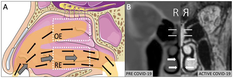
COVID-19 represents an extraordinary global health threat with multi-organ sequelae. Olfactory dysfunction (OD) has emerged in the majority of cases and is predictive of milder disease [1, 2]. However, OD in COVID-19 is unlike typical post-viral smell loss in that it occurs largely in the absence of other upper airway complaints [1, 3]. This symptom pattern is consistent with radiographic findings of severe focal olfactory epithelium (OE) inflammation with otherwise normal nasal respiratory epithelium (RE, Fig 1) [4]. The explanation for this selective olfactory involvement remains mysterious as the OE has the lowest expression of angiotensin converting enzyme 2 (ACE2), the SARS-CoV-2 binding receptor, within the entire nasal cavity [5]. A recent open letter from 239 scientists to international public health organizations called for acknowledgement of airborne spread as a potential transmission mode for SARS-CoV-2 [6] with the subsequent recognition by the World Health Organization in a scientific brief [7]. Given the increased emphasis on airborne transmission, we hypothesized that small concentrations of persistently airborne aerosols (those below 5μm) would be more prone to dispersing within the olfactory epithelium than the lower nasal airway. This selective olfactory dispersion of smaller-sized airborne particulate would therefore provide a novel mechanism linking atypical olfactory dysfunction in COVID-19 with airborne transmission. The purpose of this study was to therefore experimentally characterize whether airborne aerosols are capable of selective OE dispersal within the healthy human nasal cavity.

Fig 1.

Fig 1

A) Illustration of classic description of airborne aerosols (thin arrows) bypassing the nasal respiratory epithelium (RE) with proposed mechanism for a proportion of these aerosols penetrating into the olfactory epithelium (OE). Thick arrows depict larger-sized aerosolized droplets settling within RE (adapted from Servier Medical Art). B) Matched right sided CT and MRI (reflected) scan of same patient demonstrating new onset isolated inflammation of OE (thin arrows) during COVID-19 infection with sparing of RE (thick arrows).

Methods

The Mass General Brigham IRB approved the IRB Protocol, 2020P-001246. Written informed consent was obtained from all subjects. Participants were healthy subjects between the ages of 25 and 35 without a history of chronic rhinosinusitis, allergic rhinitis, or other rhinologic disease. At the time of data collection, subjects did not have any symptoms of acute sinusitis, rhinorrhea, or subjective nasal obstruction. A Hudson RCI 1883 nebulizer (Teleflex Medical, Morrisville, NC was used to generate smaller sized particulate in the sub 5μm range, classically characterized as a particle size capable of being persistently airborne for extended periods of time and having protracted settling rates. An optical particle sizer (OPS 3330, TSI Inc, Shoreview, MN) was utilized to measure the size distribution of nebulized airborne particulates to ensure that particles in this range were generated. Alternatively, an atomizer was used to generate larger aerosolized particulate, in the 30–100 μm range (MADomizer, Teleflex, Wayne, PA). A fluorescein solution of 1mg FUL-GLO Fluorescein Sodium (NDC17478-404-01, Akorn, Inc, Lake Forest, IL, USA) in 5mL saline was utilized in both conditions in the respective source of particle generation (nebulizer vs. atomizer). Immediately following 60 seconds of exposure to nebulization or atomization, subjects then underwent rigid nasal endoscopy equipped with a blue light liter for fluorescein visualization (Karl Storz, Tuttlingen, Germany). Digital images were captured of the OE (olfactory cleft, superior middle turbinate) and RE (nasal floor, inferior turbinate) bilaterally, randomized, and provided to a blinded reviewer for image processing using ImageJ (version 2.0.0-rc-69/1.52p). Images were systematically color-adjusted, thresholded, and transformed to 8-bit grayscale for quantification. Maximum fluorescence intensity, average-intensity of non-zero pixels, and standard error of non-zero pixels were calculated, with subtraction of non-fluorescein stained background values.

Statistics

Stata version 13 (StataCorp, College Station, TX) was used for statistical analysis using the within-program Mann-Whitney U test comparing maximum and average fluorescent values between the RE and OE (within subjects, n = 3). No outlier data was excluded, and there were no missing values for any experimental replicate. Medians and interquartile range (IQR) are reported as median (IQR spread). A p-value of <0.05 was considered significant.

Results

Nebulizer particulate analysis with an optical particle sizer confirmed reliable sub 5μm airborne aerosol generation with a peak of 2μm. Following fluorescein-labeled airborne aerosol inhalation of nebulized particulate, a significantly higher deposition of aerosols were found in the OE than the RE with minimal to no deposition observed in the RE (maximum fluorescence: OE 19.5(22.5), RE 1(3.2), p<0.001, U = 0, n = 10,6; average fluorescence: OE 2.3(4.5), RE 0.1(0.2) p<0.01, U = 3, n = 10,6, Mann-Whitney U test, n = 3 subjects, Fig 2).

Fig 2.
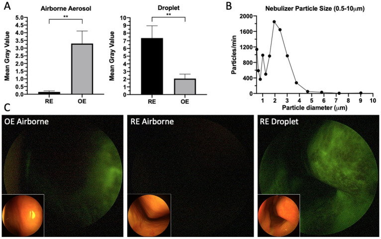
Fig 2

A) Small airborne aerosol (0.5–5μm) deposition versus aerosolized droplet (30–100 μm) deposition of fluorescein solution in the respiratory epithelium (RE) and olfactory epithelium (OE) (small airborne aerosol average fluorescence: OE 2.3(IQR 4.5), RE 0.1(IQR 0.2), p<0.01, aerosolized droplet average fluorescence: OE 1.9(IQR 2.1), RE 5.9(IQR 5.9), p<0.01). B) Size distribution of airborne aerosols in the 0.5–10μm range produced during nebulizer use. C) Blue-light filtered endoscopic nasal images (with brightfield insets) of fluorescein airborne and droplet labeled distribution to the olfactory (OE) and respiratory epithelium (RE) in human subjects.

Conversely, following fluorescein labeled aerosolized droplet (30–100 μm) inhalation, significantly greater deposition was observed in the RE as compared with the OE (maximum fluorescence: OE 13(14.3), RE 38(45.5) p<0.01, U = 2, n = 6,6; average fluorescence: OE 1.9(2.1), RE 5.9(5.9), p<0.01, U = 1, n = 6,6, Mann-Whitney U test, n = 3 subjects, Fig 2A).

Discussion

Self-reported OD has been widely described in COVID-19 through a variety of case reports [4], case series [8] and surveys [9], with a pooled prevalence of 52.7% [2]. The true rate is likely higher as formal smell testing by Moein et al. [8] revealed OD in 98% of COVID-19 positive patients, only 35% of whom self-reported. Using an olfaction survey, Yan et al. [10] found that loss of smell was in fact more highly correlated with COVID-19 positivity than any other systemic or pulmonary symptom. This correlative finding is echoed in a study utilizing a symptom tracker app in the United Kingdom [9]. Yan et al further demonstrated that self-reported OD was associated with milder disease and most often occurred in the absence of other symptoms, unlike typical post-viral OD. This finding was confirmed by Kaye et al, showing unexpectedly low rates of nasal congestion (25%) and rhinorrhea (18%) in a cohort of COVID-19 positive anosmic patients [3]. These results suggest that SARS-CoV-2 exhibits a unique predilection for impacting the OE to the relative exclusion of the remainder of the nasal airway, a feature corroborated by published reports of nasal CT and MRI scans(Fig 1B) [4].

The simplest mechanism for this distinct and isolated OE inflammation would be evidence of SARS-CoV-2 tropism for either the olfactory neurons or the OE itself. The neurotropic hypothesis appears unlikely as olfactory neurons lack both the ACE2 protein and TMPRSS2 gene required for viral spike protein binding and cellular entry [5]. Furthermore the rapidity of recovery [10] and lack of protective effect among females [8], otherwise common in neuropathic OD, suggest a non-neural etiology. In contradistinction, OE tropism could be explained by the presence of ACE2 within the sustentacular cells which support the olfactory neurons [5]. However, Brann et al. [5] demonstrated that ACE2 expression was actually higher within the ciliated and secretory cells found throughout the remainder of the RE than in the OE sustentacular cells. Therefore, ACE2 avidity alone cannot explain this phenomenon.

In the absence of a biologic explanation for atypical OD, our study explored the feasibility of a mechanism of differential particulate deposition. Small-sized, persistently airborne aerosols (less than 5–10μm) are classically understood to bypass the upper airway in favor of alveolar deposition [11]. While our results confirm this effect within the RE, they also reveal that airborne aerosols in fact deposit in appreciable concentrations within the OE. This effect is unique to small-sized airborne aerosols as we found that larger aerosolized droplets (30–100μm) were more likely to deposit in the RE. Based on these distribution patterns, we therefore surmise that against the background of widely distributed ACE2 throughout both the OE and RE, low concentrations of airborne SARS-Co-V will differentially bind to the OE resulting in localized inflammation.

As the potential for airborne transmission of SARS-CoV-2 becomes increasingly accepted by the medical community [6, 7], clues derived from olfactory physiology, objective smell testing, and imaging studies converge around airborne aerosol exposure as an explanation for the widespread, profound, and relatively isolated OD in COVID-19. Our study is limited by a small number of subjects as well as uniformity of ambient conditions; temperature and humidity changes in alternate conditions could also affect intranasal diffusion, sedimentation, or other variables contributing to deposition of particulate. However, in this small number of subjects our data experimentally shows that despite bypassing the majority of the upper airway, smaller airborne aerosols appear to differentially deposit in significant concentrations within the olfactory epithelium. This provides a compelling aerodynamic mechanism to explain the common and relatively isolated olfactory dysfunction associated with the majority of COVID-19 infections.

Supporting information

S1 Dataset. Raw data of maximum value, mean gray value of non-zero pixels, and standard error of non-zero pixels for each subject and condition.

(XLTX)

Data Availability

All relevant data is included in supplementary files.

Funding Statement

The authors received no specific funding for this work.

References

  • 1.Yan CH, Faraji F, Prajapati DP, Ostrander BT, DeConde AS. Self‐reported olfactory loss associates with outpatient clinical course in Covid‐19. Int Forum Allergy Rhinol. 2020; 10.1002/alr.22592 [DOI] [PMC free article] [PubMed] [Google Scholar]
  • 2.Tong JY, Wong A, Zhu D, Fastenberg JH, Tham T. The Prevalence of Olfactory and Gustatory Dysfunction in COVID-19 Patients: A Systematic Review and Meta-analysis. Otolaryngol Neck Surg [Internet]. 2020. May 5;0194599820926473. 10.1177/0194599820926473 [DOI] [PubMed] [Google Scholar]
  • 3.Kaye R, Chang CWD, Kazahaya K, Brereton J, Denneny JC. COVID-19 Anosmia Reporting Tool: Initial Findings. Otolaryngol Neck Surg. 2020; 10.1177/0194599820922992 [DOI] [PubMed] [Google Scholar]
  • 4.Eliezer M, Hautefort C, Hamel A-L, Verillaud B, Herman P, Houdart E, et al. Sudden and Complete Olfactory Loss Function as a Possible Symptom of COVID-19. JAMA Otolaryngol Neck Surg. 2020. April; 10.1001/jamaoto.2020.0832 [DOI] [PubMed] [Google Scholar]
  • 5.Brann DH, Tsukahara T, Weinreb C, Logan DW, Datta SR. Non-neural expression of SARS-CoV-2 entry genes in the olfactory epithelium suggests mechanisms underlying anosmia in COVID-19 patients. bioRxiv. 2020; [DOI] [PMC free article] [PubMed] [Google Scholar]
  • 6.Morawska L, Milton DK. It is Time to Address Airborne Transmission of COVID-19. Clin Infect Dis [Internet]. 2020. July 6; 10.1093/cid/ciaa939 [DOI] [PMC free article] [PubMed] [Google Scholar]
  • 7.World Health Organization(WHO). Transmission of SARS-CoV-2: implications for infection prevention precautions. Sci Br. 2020; [Google Scholar]
  • 8.Moein ST, Hashemian SMR, Mansourafshar B, Khorram-Tousi A, Tabarsi P, Doty RL. Smell dysfunction: a biomarker for COVID-19. Int Forum Allergy Rhinol. 2020. April;alr.22587. 10.1002/alr.22587 [DOI] [PMC free article] [PubMed] [Google Scholar]
  • 9.Menni C, Valdes A, Freydin MB, Ganesh S, Moustafa JE-S, Visconti A, et al. Loss of smell and taste in combination with other symptoms is a strong predictor of COVID-19 infection. medRxiv. 2020; [Google Scholar]
  • 10.Yan CH, Faraji F, Prajapati DP, Boone CE, DeConde AS. Association of chemosensory dysfunction and Covid-19 in patients presenting with influenza-like symptoms. Int Forum Allergy Rhinol. 2020. April; 10.1002/alr.22579 [DOI] [PMC free article] [PubMed] [Google Scholar]
  • 11.Jones RM, Brosseau LM. Aerosol transmission of infectious disease. J Occup Environ Med. 2015; 10.1097/JOM.0000000000000448 [DOI] [PubMed] [Google Scholar]

Decision Letter 0

Vladimir Mikheev

27 Oct 2020

PONE-D-20-22099

Airborne Aerosol Olfactory Dispersion Contributes to Anosmia in COVID-19

PLOS ONE

Dear Dr. Workman,

Thank you for submitting your manuscript to PLOS ONE. After careful consideration, we feel that it has merit but does not fully meet PLOS ONE’s publication criteria as it currently stands. Therefore, we invite you to submit a revised version of the manuscript that addresses the points raised during the review process.

Please submit your revised manuscript by November 9, 2020. If you will need more time than this to complete your revisions, please reply to this message or contact the journal office at plosone@plos.org. When you're ready to submit your revision, log on to https://www.editorialmanager.com/pone/ and select the 'Submissions Needing Revision' folder to locate your manuscript file.

Please include the following items when submitting your revised manuscript:

  • A rebuttal letter that responds to each point raised by the academic editor and reviewer(s). You should upload this letter as a separate file labeled 'Response to Reviewers'.

  • A marked-up copy of your manuscript that highlights changes made to the original version. You should upload this as a separate file labeled 'Revised Manuscript with Track Changes'.

  • An unmarked version of your revised paper without tracked changes. You should upload this as a separate file labeled 'Manuscript'.

If you would like to make changes to your financial disclosure, please include your updated statement in your cover letter. Guidelines for resubmitting your figure files are available below the reviewer comments at the end of this letter.

If applicable, we recommend that you deposit your laboratory protocols in protocols.io to enhance the reproducibility of your results. Protocols.io assigns your protocol its own identifier (DOI) so that it can be cited independently in the future. For instructions see: http://journals.plos.org/plosone/s/submission-guidelines#loc-laboratory-protocols

We look forward to receiving your revised manuscript.

Kind regards,

Vladimir Mikheev

Academic Editor

PLOS ONE

Journal Requirements:

When submitting your revision, we need you to address these additional requirements.

1. Please ensure that your manuscript meets PLOS ONE's style requirements, including those for file naming. The PLOS ONE style templates can be found at

https://journals.plos.org/plosone/s/file?id=wjVg/PLOSOne_formatting_sample_main_body.pdf and

https://journals.plos.org/plosone/s/file?id=ba62/PLOSOne_formatting_sample_title_authors_affiliations.pdf

2. In your methods section and your ethics statement in the online submission form please clarify whether consent was informed consent.

3. Please provide the source and catalog number for the FUL-GLO Fluorescein Sodium used in this study.

4. To comply with PLOS ONE submission guidelines, in your Methods section, please provide additional information regarding your statistical analyses.

For more information on PLOS ONE's expectations for statistical reporting, please see https://journals.plos.org/plosone/s/submission-guidelines.#loc-statistical-reporting

5. Please include captions for your Supporting Information files at the end of your manuscript, and update any in-text citations to match accordingly. Please see our Supporting Information guidelines for more information: http://journals.plos.org/plosone/s/supporting-information

Additional Editor Comments:

Dear Dr. Workman,

Deep apologies for this long delay.

It was really hard to find reviewers but now we have necessary minimum number of reviews.

I think this paper is important one but you need to address minor comments suggested by the reviewers.

Again sorry for the delay.

[Note: HTML markup is below. Please do not edit.]

Reviewers' comments:

Reviewer's Responses to Questions

Comments to the Author

1. Is the manuscript technically sound, and do the data support the conclusions?

The manuscript must describe a technically sound piece of scientific research with data that supports the conclusions. Experiments must have been conducted rigorously, with appropriate controls, replication, and sample sizes. The conclusions must be drawn appropriately based on the data presented.

Reviewer #1: Yes

Reviewer #2: Yes

**********

2. Has the statistical analysis been performed appropriately and rigorously?

Reviewer #1: I Don't Know

Reviewer #2: Yes

**********

3. Have the authors made all data underlying the findings in their manuscript fully available?

The PLOS Data policy requires authors to make all data underlying the findings described in their manuscript fully available without restriction, with rare exception (please refer to the Data Availability Statement in the manuscript PDF file). The data should be provided as part of the manuscript or its supporting information, or deposited to a public repository. For example, in addition to summary statistics, the data points behind means, medians and variance measures should be available. If there are restrictions on publicly sharing data—e.g. participant privacy or use of data from a third party—those must be specified.

Reviewer #1: Yes

Reviewer #2: Yes

**********

4. Is the manuscript presented in an intelligible fashion and written in standard English?

PLOS ONE does not copyedit accepted manuscripts, so the language in submitted articles must be clear, correct, and unambiguous. Any typographical or grammatical errors should be corrected at revision, so please note any specific errors here.

Reviewer #1: Yes

Reviewer #2: Yes

**********

5. Review Comments to the Author

Please use the space provided to explain your answers to the questions above. You may also include additional comments for the author, including concerns about dual publication, research ethics, or publication ethics. (Please upload your review as an attachment if it exceeds 20,000 characters)

Reviewer #1: The objective of this study is to compare aerosol deposition in the OE and RE of nasal passages. Measurements clearly show that small particles are able to reach the olfactory region (mainly by diffusion given that there is very little airflow into the region) while large particles lack diffusive properties and hence travel mainly through the RE as evidenced by its deposition in this region. COVID particles are considered fine and thus can penetrate the OE region. This reviewer has the following comments regarding the manuscript:

The Method section is confusing and lacks details. It needs to clearly describe the experimental system for each particle type, choice and reason for the selection of two classes of particles (lines 61 – 63). Droplets are aerosols too. You need to distinguish the aerosols by size and composition. Also, need to explain why you picked large particles versus small.

Lines 44 – 46: The statement is partly inaccurate and the process is well described in the aerosol literature. The inhaled air splits at the junction with the majority of the flow (and thus particles regardless of the size) pass through the RE region. However, there is little deposition due to high flow convection in the RE region (thus small gravitational settling). There are far fewer particles reaching the OE region. However, those that reach there deposit by Brownian diffusion and sedimentation. The reason for higher deposition in OE is not due to having more particles but having more deposition. Please revise your hypothesis and clearly explain how you are going to achieve your hypothesis.

Line 60: you did not validate your size distribution but measured it.

Reviewer #2: The authors should be commended for this study, which hypothesizes that an aerodynamic mechanism may help explain the link between COVID-19 and olfactory dysfunction. Specifically, they show that airborne aerosols differentially deposit in significant concentrations within the olfactory epithelium. This paper, which is brief but methodologically sound, represents a valuable addition to the literature.

I would suggest that the authors address several issues prior to potential publication. A primary concern is that there is no mention in the methods/results section of how many subjects were analyzed. The authors should be more clear that only 3 patients (based on the raw data) were analyzed. This small number limits the conclusions one can make from this data and this should be explicitly stated in the discussion section. Furthermore, there is no descriptions of the subjects other than 'healthy.' Further limitations should be added to the discussion section.

**********

6. PLOS authors have the option to publish the peer review history of their article (what does this mean?). If published, this will include your full peer review and any attached files.

If you choose “no”, your identity will remain anonymous but your review may still be made public.

Do you want your identity to be public for this peer review? For information about this choice, including consent withdrawal, please see our Privacy Policy.

Reviewer #1: Yes: Bahman Asgharian

Reviewer #2: No

[NOTE: If reviewer comments were submitted as an attachment file, they will be attached to this email and accessible via the submission site. Please log into your account, locate the manuscript record, and check for the action link "View Attachments". If this link does not appear, there are no attachment files.]

While revising your submission, please upload your figure files to the Preflight Analysis and Conversion Engine (PACE) digital diagnostic tool, https://pacev2.apexcovantage.com/. PACE helps ensure that figures meet PLOS requirements. To use PACE, you must first register as a user. Registration is free. Then, login and navigate to the UPLOAD tab, where you will find detailed instructions on how to use the tool. If you encounter any issues or have any questions when using PACE, please email PLOS at figures@plos.org. Please note that Supporting Information files do not need this step.

PLoS One. 2021 Feb 5;16(2):e0244127. doi: 10.1371/journal.pone.0244127.r002

Author response to Decision Letter 0


24 Nov 2020

Dear Editors:

Thank you very much for your consideration and response, as well as for the helpful comments of the reviewers. We have made changes to the manuscript in line with the requests. These are outlined below and are highlighted in the manuscript text. We are confident that the revised work will be of interest to the PLOS ONE readers.

Best regards,

Alan Workman, MD, MTR

Benjamin Bleier, MD, FACS, FARS

Associate Editor Comments

1. Please ensure that your manuscript meets PLOS ONE's style requirements, including those for file naming.

These style requirements have now been added to the updated manuscript.

2. In your methods section and your ethics statement in the online submission form please clarify whether consent was informed consent.

The statement, “…and informed consent was obtained from all participants was added to the first line of the methods.

3. Please provide the source and catalog number for the FUL-GLO Fluorescein Sodium used in this study.

The catalog number is now included. The source was Akorn Inc., Lake Forest, IL, USA (written in text as well).

4. To comply with PLOS ONE submission guidelines, in your Methods section, please provide additional information regarding your statistical analyses.

A separate section on statistical analysis has been added to the methods, with enough detail that utilizing our raw data as well as the statistical tests described our results would be easily reproducible.

5. Please include captions for your Supporting Information files at the end of your manuscript, and update any in-text citations to match accordingly.

The file has been renamed and the captions have been added.

Reviewer 1 Comments

1. The Method section is confusing and lacks details. It needs to clearly describe the experimental system for each particle type, choice and reason for the selection of two classes of particles (lines 61 – 63). Droplets are aerosols too. You need to distinguish the aerosols by size and composition. Also, need to explain why you picked large particles versus small. Lines 44 – 46: The statement is partly inaccurate and the process is well described in the aerosol literature. The inhaled air splits at the junction with the majority of the flow (and thus particles regardless of the size) pass through the RE region. However, there is little deposition due to high flow convection in the RE region (thus small gravitational settling). There are far fewer particles reaching the OE region. However, those that reach there deposit by Brownian diffusion and sedimentation. The reason for higher deposition in OE is not due to having more particles but having more deposition. Please revise your hypothesis and clearly explain how you are going to achieve your hypothesis.

The hypothesis has now been revised to be significantly more clear throughout all sections of the manuscript. As you discussed, we have now distinguished between “persistently aerosolized particulate” of smaller size (sub 5-10�m) and larger aerosolized droplets (30-100�m), with the former generated by the nebulizer and the latter generated by the atomizer. This distinction between the size of particulate forms the basis of our hypothesis, in that the smaller particles will not deposit in the RE but do deposit in the OE by diffusion, while larger particulate does indeed settle in the RE. The composition of the particulate remains the same (identical solutions). We have additionally changed the text to highlight that our hypothesis is that small airborne aerosols selectively deposit in the OE, not that disperse to the OE.

2. Line 60: you did not validate your size distribution but measured it.

This has been corrected to utilize the word “measure.”

Reviewer Two Comments

1. A primary concern is that there is no mention in the methods/results section of how many subjects were analyzed. The authors should be more clear that only 3 patients (based on the raw data) were analyzed. This small number limits the conclusions one can make from this data and this should be explicitly stated in the discussion section. Furthermore, there is no descriptions of the subjects other than 'healthy.' Further limitations should be added to the discussion section.

We have now listed “n=3 subjects” in the description of of the raw data and statistics within the results section. Within the methods, we have added the sentences “Participants were healthy subjects between the ages of 25 and 35 without a history of chronic rhinosinusitis, allergic rhinitis, or other rhinologic disease. At the time of data collection, subjects did not have any symptoms of acute sinusitis, rhinorrhea, or subjective nasal obstruction.” In the discussion section, we have now noted additional limitations, and have added the sentence “our study is limited by a small number of subjects as well as uniformity of ambient conditions; temperature and humidity changes in alternate conditions could also affect intranasal diffusion, sedimentation, or other variables contributing to deposition of particulate.”

Decision Letter 1

Vladimir Mikheev

1 Dec 2020

PONE-D-20-22099R1

Airborne Aerosol Olfactory Deposition Contributes to Anosmia in COVID-19

PLOS ONE

Dear Dr. Workman,

Thank you for submitting your manuscript to PLOS ONE. After careful consideration, we feel that it has merit but does not fully meet PLOS ONE’s publication criteria as it currently stands. Therefore, we invite you to submit a revised version of the manuscript that addresses the points raised during the review process.

==============================

Dear Authors,

Thank you for revision of your manuscript.

I believe you properly addressed Reviewers comments.

I think your manuscript is almost ready for publication but I would like you to address couple of my comments as well.

Reviewer 1 stated: "There are far fewer particles reaching the OE region. However, those that reach there deposit by Brownian diffusion and sedimentation."

Please note that although in general its true (both Brownian diffusion and sedimentation play role) but talking specifically about COVID-19 it has to be noted that single COVID-19 particles (containing only one virion) could be of ~100 nm (60-150 nm) size but peak concentrations found in hospitals were within ~250-500 nm size (please see reference below). Particles of this size range should not deposit by Brownian diffusion (rather particles below 100 nm are subject to Brownian diffusion).

Therefore I would strongly suggest to edit your statement made on L56-57 (Caption to Figure 1) "proportion of these aerosols diffusing through Brownian motion into the olfactory epithelium (OE)" to "proportion of these aerosols penetrating into the olfactory epithelium (OE)".

Also, L67 - please specify what type of nebulizer was used to generate smaller sized particulate in the sub 5 micron range.

Reference:

Liu Y, Ning Z, Chen Y, Guo M, Liu Y, Gali NK, et al. Aerodynamic analysis of 193 SARS-CoV-2 in two Wuhan hospitals. Nature. 2020; https://doi.org/10.1038/s41586-194 020-2271-3

==============================

Please submit your revised manuscript by December 14, 2020. If you will need more time than this to complete your revisions, please reply to this message or contact the journal office at plosone@plos.org. When you're ready to submit your revision, log on to https://www.editorialmanager.com/pone/ and select the 'Submissions Needing Revision' folder to locate your manuscript file.

Please include the following items when submitting your revised manuscript:

  • A rebuttal letter that responds to each point raised by the academic editor and reviewer(s). You should upload this letter as a separate file labeled 'Response to Reviewers'.

  • A marked-up copy of your manuscript that highlights changes made to the original version. You should upload this as a separate file labeled 'Revised Manuscript with Track Changes'.

  • An unmarked version of your revised paper without tracked changes. You should upload this as a separate file labeled 'Manuscript'.

If you would like to make changes to your financial disclosure, please include your updated statement in your cover letter. Guidelines for resubmitting your figure files are available below the reviewer comments at the end of this letter.

If applicable, we recommend that you deposit your laboratory protocols in protocols.io to enhance the reproducibility of your results. Protocols.io assigns your protocol its own identifier (DOI) so that it can be cited independently in the future. For instructions see: http://journals.plos.org/plosone/s/submission-guidelines#loc-laboratory-protocols

We look forward to receiving your revised manuscript.

Kind regards,

Vladimir Mikheev

Academic Editor

PLOS ONE

[Note: HTML markup is below. Please do not edit.]

[NOTE: If reviewer comments were submitted as an attachment file, they will be attached to this email and accessible via the submission site. Please log into your account, locate the manuscript record, and check for the action link "View Attachments". If this link does not appear, there are no attachment files.]

While revising your submission, please upload your figure files to the Preflight Analysis and Conversion Engine (PACE) digital diagnostic tool, https://pacev2.apexcovantage.com/. PACE helps ensure that figures meet PLOS requirements. To use PACE, you must first register as a user. Registration is free. Then, login and navigate to the UPLOAD tab, where you will find detailed instructions on how to use the tool. If you encounter any issues or have any questions when using PACE, please email PLOS at figures@plos.org. Please note that Supporting Information files do not need this step.

PLoS One. 2021 Feb 5;16(2):e0244127. doi: 10.1371/journal.pone.0244127.r004

Author response to Decision Letter 1


3 Dec 2020

Dr. Mikheev,

Thank you again for your ongoing consideration of our manuscript. We are happy to address your comments and agree with you that the requested changes make the manuscript clearer and are more harmonious with the existing literature on SARS-CoV-2. The changes have been made in the text (line 56-57 for the first comment) and addition of the nebulizer model to line 67.

Best regards,

Alan Workman, MD, MTR

Benjamin Bleier, MD, FACS, FARS

Decision Letter 2

Vladimir Mikheev

4 Dec 2020

Airborne Aerosol Olfactory Deposition Contributes to Anosmia in COVID-19

PONE-D-20-22099R2

Dear Dr. Workman,

We’re pleased to inform you that your manuscript has been judged scientifically suitable for publication and will be formally accepted for publication once it meets all outstanding technical requirements.

Within one week, you’ll receive an e-mail detailing the required amendments. When these have been addressed, you’ll receive a formal acceptance letter and your manuscript will be scheduled for publication.

An invoice for payment will follow shortly after the formal acceptance. To ensure an efficient process, please log into Editorial Manager at http://www.editorialmanager.com/pone/, click the 'Update My Information' link at the top of the page, and double check that your user information is up-to-date. If you have any billing related questions, please contact our Author Billing department directly at authorbilling@plos.org.

If your institution or institutions have a press office, please notify them about your upcoming paper to help maximize its impact. If they’ll be preparing press materials, please inform our press team as soon as possible -- no later than 48 hours after receiving the formal acceptance. Your manuscript will remain under strict press embargo until 2 pm Eastern Time on the date of publication. For more information, please contact onepress@plos.org.

Kind regards,

Vladimir Mikheev

Academic Editor

PLOS ONE

Additional Editor Comments (optional):

Reviewers' comments:

Acceptance letter

Vladimir Mikheev

27 Jan 2021

PONE-D-20-22099R2

Airborne Aerosol Olfactory Deposition Contributes to Anosmia in COVID-19

Dear Dr. Workman:

I'm pleased to inform you that your manuscript has been deemed suitable for publication in PLOS ONE. Congratulations! Your manuscript is now with our production department.

If your institution or institutions have a press office, please let them know about your upcoming paper now to help maximize its impact. If they'll be preparing press materials, please inform our press team within the next 48 hours. Your manuscript will remain under strict press embargo until 2 pm Eastern Time on the date of publication. For more information please contact onepress@plos.org.

If we can help with anything else, please email us at plosone@plos.org.

Thank you for submitting your work to PLOS ONE and supporting open access.

Kind regards,

PLOS ONE Editorial Office Staff

on behalf of

Dr. Vladimir Mikheev

Academic Editor

PLOS ONE

Associated Data

    This section collects any data citations, data availability statements, or supplementary materials included in this article.

    Supplementary Materials

    S1 Dataset. Raw data of maximum value, mean gray value of non-zero pixels, and standard error of non-zero pixels for each subject and condition.

    (XLTX)

    Data Availability Statement

    All relevant data is included in supplementary files.


    Articles from PLoS ONE are provided here courtesy of PLOS

    RESOURCES