Abstract
Hypercholesterolemia is an independent risk factor of cardiovascular disease, which is among the major causes of death worldwide. The aim of this study was to explore whether Bifidobacterium longum strains exerted intra-species differences in cholesterol-lowering effects in hypercholesterolemic rats and to investigate the potential mechanisms. SD rats underwent gavage with each B. longum strain (CCFM 1077, I3, J3 and B3) daily for 28 days. B. longum CCFM 1077 exerted the most potent cholesterol-lowering effect, followed by B. longum I3 and B3, whereas B. longum B3 had no effect in alleviating hypercholesterolemia. Divergent alleviation of different B. longum strains on hypercholesterolemia can be attributed to the differences in bile salt deconjugation ability and cholesterol assimilation ability in vitro. By 16S rRNA metagenomics analysis, the relative abundance of beneficial genus increased in the B. longum CCFM 1077 treatment group. The expression of key genes involved in cholesterol metabolism were also altered after the B. longum CCFM 1077 treatment. In conclusion, B. longum exhibits strain-specific effects in the alleviation of hypercholesterolemia, mainly due to differences in bacterial characteristics, bile salt deconjugation ability, cholesterol assimilation ability, expressions of key genes involved in cholesterol metabolism and alterations of gut microbiota.
Keywords: B. longum strains, hypercholesterolemia, strain-specific, bile salt deconjugation, cholesterol assimilation, gut microbiota
1. Introduction
Hypercholesterolemia is among the major causative factors for cardiovascular disease (CVD) [1]. CVD is regarded as a major health issue, accounting for 40% of all deaths in China in the past ten years; thus, hypercholesterolemia has become a serious challenge to the Chinese government for the prevention and control of these diseases [2,3]. In recent years, it has been widely reported that the reduction in total cholesterol and low-density lipoprotein cholesterol (LDL-C) in patients with hypercholesterolemia may reduce their CVD risk [4,5,6]. Previous studies have shown three-fold differences in the risk of CVD between people with hypercholesterolemia and those without [7]. Moreover, a 1% elevation in the serum cholesterol concentration has been found to cause a 2% to 3% increase in the incidence of CVD [8]. Among the main causes for hypercholesterolemia-related CVD is unhealthy changes in eating habits, such as increased intake of cholesterol and saturated fat, which disrupts the blood cholesterol metabolism and alters the composition and abundance of gut microbiota [9].
Currently, both pharmacological and non-pharmacological approaches, including drug treatments, dietary interventions and exercise, are clinically prescribed to control the serum cholesterol level [10]. However, due to the side effects of lipid-reducing drugs, contraindications for such medications, or personal preferences, many people prefer other functional foods to combat hypercholesterolemia. Therefore, it is crucial to find a safe and effective approach to alleviate hypercholesterolemia. The gut microbiota is reported to be related to metabolic diseases such as obesity, diabetes mellitus and hypercholesterolemia. Several studies have indicated that the composition of gut microbiota in hypercholesterolemic patients differs from that in healthy people, which is mainly characterized by the decreased relative levels of beneficial microbes, such as Bifidobacterium and Lactobacillus, and increased relative levels of pathogenic bacteria [11,12,13]. Therefore, probiotics are a new option for treatment of cholesterolemia.
Several animal and clinical trial studies have indicated the effectiveness of probiotics to treat hypercholesterolemia, either with single strain (L. plantarum ECGC 13110402, L. reuteri NCIMB 30242, or B. longum BB536) [14,15,16] or multiple strains (L.plantarum (CECT 7527,7528,7529), L. plantarum (ECGC 13, 110, 402), Bifidobacterium (B. animalis subsp. lactis MB 2409, B. bifidum MB 109 and B. longum subsp. longum BL04)) [14,17,18]. However, some strains (L. rhamnosus LC705, Pediococcus pentosaceus LP28) have been reported to be ineffective in alleviating hypercholesterolemia [19,20]. B. longum, as the most prevalent Bifidobacterium species in the human gastrointestinal tract, has been proven to be effective in protecting against CVDs, such as hypertension [21] and brain–gut-related diseases, such as chronic colitis and irritable bowel syndrome [22]. The underlying mechanism of the cholesterol-lowering effect of probiotics has been reported to be related to the expression of several genes involved in cholesterol metabolism [23,24,25]. FXR, CYP7A1 and SHP play important roles in the synthesis of bile acids from cholesterol, and LXR plays a role in reverse cholesterol transport [26,27]. Many hypotheses suggest that FXR is influenced by changes in the bile acid pool and that a decreased FXR expression downregulates SHP expression and upregulates CYP7A1 expression, both of which are bile acid synthesis rate-limiting enzymes [28]. Based on this background, we hypothesized that B. longum could prevent and treat hypercholesterolemia and that inter-strain differences exist in the treatment of hypercholesterolemia induced by a cholesterol-enriched diet in SD rats.
Therefore, we sought to explore whether B. longum presents strain-specific differences in the alleviation of hypercholesterolemia induced by a cholesterol-enriched diet in rats. Furthermore, the potential mechanisms of the inter-strain differences were evaluated, such as the basic physiological properties, the capacities of bile salt deconjugation and cholesterol assimilation, the mediated effects of key genes involved in cholesterol metabolism and the alteration of gut microbiota.
2. Results
2.1. Subsection Growth Characteristics of B. longum Strains in Vitro
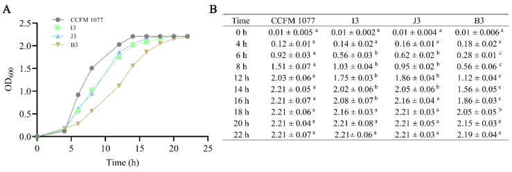
For determination of the growth characteristics of B. longum strains in vitro, the growth curve of the four strains was drawn. As is shown in Figure 1A, compared with other strains, B. longum CCFM 1077 is fastest in entering the logarithmic phase, followed by B. longum J3 and B. longum I3, whereas B. longum B3 is the slowest in entering the logarithmic phase. In addition, the statistical differences between different strains at different growth time are shown in Figure 1B.
Figure 1.
Growth status of four B. longum strains. (A) Growth curves of four B. longum strains. The X-axis is the culture time of four B. longum strains, and the Y-axis is the absorbance of culture liquid at 600 nm. The OD600 was tested three times at different time points (0, 4, 6, 8, 12, 14, 16, 18, 20 and 22 h). The data are expressed with the average of three OD600 values at each time point. (B) All OD600 values are means ± standard error of the mean. a, b, c: Means in the same row with different superscript letters are significantly different (p < 0.01) based on determination using a one-way ANOVA followed by Tukey multiple comparison test.
2.2. Tolerance Ability of B. longum to Simulated Gastroenteric Fluid
The tolerance ability to gastric acid and bile salts of the four B. longum strains (CCFM 1077, I3, J3 and B3) was measured after cultivation at 37 °C in Whitley DG250 Anaerobic Workstation. As shown in Table 1, each of the four B. longum strains (B. longum CCFM 1077, B. longum I3, B. longum J3 and B. longum B3) showed high tolerance to the simulated intestinal environment of pH 2.5 and containing 0.3% bile acid (p < 0.01).
Table 1.
Tolerance of four B.longum strains to simulated gastroenteric environments.
| Strain | Initial Survival in PBS | Survival after 2 h at pH 2.5 in SGJ |
Survival after 3 h at pH 8.0 in 0.3% Oxigall |
||
|---|---|---|---|---|---|
| Mean Counts (Log CFU/mL) |
Mean Counts (Log CFU/mL) |
Survival Rate | Mean Counts (Log CFU/mL) |
Survival Rate | |
| B. longum CCFM 1077 | 9.76 ± 0.06 a | 7.97 ± 0.18 b | 81.66% | 6.36 ± 0.12c | 65.16% |
| B. longum I3 | 9.23 ± 0.24 a | 7.56 ± 0.14 b | 81.91% | 6.08 ± 0.08 c | 65.87% |
| B. longum J3 | 9.32 ± 0.18 a | 7.86 ± 0.26 b | 84.33% | 5.95 ± 0.12 c | 63.84% |
| B. longum B3 | 9.58 ± 0.08 a | 6.98 ± 0.23 b | 72.86% | 6.24 ± 0.18 c | 65.14% |
All values are means ± standard error of the mean. a,b: Means in the same row with different superscript letters are significantly different (p < 0.01) based on determination using a one-way ANOVA followed by Tukey multiple comparison test. % survival = final (CFU/mL)/control (CFU/mL) × 100% survival indicates that the growth rate of the strain was not affected by the treatment. PBS: Phosphate Buffer solution. SGJ: Simulated gastric juice.
2.3. Bile Salt Deconjugation and Cholesterol Assimilation Abilities of the B. longum Strains
Two properties of B. longum strains (bile salt deconjugation ability and cholesterol assimilation ability) were used to evaluate the cholesterol-lowering effect in vitro. As shown in Table 2, different properties were found in these four B. longum strains: B. longum CCFM 1077 shows both high abilities of bile salt deconjugation and cholesterol assimilation; B. longum I3 shows only high ability of bile salt deconjugation; B. longum J3 shows only high ability of cholesterol assimilation; B. longum B3 shows neither abilities of bile salt deconjugation or cholesterol assimilation.
| (1) |
| (2) |
Table 2.
Bile salt hydrolysis ability and cholesterol assimilation ability of B. longum strains.
| Strain | Bile Salt Hydrolysis Ability (%) | Cholesterol Assimilation Ability (%) |
|---|---|---|
| B. longum CCFM 1077 | 98.66 ± 0.65 a | 97.68 ± 1.03 a |
| B. longum I3 | 97.36 ± 0.36 a | 0.96 ± 0.16 b |
| B. longum J3 | 1.01 ± 0.02 b | 99.36 ± 0.32 a |
| B. longum B3 | 1.12 ± 0.06 b | 0.82 ± 0.15 b |
All values are means ± standard error of the mean. Means in the same column with different superscript letters are significantly different (p < 0.01) based on the analysis using a one-way ANOVA followed by a Tukey multiple comparison test. C0 is the GCA concentration of the control sample, and C1 is the GCA concentration of the B. longum sample.
2.4. The Effects of B. longum Strains on the Serum Lipids
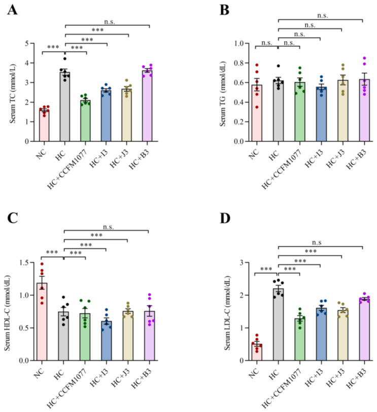
In terms of body weight, no significant differences were found among the three B. longum groups, HC group and NC group (Supplementary Figure S1). The serum lipid levels of the six groups are shown in Figure 2. Compared with the NC group, the HC group showed significantly higher levels in the serum TC and LDL-C levels. A significant decrease in the serum lipid levels was also observed in the CCFM 1077, I3 and J3 groups compared with the HC group (p < 0.001, Figure 1). Specifically, compared with the HC group, in the HC-CCFM 1077, HC-I3 and HC-J3 groups, the serum TC levels decreased by 44.44%, 30.56% and 27.77%; the serum LDL-C levels decreased by 40.91%, 31.82% and 27.27%, respectively. However, the serum HDL-C and TG levels showed no significant difference between the experimental groups. In addition, the serum lipid levels showed no difference between the HC-B3 and HC groups.
Figure 2.
The effects of four B. longum strains on serum lipid levels of (A) TC, (B) TG, (C) HDL-C and (D) LDL-C in rats with hypercholesterolemia. *** represents p < 0.001, n.s represent non-significant with p > 0.05.
2.5. Effects of B. longum Strains on Fecal Bile Acid and Cholesterol Levels
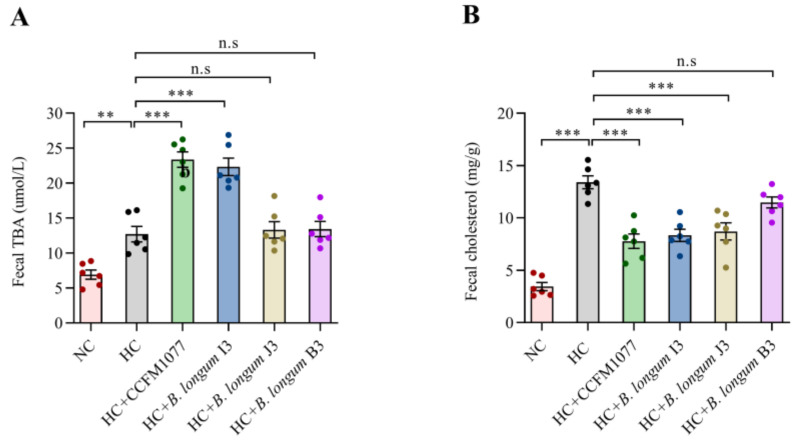
The fecal bile acid and cholesterol levels in the different groups of rats on the last day are shown in Figure 3. The HC-CCFM 1077 and HC-I3 groups showed significantly higher levels of fecal total bile acid than the HC group (76.92% and 69.23%, p < 0.001, Figure 3A), and the HC-CCFM 1077 and HC-J3 groups showed significantly higher levels of fecal cholesterol (73.33% and 60.32%, p < 0.001, Figure 3B). However, B. longum B3 had no effect on the fecal excretion of bile acid and cholesterol compared with other strains.
Figure 3.
The effects of four B. longum strains on the levels of fecal bile acid (A) and fecal cholesterol (B). Data are expressed as means ± standard error of the mean in each group (n = 6). *** represents p < 0.001, ** represents p < 0.01, n.s represent non-significant with p > 0.05. p-values were determined using a one-way ANOVA followed by Tukey multiple comparison test.
2.6. Effects of B. longum Strains on Liver Gene Expression
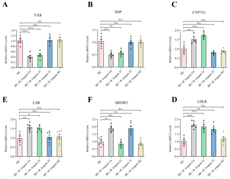
As shown in Figure 4, compared with the HC group, HC-CCFM 1077 and HC-I3 groups showed significantly downregulated FXR and SHP expression and upregulated CYP7A1 and LXR expression (p < 0.001, Figure 4A–D). SREBP2 expression was significantly upregulated by nearly two times in the HC-CCFM 1077 and HC-J3 groups (p < 0.01, Figure 4E); LDLR expression was also significantly upregulated in the HC-CCFM 1077, HC-I3 and HC-J3 groups (p < 0.001, Figure 4F).
Figure 4.
The effects of four B. longum strains on liver expression of genes involved in cholesterol and bile acid metabolism by RT-PCR. (A) FXR; (B) SHP; (C) CYP7A1; (D) LXR; (E) SREBP2; (F) LDLR. Each column represents the means ± standard error of the mean (n = 6). *** represents p < 0.001, ** represents p < 0.01, * represents p < 0.05, n.s represent non-significant with p > 0.05.
2.7. Effects of the Four B. longum Strains on Gut Microbiota
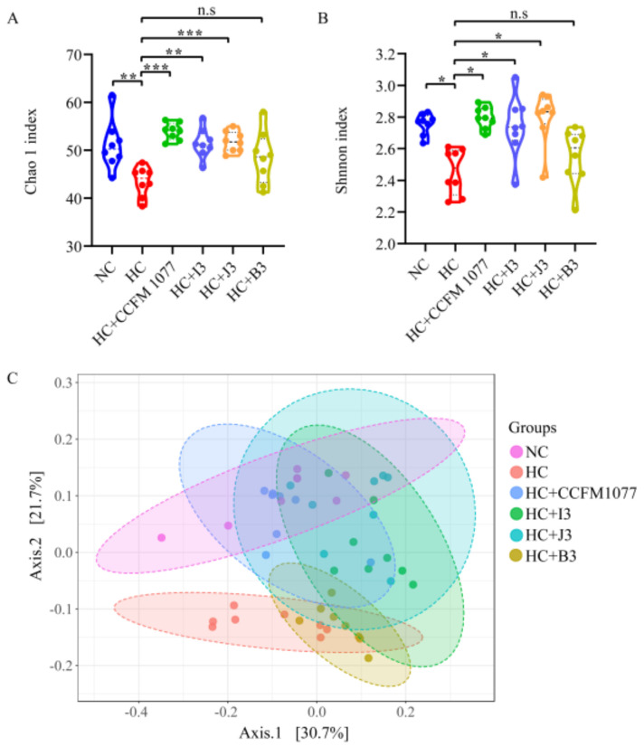
In our study, alpha diversity of gut microbiota was measured by Chao 1 and Shannon indexes. Both Chao 1 index and Shannon index of B. longum 1077, I3 and J3 treated rats were significantly higher than rats fed with a cholesterol-enriched diet, whereas B. longum B3 had no effect in these two indexes (Figure 5A,B). In terms of β-diversity, the gut microbiota between NC group and HC group were remarkably different; three B. longum groups (B. longum CCFM 1077, I3 and J3) improved the shift caused by the cholesterol-enriched diet, while B. longum B3 did not alter this shift (Figure 5C).
Figure 5.
Effects of four B. longum strains (CCFM 1077, I3, J3, B3) on the overall structure of fecal microbiota. Alpha diversity was represented by (A) Chao 1 index and (B) Shannon index. Beta diversity was represented by (C) PCoA, based on weighted UniFrac distances. *** represents p < 0.001, ** represents p < 0.01, * represents p < 0.05, n.s represent non-significant with p > 0.05.
Through MiSeq sequencing analysis, we obtained 1,780,569 high-quality, classifiable 16S rRNA gene sequences in 36 fecal samples. The average sequence read was 12,864 bp per sample. Typical sequences were clustered, and a sequence similarity of 97% was considered as the cut-off. The number of OTUs per sample ranged from 966 to 8529.
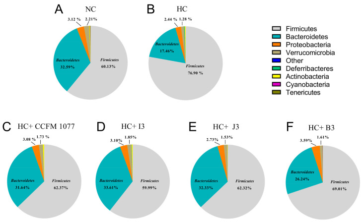
In terms of the phylum level (Figure 6), the individual OTUs showed that the gut microbiota were mainly dominated by Firmicutes, Bacteroidetes, Proteobacteria and Verrucomicrobia, which accounted for 60.13%, 32.59%, 3.12% and 2.21% of the total gut microbiota, respectively. The composition of gut microbiota varied between different groups after feeding high-cholesterol diets to the groups for 28 days. Compared with the HC group, the HC-CCFM 1077, HC-I3 and HC-J3 groups showed a similar alteration, with a significant decrease in the relative abundance of Firmicutes (76.89% vs. 62.37%, 59.99% and 62.32%, respectively) and a significant increase in that of Bacteroidetes (17.46% vs. 31.64%, 33.60% and 32.33%, respectively) (p < 0.05). However, the HC+B3 group did not remarkably change the composition of gut microbiota at the phylum level.
Figure 6.
The relative abundance of the main phyla after consumption of high-cholesterol diet and administration of B. longum strains. (A) NC: non-cholesterol added diet; (B) HC: high-cholesterol diet; (C) HC+CCFM 1077: high-cholesterol diet + B. longum CCFM 1077; (D) HC+I3 high-cholesterol diet + B. longum I3; (E) HC+J3: high-cholesterol diet + B. longum J3; (F) HC+B3: high-cholesterol diet + B. longum B3.
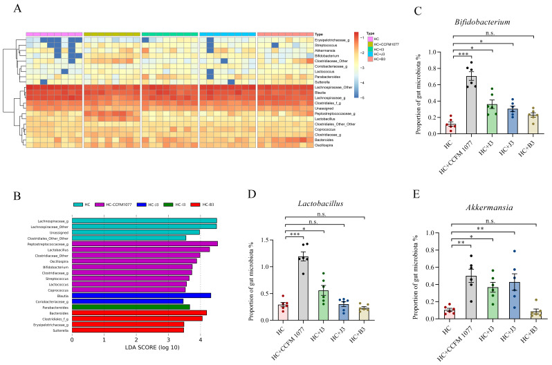
At the genus level, genera with relative abundances over 0.1% are shown in Figure 7A. We categorized these genera into three main cluster groups (Cluster 1: low abundance; Cluster 2: high abundance; Cluster 3: medium abundance). Cluster 1 was dominated by Streptococcus and Bifidobacterium; Cluster 2 was dominated by Lachnospiraceae and Blautia; Cluster 3 was dominated by Bacteroides and Lactobacillus. In Cluster 1, compared with the HC group, Streptococcus, Akkermansia, Bifidobacterium and Lactococcus were more enriched in the HC-CCFM 1077 group. In Cluster 2, no different alterations were observed in these five groups. In Cluster 3, compared with the HC group, Petostreptococcaceae and Lactobacillus were observed to be increased in the HC-CCFM 1077 group. To further investigate the specific composition of gut microbita in different groups, the results of the LEfSe analysis are shown in Figure 7B. Compared with the HC group, the HC-CCFM 1077 group showed the greatest alterations in specific genera; among them, Lactobacillus and Bifidobacterium were the main contributors. Blautia, Parabacteroides and Bacteroides were also observed as the most specific in the HC-J3, HC-I3 and HC-B3 groups, respectively. In addition, we further analyzed the abundance of reported beneficial microbes (Bifidobacterium, Lactobacillus and Akkermansia) in the feces among these groups (Figure 7C–E). Compared with the HC group, these three genera in the HC-CCFM 1077 group and HC-I3 group were observed to be significantly increased (p < 0.001, p < 0.05); only Bifidobacterium and Akkermansia in the HC-B3 group were found to be significantly increased (p < 0.05), whereas these three genera in the HC-J3 group exerted no significant difference. Taken together, the cholesterol-lowering effect may be related to the alteration of the gut compositions and increase in the relative abundance of some genera, which is beneficial to human health.
Figure 7.
The relative abundance of the main genus after consumption of high-cholesterol diet and administration of B. longum strains. (A) Relative abundance (%) heatmap including all fecal samples. Sample clustering is shown on the left-hand side. (B) LEfSe analysis of the specific difference in the genus level in different groups. (C) The abundance of Bacterium in the feces. (D) The abundance of Lactobacillus in the feces. (E) The abundance of Akkermansia in the feces. Data are presented as means ± standard error of the mean, n = 6. *** represents p < 0.001, ** represents p < 0.01, * represents p < 0.05, n.s represent non-significant with p > 0.05.
2.8. Relationship between the Hypercholesterolemia-Alleviation Effects of B. longum Strains and Their Properties In Vitro
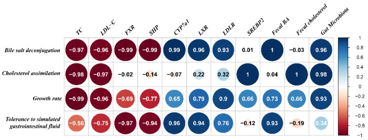
In Figure 8, the Pearson correlation coefficients showed that the bile salt deconjugation ability, cholesterol assimilation ability and growth rate of strains were the most relevant indicators of the hypercholesterolemia-alleviation effects of B. longum strains. The serum lipid profiles (TC and LDL-C) and the expressions of some key genes involved in lipid metabolism (including FXR, SHP) were significantly negatively related to the bile salt deconjugation ability, with Pearson correlation coefficients below −0.95. In contrast, with the enhancement of bile salt deconjugation ability, the expressions of CYP7A1 and LXR, the fecal bile acid levels and the composition of gut microbiota exerted an upward trend. In terms of cholesterol assimilation ability, the TC and LDL-C levels showed a remarkable negative correlation (−0.98 and −0.97); however, the expression of SREBP2, the fecal cholesterol level and the composition of gut microbiota showed a significantly positive correlation (1, 1 and 0.98). Furthermore, the growth rate only had a positive correlation with the serum TC and LDL-C levels.
Figure 8.
The correlation between the hypercholesterolemia-alleviation effects of B. longum strains and their properties in vitro.
3. Discussion
Hypercholesterolemia is among the major risk factors for CVD, which is the leading cause of mortality worldwide [29,30]. To combat hypercholesterolemia, many companies and research institutions have developed lipid-lowering drugs, such as statins, fibrates, ezetimibe and berberine, via clinical research [31]. Despite the high therapeutic efficacies of these drugs, they all have adverse effects in humans, such as gastrointestinal symptoms and rashes [32]. Therefore, probiotics, which are microorganisms with many beneficial effects in humans, are being widely investigated for cholesterol-reducing functions in both animal experiments and clinic trials.
B. longum has been reported as the most prevalent Bifidobacterium species in the human gastrointestinal tract [33]. As a common resident of the gut, B. longum has been shown to possess a high intestinal colonization ability in human trials. The colonization ability of B. longum in the gut is related to the age of the people and the residential area [34]. For example, Fang and colleagues found that the abundance of B. longum in superlongevity people (aged over 90 years) from a longevity village (Bama, China) was higher than that in people from a normal area (Nanning, China) [35]. In addition, B. longum has been reported as the most universal species of the Bifidobacterium genus in the feces of Italian centenarians [36]. Several studies have investigated the cholesterol-lowering effect of B. longum strains, for example, B. longum BB536 was reported to significantly lower serum TC, LDL-C, VLDL and MDA [37]; however, B. longum BL 04 slightly altered the TC and LDL-C levels [18]. Although these B. longum strains were confirmed to alleviate hypercholesterolemia to some extent, the specific cholesterol-lowering effects were different. Therefore, the aim of our study was to explore whether B. longum presents inter-strain differences in the alleviation of hypercholesterolemia in vivo.
In our study, four B. longum strains (CCFM 1077, I3, J3 and B3) isolated from different superlongevity people (aged over 90 years old) were selected. Each of these four strains can survive adequately in the simulated intestinal conditions and can thus exert their effects in the human gut. Our results show that the B. longum strain exerted a strain-specific effect on cholesterol lowering. The precise cholesterol-lowering mechanism of probiotics is not fully understood, and many hypotheses have been proposed, such as bile salt deconjugation [38], cholesterol assimilation [39] and antioxidant activity of probiotics [40]. In our study, after intragastric administration of B. longum CCFM 1077, the content of both total bile acids and total cholesterol in the rat feces increased significantly, B. longum I3 only affected the fecal bile acid content and B. longum J3 only affected the fecal cholesterol content, whereas B. longum B3 had no effects on these two contents in feces. This finding indicates that the most significant alleviation effect of cholesterolemia by B. longum CCFM 1077 was related to the combination of high abilities of bile salt deconjugation and cholesterol assimilation in vitro.
Furthermore, to evaluate the underlying mechanism of the cholesterol-lowering effect of B. longum, we also studied the expression of several genes involved in cholesterol metabolism [23,24,25]. FXR, CYP7A1 and SHP play important roles in the synthesis of bile acids from cholesterol, and LXR plays a role in reverse cholesterol transport [26,27]. Many hypotheses suggest that FXR is influenced by changes in the bile acid pool and that a decreased FXR expression downregulates SHP expression and upregulates CYP7A1 expression, both of which are bile acid synthesis rate-limiting enzymes [28]. In this study, we found that compared with B. longum J3 and B. longum B3 administration, B. longum CCFM 1077 and B. longum I3 administration downregulated FXR and SHP expression and upregulated CYP7A1 and LXR expression (Figure 4A–D). This may be due to the high bile salt deconjugation ability of B. longum CCFM 1077 and B. longum I3, which hydrolyzed conjugated bile acids to free primary bile acids that are less efficiently reabsorbed in the intestine, thus increasing the fecal excretion of bile salts. This alteration in the bile acid pool downregulated FXR expression that in turn downregulated SHP expression and upregulated CYP7A1 expression, thus increasing cholesterol catabolism and bile synthesis. Consistent with this finding, GQ Wang et al. reported that FXR and SHP expression was decreased and CYP7A1 expression was increased in hypercholesterolemia mice after the oral administration of L. plantarum AR113 [41]. LDLR and SREBP2 are also important factors in the metabolism of cholesterol and other lipids [42]. In this study, LDLR expression in the B. longum CCFM 1077, B. longum I3 and B. longum J3 groups was upregulated compared with that in the B. longum B3 group, indicating that both bile salt deconjugation ability and cholesterol assimilation ability play an important role in reducing the serum cholesterol level by upregulating LDLR expression. However, we found that only B. longum CCFM 1077 and B. longum J3 increased SREBP2 expression, suggesting that cholesterol assimilation leads to cholesterol reduction via the upregulation of SREBP2 expression. This result is consistent with that of an earlier study that reported mediation of the cholesterol-lowering effect of soybean protein via the upregulation of LDLR expression [29,43].
Many studies have demonstrated that a high-cholesterol diet intervention disrupts the gut microbial balance [44,45]. In this study, we found that a high-cholesterol diet decreased the diversity and abundance of the gut microbiota in rats; in particular, the abundance of Firmicutes was higher and that of Bacteroidetes was lower in the HC group than in the B. longum groups. Therefore, we hypothesized that B. longum strains indirectly alleviate hypercholesterolemia by altering the gut microbiota. Previous studies have suggested that certain bacterial genera in the gut improve the health of the host by maintaining the gut microbial balance. Bifidobacterium and Lactobacillus were reported to lower the risk of CVD by reducing the liver cholesterol level [46], and Akkermansia could improve cholesterol levels in cases of metabolic syndrome [47]. Consistent with these studies, after intragastrically administering B. longum strains (CCFM 1077, I3 and J3) for 28 days, the abundance of Bifidobacterium, Lactobacillus and Akkermansia increased in the rat feces, thereby alleviating the adverse effects of high-cholesterol diet in vivo. However, this alteration was not observed in the B. longum B3 group. Taken together, our results show that only B. longum strains with either bile salt deconjugation ability or cholesterol assimilation ability can alleviate hypercholesterolemia by improving the composition of gut microbiota and increasing the abundance of health-promoting bacterial genera.
Physiological properties of strains in vitro are important for probiotic strains to take effect in human health. Correlation analysis showed a significant correlation of properties of strains in vitro (growth rate, bile salt deconjugation ability and cholesterol assimilation ability) with the cholesterol-alleviating effects. It has been reported that a strain with high bile salt deconjugation ability exerts the cholesterol-lowering effect by increasing the excretion of bile acid [48]. Chun-Feng et al. found that BSH-active Lactobacillus casei F0422 combined with Tween 80 with Cacl2 could increase the hypocholesterolemic effect in rats [49]. Guangqiang et al. found that L. casei pWQH01 (overexpression of bsh1) could significantly decreased the serum TC and LDL-C [41]. These results suggested that increased bile salt deconjugation may be among the factors of cholesterolemia mitigation. The high cholesterol assimilation ability of a strain means that more cholesterol is absorbed by the strain in the gut, and the assimilated cholesterol is excreted with large quantities of probiotic strain; for example, B. longum 5022 and L. fermentum LP4 with high cholesterol assimilation abilities have been reported to alleviate hypercholesterolemia [50,51]. The growth rate, as an indicator to evaluate how fast a strain grows, is also related to the hypercholesterolemia-alleviating effect. A fast growth rate contributes to the metabolism and proliferation of a strain and also increases the bile salt deconjugation ability and cholesterol assimilation ability.
In this study, remarkable differences in the bile salt deconjugation ability and cholesterol assimilation ability existed between the four B. longum strains. Among the B. longum strains, CCFM 1077 had both high abilities of bile salt deconjugation and cholesterol assimilation, I3 had only high bile salt deconjugation ability, J3 had only high cholesterol assimilation ability, whereas B3 had neither of these two abilities. Interestingly, in the animal experiments, B. longum CCFM 1077 exerted the most potent cholesterol-lowering effect, followed by B. longum I3 and B3, whereas B. longum B3 had no effect in alleviating hypercholesterolemia. This result supported that the strain-specific effects of B. longum strains on hypercholesterolemia rats mainly correlated with bile salt deconjugation and cholesterol assimilation abilities in vitro. In future, we will conduct clinical studies to further investigate the strain-specific effects of different probiotics on hypercholesterolemia.
4. Materials and Methods
4.1. The Culture Conditions and Growth Curve of Bacterial Strains
Four B. longum strains (CCFM 1077, I3, J3, B3) isolated from different superlongevity people (aged over 90 years old) were obtained from Culture Collections of Food Microbiology, Jiangnan University (Wuxi, China). To ensure the viable count of the probiotics before use, all four B. longum strains were reactivated three times by using 2% (v/v) inoculum in modified MRS (mMRS) broth supplemented with 0.5 ‰ (w/v) L-cysteine for 48 h in Whitley DG250 Anaerobic Workstation (37 °C). About 107 CFU/mL of each B. longum strains was inoculated into fresh mMRS broth, then grown at 37 °C in anaerobic incubation workstation. OD600 values were measured every two hours. The horizontal axis in the diagram of growth curve shows the incubation time, and the vertical axis shows the corresponding absorbance values.
4.2. The Culture Conditions and Growth Curve of Bacterial Strains
The method used for testing the acid and bile tolerance of four B. longum strains has been previously described [50]. Briefly, B. longum was cultivated in mMRS broth at 37 °C for 18 h and then harvested by centrifugation (10,000× g, 4 °C, 10 min). After this, the cell pellets were suspended in phosphate-buffered saline solution, simulated bile salts (0.3% Oxigall dissolved in PBS, pH 8.0) or simulated gastric juice (3 mg of pepsin dissolved in 1 mL of 0.5% saline buffer, pH 2.5). The suspensions were incubated at 37 °C in simulated gastric juice for 1 h and then in simulated bile salts for 3 h. Finally, cells in the suspensions were harvested by centrifugation (10,000× g, 4 °C, 5 min) and resuspended in mMRS broth, followed by incubation at 37 °C for 48 h.
4.3. Quantitative Determination of Bile Salt Deconjugation Ability of B. longum Strains by HPLC
The bile salt deconjugation ability measurement of four B. longum strains was performed as previously described [52], with slight modifications. Briefly, cultures were grown in 10 mL of MRS broth (pH 6.5) by incubation at 37 °C for 18 h in the Whitley DG250 Anaerobic Workstation until a density of 1 × 10⁹ CFU/mL was reached. MRS broth (pH 6.5) containing 10 mM glycocholic acid (GCA; Sigma Chemical Co., St. Louis, MO, USA) was diluted 1:1 to a GCA concentration of 2.3 mg/mL, and 0.5 mL was added to the B. longum cultures. The cultures were then incubated for an additional 6 h under anaerobic conditions at 37 °C. The pH of the culture was then adjusted to 7.5 with KOH to stop the bile salt deconjugation ability. For HPLC quantification of GCA, the whole culture samples were first diluted 20 times in HPLC-grade methanol, 10 mL of which was injected into a C18 column (250 mm × 4.6 mm × 5 μm). In total, 30% 0.07-M sodium acetate (pH 3.0) and 70% methanol were used as the running buffer, and the flow rate was set at 1 mL/min. A Shimadzu SPD-10A UV-Vis detector was used to detect GCA at 205 nm. A standard stock solution of GCA was prepared by dissolving 10 mg GCA in 10 mL methanol. The concentration of stock solution was 100 μg/mL by diluting GCA in methanol. The standard was used as the reference control for GCA quantification. To plot the curve of bile salt deconjugation, the means of triplicate samples were calculated. Bile salt deconjugation ability was calculated as follows:
| (3) |
where C0 is the GCA concentration of the control sample, and C1 is the GCA concentration of the B. longum sample.
4.4. Cholesterol Assimilation by B. longum Strains
The cholesterol concentration was determined by HPLC using the method described in a previous study [53], with slight modifications. After centrifuging the bacterial cultures at 7500g and 4 °C for 10 min, 1 μL of the culture supernatant was filtered and injected into the HPLC device (Agilent 1100, Agilent Technologies, Chandler, AZ, USA). The analysis was performed on a ZORBAX Eclipse XDB-C18 column (rapid resolution, 1.8 μm particle size, 4.6 × 50 mm, Agilent) with a variable wavelength UV/Vis detector. Acetonitrile at 2.5 mL/min was used for isocratic elution. Cholesterol was finally confirmed by the retention time of 4 min at 210 nm and was quantified by interpolation of the calibration curve. Standard solutions of various concentrations were prepared by diluting the cholesterol stock solution in MRS broth. Linearity was demonstrated from 0.01 to 0.1 g/L (r2 = 0.996). The limit of detection was 0.01 g/L.
| (4) |
4.5. Animals and Diets
Forty-eight SD rats (body weight, 220–240 g) were purchased from Shanghai SLAC Laboratory Animal Co., Ltd. (Shanghai, China). The rats were housed in stainless metal cages (two rats per cage) in a room maintained at 55% ± 5% relative humidity and 22 ± 2 °C under a 12 h light/dark cycle. After a 7 day adaptation to the new environment, the rats were randomly divided into the following six groups according to the diets: (1) non-added cholesterol diet (NC); (2) high-cholesterol diet (HC); (3) HC-diet plus B. longum CCFM 1077 (HC-CCFM 1077); (4) HC-diet plus B. longum I3 (HC-I3); (5) HC-diet plus B. longum J3 (HC-J3); and (6) HC-diet plus B. longum B3 (HC-B3). The experiment lasted for 28 days, during which the rats had free access to water and their group-specific diet. Each day, 2 mL of saline solution (0.85%) was administered to the NC and HC groups, whereas the HC-CCFM 1077, HC-I3, HC-J3 and HC-B3 groups received 2 mL (109 cfu/mL) of their specific B. longum strain dissolved in saline solution (0.85%) by gavage. Dietary intake was monitored daily, and body weights were recorded at the end of the experiment (Figure S1). The components of diets are shown in Table 3. All experimental procedures complied with the Animal Care Committee of Jiangnan University (Identification Number: JN No. 20160823-20160929, Approved date: 25th July 2016), and were carried out under the guidelines set by the European Community (Directive 2010/63/EU).
Table 3.
Composition of the experimental diets.
| Ingredient | Cholesterol-Free Diet (g/kg) |
Cholesterol-Enriched Diet (g/kg) |
|---|---|---|
| Cornstarch | 465.692 | 459.442 |
| Dextrinized cornstarch | 155 | 155 |
| Casein | 140 | 140 |
| Sucrose | 100 | 100 |
| Soybean oil | 40 | 40 |
| Cellulose | 50 | 50 |
| Choline biartrate | 2.5 | 2.5 |
| L-Systine | 1.8 | 1.8 |
| t-Butylhydroquinone | 0.008 | 0.008 |
| Mineral | 35 | 35 |
| Vitamin | 10 | 10 |
| Cholesterol | - | 5 |
| Sodium cholate | - | 1.25 |
Cholesterol-free and cholesterol-enriched diets were both bought from Trophic Animal Feed High-Tech Co., Ltd., Nantong, China.
4.6. Analysis of Serum Lipid Levels
After the 28 days of experiment, the rats were fasted overnight (12 h) and sacrificed under isoflurane. Serum was collected from their abdominal aorta using blood collection tubes (SLAC Laboratory Animal Co., Ltd., Shanghai, China) containing heparin as an anticoagulant. The tubes were then centrifuged at 1400 g at 4 °C for 15 min for serum separation, and the serum samples were stored at −80 °C until analysis. The serum total cholesterol (TC), triglyceride (TG), high-density lipoprotein cholesterol (HDL-C) and LDL-C levels were measured using a Biochemical Analyzer (Beckman Coulter, Brea, CA, USA) [54].
4.7. Analysis of Fecal Cholesterol and Bile Acid Contents
On the last day of the experiment, the feces of each rat were collected and immediately stored at −80 °C until further analysis. Fecal cholesterol and bile acid contents were measured using commercial kits (Sangon Biotech, Shanghai, China).
4.8. Extraction of Liver RNA and RT-PCR Analysis
The total RNA samples from rat livers were extracted according to the manufacturer’s protocol (TaKaRa Bio, Otsu, Japan). The mRNA levels of sterol regulatory element-binding protein 2 (SREBP2), cholesterol 7α-hydroxylase (CYP7A1), liver X receptor (LXR), farnesoid X receptor (FXR) and low-density lipoprotein receptor (LDLR) were measured as described previously [50]. Total RNAs were extracted with TRIzol (Thermo Scientific, Wilmington, DE, USA), and cDNAs were synthesized by reverse transcription (TaKaRa Bio, Otsu, Japan). The quantitative real-time polymerase chain reaction (RT-qPCR) used the SYBR Premix Ex TaqII (TaKaRa Bio, Otsu, Japan) and the appropriate primers. The primer sequences which were obtained from Sangon Biological Engineering (Shanghai, China) are shown in Table 4. The thermocycler conditions used were 95 °C for 30 s, followed by 45 cycles of 95 °C for 5 s and 60 °C for 30 s.
Table 4.
Primer sequences used for RT-PCR in this study.
| Gene | Forward Primers (5′-3′) | Reverse Primers (5′-3′) |
|---|---|---|
| FXR | CCAACCTGGGCTTCTACCC | CACACAGCTCATCCCCTTT |
| SHP | TCTGCAGGTCGTCCGACTATTC | AGGCAGTGGCTGTGAGATGC |
| CYP7A1 | ATTCCATACCTGGGCTGTGC | ATGTTTTCAGTGGTATTTCC |
| LXR | CTCTTCTTGCCGCTTCAGTT | AGGAGTGTCGACTTCGCAAA |
| Srebp 2 | AGCAGCAGGTGCAGACGGTA | CATCTGTCTTCAGCGTGGTC |
| LDLR | AGCAGTGAGTGTATCCATCG | AATGCAGGAGCCATCTGCAC |
| β-actin | GGCTGTATTCCCCTCCATCG | CCAGTTGGTAACAATGCCATGT |
FXR = farnesoid X receptor; SHP= small heterodimer partener; CYP7A1 = cholesterol 7α-hydroxylase; LXR = liver X receptor; Srebp2 = sterol regulatory element-binding protein 2; LDLR = low-density lipoprotein receptor.
4.9. MiSeq Genome Sequencing Analysis of Community Structures
Fecal microbial DNA was extracted using a FastDNA SPIN Kit for Soil (MP Biomedical, catalog No.6560-200) in accordance with the manufacturer’s instructions. Subsequently, 16S rRNA sequencing was performed, and the reads were analyzed using the QIIME pipeline [55]. Reads shorter than 200 bp were removed after screening the raw sequences, and sequenced pair-end reads overlapping by longer than 10 bp and without any mismatch were assembled. The sequences with similarity of more than 97% were defined as operational taxonomical units (OTUs) for assembly of high-quality sequences using the QIIME software (http://qiime.sourceforge.net/).
4.10. Correlation Test
R language was used to evaluate the correlation between the significantly altered indicators in the hypercholesterolemia-alleviating effect and the properties of strains in vitro. The data were divided into two matrices: an in vitro matrix and in vivo matrix. The corrplot package was used to analyze the correlations between these two matrices. Pearson’s correlation coefficients were calculated to quantify the correlations between the indicators.
4.11. Statistical Analysis
All of the data are expressed as the mean ± standard error of the mean. Statistical significance was determined by one-way analysis of variance followed by a Tukey multiple comparison test using GraphPad Prism 8.0 (GraphPad Software Inc., San Diego, CA, USA) and SPSS version 16.0 software (SPSS, Inc., Chicago, IL, USA).
5. Conclusions
By measuring serum lipid profiles (TC, TG, LDL-C and HDL-C), it was found that B. longum CCFM 1077 exerted the most potent cholesterol-lowering effect, followed by B. longum I3 and B3, whereas B. longum B3 had no effect in alleviating hypercholesterolemia. Furthermore, B. longum CCFM 1077, I3 and B3 significantly altered the diversity and composition of gut microbiota. These results suggested that B. longum (CCFM 1077, I3, J3 and B3) exerted strain-specific effects in alleviation of hypercholesterolemia. The potential causes of the strain-specific effects in alleviation of hypercholesterolemia might be the differences in the bile salt deconjugation ability, cholesterol assimilation ability and growth rate of different strains, which had different effects on the intestinal microenvironment and expressions of key genes involved in the lipid metabolism.
Acknowledgments
We would like to thank the National Engineering Research Center for Functional Food, Jiangnan University, Wuxi, for technical support.
Abbreviations
| B. longum | Bifidobacterium longum |
| L. plantarum | Lactobacillus plantarum |
| B. bifidum | Bifidobacterium bifidum |
| L. rhamnosus | Lactobacillus rhamnosus |
| BSH | Bile salt hydrolysis |
| FXR | farnesoid X receptor |
| SHP | small heterodimer partner |
| CYP7A1 | cholesterol 7α-hydroxylase |
| LXR | liver X receptor |
| SREBP2 | sterol regulatory element binding protein 2 |
| LDLR | low density lipoprotein receptor |
Supplementary Materials
The following are available online at https://www.mdpi.com/1422-0067/22/3/1305/s1, Figure S1: Body weight on the final day.
Author Contributions
Conceptualization, J.J. and Q.Z. (Qixiao Zhai); Methodology, J.J. and C.W.; Software, J.J., C.Z.; Validation, Q.Z. (Qingsong Zhang), L.Y. and J.Z.; Formal Analysis, J.J. and Q.Z. (Qixiao Zhai); Investigation, J.J. and C.W.; Resources, H.Z., A.N., W.C., Q.Z. (Qixiao Zhai); Data Curation, H.Z. and Q.Z. (Qixiao Zhai); Writing—Original Draft Preparation, J.J.; Writing—Review & Editing, J.J. and Q.Z. (Qixiao Zhai); Visualization, J.J. and Q.Z. (Qixiao Zhai); Supervision, H.Z., W.C., Q.Z. (Qixiao Zhai); Project Administration, H.Z., A.N., W.C. and Q.Z. (Qixiao Zhai); Funding Acquisition, H.Z., A.N., J.Z., W.C. and Q.Z. (Qixiao Zhai). All authors have read and agreed to the published version of the manuscript.
Funding
This work was supported by the National Natural Science Foundation of China Program [No.31820103010 and No. 31871773]; Projects of Innovation and Development Pillar Program for Key Industries in Southern Xinjiang of Xinjiang Production and Construction Corps [2018DB002]; National Key Research and Development Project [No. 2018YFC1604206]; National First-Class Discipline Program of Food Science and Technology [JUFSTR20180102]; the BBSRC Newton Fund Joint Centre Award [BBS/OS/NW/000006]; and Collaborative Innovation Center of Food Safety and Quality Control in Jiangsu Province.
Institutional Review Board Statement
The study was conducted according to the guidelines of the Declaration of Helsinki, and approved by the Animal Care Committee of Jiangnan University on 25th July 2016 (protocol code: JN No. 20160823-20160929).
Informed Consent Statement
Not applicable for studies not involving humans.
Data Availability Statement
No new data were created or analyzed in this study. Data sharing is not applicable to this article.
Conflicts of Interest
The authors declare no conflict of interest.
Footnotes
Publisher’s Note: MDPI stays neutral with regard to jurisdictional claims in published maps and institutional affiliations.
References
- 1.Gielen S., Landmesser U. The Year in Cardiology 2013: Cardiovascular disease prevention. Eur. Heart J. 2014;35:307–312. doi: 10.1093/eurheartj/eht551. [DOI] [PubMed] [Google Scholar]
- 2.Hu S.S., Kong L.Z., Gao R.L., Zhu M.L., Wang W., Wang Y.J., Wu Z.S., Chen W.W., Liu M.B. Outline of the Report on Cardiovascular Disease in China, 2010. Biomed. Environ. Sci. 2012;25:251–256. doi: 10.3967/0895-3988.2012.03.001. [DOI] [PubMed] [Google Scholar]
- 3.Zhao D., Liu J., Wang M., Zhang X., Zhou M. Epidemiology of cardiovascular disease in China: Current features and implications. Nat. Rev. Cardiol. 2019;16:203–212. doi: 10.1038/s41569-018-0119-4. [DOI] [PubMed] [Google Scholar]
- 4.Sayin S.I., Wahlstrom A., Felin J., Jantti S., Marschall H.U., Bamberg K., Angelin B., Hyotylainen T., Oresic M., Backhed F. Gut microbiota regulates bile acid metabolism by reducing the levels of tauro-beta-muricholic acid, a naturally occurring FXR antagonist. Cell Metab. 2013;17:225–235. doi: 10.1016/j.cmet.2013.01.003. [DOI] [PubMed] [Google Scholar]
- 5.Houston D.K., Ding J., Lee J.S., Garcia M., Kanaya A.M., Tylavsky F.A., Newman A.B., Visser M., Kritchevsky S.B. Dietary fat and cholesterol and risk of cardiovascular disease in older adults: The Health ABC Study. Nutr. Metab. Cardiovasc. 2011;21:430–437. doi: 10.1016/j.numecd.2009.11.007. [DOI] [PMC free article] [PubMed] [Google Scholar]
- 6.Larsson S.C., Virtamo J., Wolk A. Dietary fats and dietary cholesterol and risk of stroke in women. Atherosclerosis. 2012;221:282–286. doi: 10.1016/j.atherosclerosis.2011.12.043. [DOI] [PubMed] [Google Scholar]
- 7.Davis C.E., Rifkind B.M., Brenner H., Gordon D.J. A single cholesterol measurement underestimates the risk of coronary heart disease. An empirical example from the Lipid Research Clinics Mortality Follow-up Study. JAMA. 1990;264:3044–3046. doi: 10.1001/jama.1990.03450230080033. [DOI] [PubMed] [Google Scholar]
- 8.Grundy S.M. Atherosclerosis imaging and the future of lipid management. Circulation. 2004;110:3509–3511. doi: 10.1161/01.CIR.0000151100.28000.B3. [DOI] [PubMed] [Google Scholar]
- 9.Kim H., Kim D.-H., Seo K.-H., Chon J.-W., Nah S.-Y., Bartley G.E., Arvik T., Lipson R., Yokoyama W. Modulation of the Intestinal Microbiota Is Associated with Lower Plasma Cholesterol and Weight Gain in Hamsters Fed Chardonnay Grape Seed Flour. J. Agric. Food Chem. 2015;63:1460–1467. doi: 10.1021/jf5026373. [DOI] [PubMed] [Google Scholar]
- 10.Dunn-Emke S., Weidner G., Ornish D. Benefits of a low-fat plant-based diet. Obes. Res. 2001;9:731. doi: 10.1038/oby.2001.100. [DOI] [PubMed] [Google Scholar]
- 11.Tong X., Xu J., Lian F., Yu X., Zhao Y., Xu L., Zhang M., Zhao X., Shen J., Wu S., et al. Structural Alteration of Gut Microbiota during the Amelioration of Human Type 2 Diabetes with Hyperlipidemia by Metformin and a Traditional Chinese Herbal Formula: A Multicenter, Randomized, Open Label Clinical Trial. mBio. 2018;9:e0239217. doi: 10.1128/mBio.02392-17. [DOI] [PMC free article] [PubMed] [Google Scholar]
- 12.Ma H., Zhang B., Hu Y., Wang J., Liu J., Qin R., Lv S., Wang S. Correlation Analysis of Intestinal Redox State with the Gut Microbiota Reveals the Positive Intervention of Tea Polyphenols on Hyperlipidemia in High Fat Diet Fed Mice. J. Agric. Food Chem. 2019;67:7325–7335. doi: 10.1021/acs.jafc.9b02211. [DOI] [PubMed] [Google Scholar]
- 13.Liu H., Pan L.L., Lv S., Yang Q., Zhang H., Chen W., Lv Z., Sun J. Alterations of Gut Microbiota and Blood Lipidome in Gestational Diabetes Mellitus with Hyperlipidemia. Front. Physiol. 2019;10:1015. doi: 10.3389/fphys.2019.01015. [DOI] [PMC free article] [PubMed] [Google Scholar]
- 14.Costabile A., Buttarazzi I., Kolida S., Quercia S., Baldini J., Swann J.R., Brigidi P., Gibson G.R. An in vivo assessment of the cholesterol-lowering efficacy of Lactobacillus plantarum ECGC 13110402 in normal to mildly hypercholesterolaemic adults. PLoS ONE. 2017;12:e0187964. doi: 10.1371/journal.pone.0187964. [DOI] [PMC free article] [PubMed] [Google Scholar]
- 15.Jones M.L., Martoni C.J., Parent M., Prakash S. Cholesterol-lowering efficacy of a microencapsulated bile salt hydrolase-active Lactobacillus reuteri NCIMB 30242 yoghurt formulation in hypercholesterolaemic adults. Br. J. Nutr. 2012;107:1505–1513. doi: 10.1017/S0007114511004703. [DOI] [PubMed] [Google Scholar]
- 16.Al-Sheraji S.H., Amin I., Azlan A., Manap M.Y., Hassan F.A. Effects of Bifidobacterium longum BB536 on lipid profile and histopathological changes in hypercholesterolaemic rats. Benef. Microbes. 2015;6:661–668. doi: 10.3920/BM2014.0032. [DOI] [PubMed] [Google Scholar]
- 17.Fuentes M.C., Lajo T., Carrion J.M., Cune J. Cholesterol-lowering efficacy of Lactobacillus plantarum CECT 7527, 7528 and 7529 in hypercholesterolaemic adults. Br. J. Nutr. 2013;109:1866–1872. doi: 10.1017/S000711451200373X. [DOI] [PubMed] [Google Scholar]
- 18.Guardamagna O., Amaretti A., Puddu P.E., Raimondi S., Abello F., Cagliero P., Rossi M. Bifidobacteria supplementation: Effects on plasma lipid profiles in dyslipidemic children. Nutrition. 2014;30:831–836. doi: 10.1016/j.nut.2014.01.014. [DOI] [PubMed] [Google Scholar]
- 19.Hatakka K., Mutanen M., Holma R., Saxelin M., Korpela R. Lactobacillus rhamnosus LC705 Together with Propionibacterium freudenreichii ssp shermanii JS Administered in Capsules Is Ineffective in Lowering Serum Lipids. J. Am. Coll. Nutr. 2008;27:441–447. doi: 10.1080/07315724.2008.10719723. [DOI] [PubMed] [Google Scholar]
- 20.Zhao X., Higashikawa F., Noda M., Kawamura Y., Matoba Y., Kumagai T., Sugiyama M. The Obesity and Fatty Liver Are Reduced by Plant-Derived Pediococcus pentosaceus LP28 in High Fat Diet-Induced Obese Mice. PLoS ONE. 2012;7:0030696. doi: 10.1371/journal.pone.0030696. [DOI] [PMC free article] [PubMed] [Google Scholar]
- 21.Ruscica M., Pavanello C., Gandini S., Macchi C., Botta M., Dall’Orto D., del Puppo M., Bertolotti M., Bosisio R., Mombelli G., et al. Nutraceutical approach for the management of cardiovascular risk—A combination containing the probiotic Bifidobacterium longum BB536 and red yeast rice extract: Results from a randomized, double-blind, placebo-controlled study. Nutr. J. 2019;18:13. doi: 10.1186/s12937-019-0438-2. [DOI] [PMC free article] [PubMed] [Google Scholar]
- 22.Park A.J., Bercik P., Huang X., Blennerhassett P., Sinclair D.D., Lu J., Deng Y., Bergonzelli G., McLean P., Collins S.M., et al. The anxiolytic effect of Bifidobacterium longum NCC3001 involves vagal pathways for gut–brain communication. Gastroenterology. 2011;140:S18–S19. doi: 10.1016/S0016-5085(11)60072-3. [DOI] [PMC free article] [PubMed] [Google Scholar]
- 23.Wang B.T., Ducker G.S., Barczak A.J., Barbeau R., Erle D.J., Shokat K.M. The mammalian target of rapamycin regulates cholesterol biosynthetic gene expression and exhibits a rapamycin-resistant transcriptional profile. Proc. Natl. Acad. Sci. USA. 2011;108:15201–15206. doi: 10.1073/pnas.1103746108. [DOI] [PMC free article] [PubMed] [Google Scholar]
- 24.Carstea E.D., Morris J.A., Coleman K.G., Loftus S.K., Zhang D., Cummings C., Gu J.Z., Rosenfeld M.A., Pavan W.J., Krizman D.B. Niemann-Pick C1 disease gene: Homology to mediators of cholesterol homeostasis. Science. 1997;277:228–231. doi: 10.1126/science.277.5323.228. [DOI] [PubMed] [Google Scholar]
- 25.Yang L., Zhu Y., Zhong S., Zheng G. Astilbin lowers the effective caffeine dose for decreasing lipid accumulation via activating AMPK in high-fat diet-induced obese mice. J. Sci. Food Agric. 2020:573–581. doi: 10.1002/jsfa.10669. [DOI] [PubMed] [Google Scholar]
- 26.Schneider K.M., Albers S., Trautwein C. Role of bile acids in the gut-liver axis. J. Hepatol. 2018;68:1083–1085. doi: 10.1016/j.jhep.2017.11.025. [DOI] [PubMed] [Google Scholar]
- 27.Michael D.R., Davies T.S., Moss J.W.E., Calvente D.L., Ramji D.P., Marchesi J.R., Pechlivanis A., Plummer S.F., Hughes T.R. The anti-cholesterolaemic effect of a consortium of probiotics: An acute study in C57BL/6J mice. Sci. Rep. 2017;7:2883. doi: 10.1038/s41598-017-02889-5. [DOI] [PMC free article] [PubMed] [Google Scholar]
- 28.Gruber J., Kennedy B.K. Microbiome and Longevity: Gut Microbes Send Signals to Host Mitochondria. Cell. 2017;169:1168–1169. doi: 10.1016/j.cell.2017.05.048. [DOI] [PubMed] [Google Scholar]
- 29.George K.S., Munoz J., Akhavan N.S., Foley E.M., Siebert S.C., Tenenbaum G., Khalil D.A., Chai S.C., Arjmandi B.H. Is soy protein effective in reducing cholesterol and improving bone health? Food Funct. 2020;11:544–551. doi: 10.1039/C9FO01081E. [DOI] [PubMed] [Google Scholar]
- 30.Chen X.C., Huang L.L., Chang T.H.A., Ong B.L., Ong S.L., Hu J.Y. Plant Traits for Phytoremediation in the Tropics. Engineering. 2019;5:841–848. doi: 10.1016/j.eng.2019.07.019. [DOI] [Google Scholar]
- 31.Bove M., Cicero A.F.G., Borghi C. Emerging drugs for the treatment of hypercholesterolemia. Expert Opin. Emerg. Drugs. 2019;24:63–69. doi: 10.1080/14728214.2019.1591372. [DOI] [PubMed] [Google Scholar]
- 32.Araki E., Yamashita S., Arai H., Yokote K., Satoh J., Inoguchi T., Nakamura J., Maegawa H., Yoshioka N., Tanizawa Y., et al. Effects of Pemafibrate, a Novel Selective PPARα Modulator, on Lipid and Glucose Metabolism in Patients with Type 2 Diabetes and Hypertriglyceridemia: A Randomized, Double-Blind, Placebo-Controlled, Phase 3 Trial. Diabetes Care. 2018;41:538–546. doi: 10.2337/dc17-1589. [DOI] [PubMed] [Google Scholar]
- 33.Zhao D., Zhu H., Gao F., Qian Z., Mao W., Yin Y., Tan J., Chen D. Antidiabetic effects of selenium-enriched Bifidobacterium longum DD98 in type 2 diabetes model of mice. Food Funct. 2020;11:6528–6541. doi: 10.1039/D0FO00180E. [DOI] [PubMed] [Google Scholar]
- 34.Ballantyne C.M., Banach M., Mancini G.B.J., Lepor N.E., Hanselman J.C., Zhao X., Leiter L.A. Efficacy and safety of bempedoic acid added to ezetimibe in statin-intolerant patients with hypercholesterolemia: A randomized, placebo-controlled study. Atherosclerosis. 2018;277:195–203. doi: 10.1016/j.atherosclerosis.2018.06.002. [DOI] [PubMed] [Google Scholar]
- 35.Wang F., Huang G.H., Cai D., Li D.L., Liang X.L., Yu T., Shen P.H., Su H.Y., Liu J.D., Gu H.C., et al. Qualitative and Semiquantitative Analysis of Fecal Bifidobacterium Species in Centenarians Living in Bama, Guangxi, China. Curr. Microbiol. 2015;71:143–149. doi: 10.1007/s00284-015-0804-z. [DOI] [PubMed] [Google Scholar]
- 36.Drago L., Toscano M., Rodighiero V., de Vecchi E., Mogna G. Cultivable and Pyrosequenced Fecal Microflora in Centenarians and Young Subjects. J. Clin. Gastroenterol. 2012;46:S81–S84. doi: 10.1097/MCG.0b013e3182693982. [DOI] [PubMed] [Google Scholar]
- 37.Jiang J.C., Wu C.E., Zhang C.C., Zhao J.X., Yu L.L., Zhang H., Arjan N., Chen W., Zhai Q.X. Effects of probiotic supplementation on cardiovascular risk factors in hypercholesterolemia: A systematic review and meta-analysis of randomized clinical trial. J. Funct. Foods. 2020;74:104177. doi: 10.1016/j.jff.2020.104177. [DOI] [Google Scholar]
- 38.Jones M.L., Tomaro-Duchesneau C., Martoni C.J., Prakash S. Cholesterol lowering with bile salt hydrolase-active probiotic bacteria, mechanism of action, clinical evidence, and future direction for heart health applications. Expert Opin. Biol. Ther. 2013;13:631–642. doi: 10.1517/14712598.2013.758706. [DOI] [PubMed] [Google Scholar]
- 39.Choi E.A., Chang H.C. Cholesterol-lowering effects of a putative probiotic strain Lactobacillus plantarum EM isolated from kimchi. LWT Food Sci. Technol. 2015;62:210–217. doi: 10.1016/j.lwt.2015.01.019. [DOI] [Google Scholar]
- 40.Song W., Song C., Shan Y., Lu W., Zhang J., Hu P., Wu X., Li L., Guo S. The antioxidative effects of three lactobacilli on high-fat diet induced obese mice. RSC Adv. 2016;6:65808–65815. doi: 10.1039/C6RA06389F. [DOI] [Google Scholar]
- 41.Wang G., Huang W., Xia Y., Xiong Z., Ai L. Cholesterol-lowering potentials of Lactobacillus strain overexpression of bile salt hydrolase on high cholesterol diet-induced hypercholesterolemic mice. Food Funct. 2019;10:1684–1695. doi: 10.1039/C8FO02181C. [DOI] [PubMed] [Google Scholar]
- 42.Horie T., Ono K., Horiguchi M., Nishi H., Nakamura T., Nagao K., Kinoshita M., Kuwabara Y., Marusawa H., Iwanaga Y., et al. MicroRNA-33 encoded by an intron of sterol regulatory element-binding protein 2 (Srebp2) regulates HDL in vivo. Proc. Natl. Acad. Sci. USA. 2010;107:17321–17326. doi: 10.1073/pnas.1008499107. [DOI] [PMC free article] [PubMed] [Google Scholar]
- 43.Zhang H.L., Wu Q.X., Qin X.M. Camellia nitidissima Chi flower extract alleviates obesity and related complications and modulates gut microbiota composition in rats with high-fat-diet-induced obesity. J. Sci. Food Agric. 2020;100:4378–4389. doi: 10.1002/jsfa.10471. [DOI] [PubMed] [Google Scholar]
- 44.Wang J., Tang H., Zhang C., Zhao Y., Derrien M., Rocher E., van Hylckama J.E.V., Strissel K., Zhao L., Obin M., et al. Modulation of gut microbiota during probiotic-mediated attenuation of metabolic syndrome in high fat diet-fed mice. ISME J. 2015;9:1–15. doi: 10.1038/ismej.2014.99. [DOI] [PMC free article] [PubMed] [Google Scholar]
- 45.Martinez I., Perdicaro D.J., Brown A.W., Hammons S., Carden T.J., Carr T.P., Eskridge K.M., Walter J. Diet-Induced Alterations of Host Cholesterol Metabolism Are Likely To Affect the Gut Microbiota Composition in Hamsters. Appl. Environ. Microb. 2013;79:516–524. doi: 10.1128/AEM.03046-12. [DOI] [PMC free article] [PubMed] [Google Scholar]
- 46.Jiang J., Feng N., Zhang C., Liu F., Zhao J., Zhang H., Zhai Q., Chen W. Lactobacillus reuteri A9 and Lactobacillus mucosae A13 isolated from Chinese superlongevity people modulate lipid metabolism in a hypercholesterolemia rat model. FEMS Microbiol. Lett. 2019;366:24. doi: 10.1093/femsle/fnz254. [DOI] [PubMed] [Google Scholar]
- 47.Everard A., Belzer C., Geurts L., Ouwerkerk J.P., Druart C., Bindels L.B., Guiot Y., Derrien M., Muccioli G.G., Delzenne N.M., et al. Cross-talk between Akkermansia muciniphila and intestinal epithelium controls diet-induced obesity. Proc. Natl. Acad. Sci. USA. 2013;110:9066–9071. doi: 10.1073/pnas.1219451110. [DOI] [PMC free article] [PubMed] [Google Scholar]
- 48.Dennis-Wall J.C., Culpepper T., Nieves C., Jr., Rowe C.C., Burns A.M., Rusch C.T., Federico A., Ukhanova M., Waugh S., Mai V., et al. Probiotics (Lactobacillus gasseri KS-13, Bifidobacterium bifidum G9-1, and Bifidobacterium longum MM-2) improve rhinoconjunctivitis-specific quality of life in individuals with seasonal allergies: A double-blind, placebo-controlled, randomized trial. Am. J. Clin. Nutr. 2017;105:758–767. doi: 10.3945/ajcn.116.140012. [DOI] [PubMed] [Google Scholar]
- 49.Guo C.F., Li J.Y. A combination of Tween 80 with CaCl2 enhances the hypocholesterolemic activity of bile salt hydrolase-active Lactobacillus casei F0422 in rats fed a cholesterol-rich diet. J. Funct. Foods. 2014;9:131–140. doi: 10.1016/j.jff.2014.04.014. [DOI] [Google Scholar]
- 50.Miremadi F., Ayyash M., Sherkat F., Stojanovska L. Cholesterol reduction mechanisms and fatty acid composition of cellular membranes of probiotic Lactobacilli and Bifidobacteria. J. Funct. Foods. 2014;9:295–305. doi: 10.1016/j.jff.2014.05.002. [DOI] [Google Scholar]
- 51.Tulumoglu S., Kaya H.I., Simsek O. Probiotic characteristics of Lactobacillus fermentum strains isolated from tulum cheese. Anaerobe. 2014;30:120–125. doi: 10.1016/j.anaerobe.2014.09.015. [DOI] [PubMed] [Google Scholar]
- 52.Franz C.M., Specht I., Haberer P., Holzapfel W.H. Bile salt hydrolase activity of Enterococci isolated from food: Screening and quantitative determination. J. Food Prot. 2001;64:725–729. doi: 10.4315/0362-028X-64.5.725. [DOI] [PubMed] [Google Scholar]
- 53.Elizabeth Hansbury T.J.S. Resolution of desmosterol, cholesterol, and other sterol intermediates by reverse-phase highpressure liquid chromatography. J. Lipid Res. 1978;19:742–746. doi: 10.1016/S0022-2275(20)41275-1. [DOI] [PubMed] [Google Scholar]
- 54.Kuo S.-M., Merhige P.M., Hagey L.R. The Effect of Dietary Prebiotics and Probiotics on Body Weight, Large Intestine Indices, and Fecal Bile Acid Profile in Wild Type and IL10-/- Mice. PLoS ONE. 2013;8:0060270. doi: 10.1371/journal.pone.0060270. [DOI] [PMC free article] [PubMed] [Google Scholar]
- 55.Caporaso J.G., Kuczynski J., Stombaugh J., Bittinger K., Bushman F.D., Costello E.K., Fierer N., Peña A.G., Goodrich J.K., Gordon J.I., et al. QIIME allows analysis of high-throughput community sequencing data. Nat. Methods. 2010;7:335. doi: 10.1038/nmeth.f.303. [DOI] [PMC free article] [PubMed] [Google Scholar]
Associated Data
This section collects any data citations, data availability statements, or supplementary materials included in this article.
Supplementary Materials
Data Availability Statement
No new data were created or analyzed in this study. Data sharing is not applicable to this article.


