Abstract.
Talaromyces marneffei (T. marneffei), formerly Penicillium marneffei, is a dimorphic fungus prevalent in Southeast Asia that can cause severe systemic infection, especially in immunocompromised patients. There are few reports about the use of posaconazole in T. marneffei infection. Here, we present a case of pulmonary T. marneffei infection in a renal transplant recipient. The patient responded rapidly to oral posaconazole administration but experienced serum creatinine fluctuation because of the interaction between posaconazole and immunosuppressants. Seven months after adjusting the dose of immunosuppressants, the patient recovered completely. Posaconazole is a potentially promising therapy for T. marneffei infection, but it should be administered under close monitoring.
INTRODUCTION
Talaromyces marneffei (T. marneffei), formerly Penicillium marneffei, is a thermally dimorphic fungus in Southeast Asia that can cause fatal opportunistic infections especially in immunocompromised patients.1 According to guidelines of the American Society of Transplantation Infectious Diseases Community of Practice, induction therapy with liposome amphotericin B followed by maintenance treatment with itraconazole is recommended.2 For patients with renal dysfunction, the application of liposomal amphotericin B is contraindicated.3 Evidences have shown the promising therapeutic activity of posaconazole against T. marneffei in vitro.4,5 However, there is a lack of published data regarding the clinical role of posaconazole in T. marneffei infection. Here, we present a case of localized pulmonary T. marneffei infection in a renal transplant recipient who was successfully treated with posaconazole.
CASE REPORT
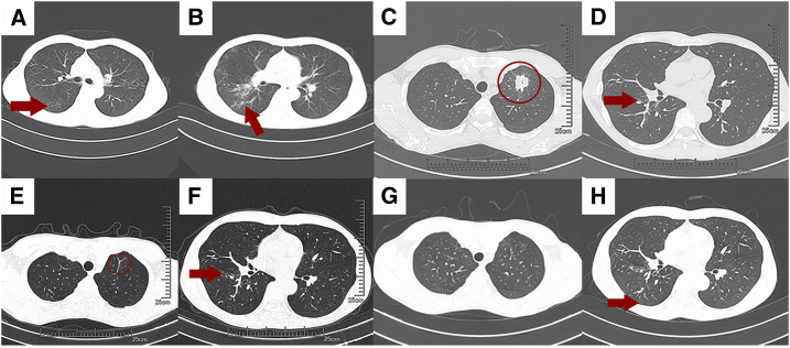
In 2015, a 34-year-old Chinese man underwent renal transplantation at Sichuan Provincial People’s Hospital because of chronic renal failure caused by IgA nephropathy. The donor (male, aged 34 years), a resident of Boshuixiang, Dazhou city, Sichuan Province of China, died of intracerebral hemorrhage caused by rupture of cerebral arteriovenous malformation. The patient received maintenance immunosuppression with mycophenolate mofetil and tacrolimus with regular therapeutic drug monitoring. In September 2017, he presented with cough and hemoptysis. Pulmonary tuberculosis was confirmed by positive acid-fast bacilli (AFB) smears and computer tomography (CT) features (Figure 1A and B). After 9 months of antituberculosis treatment at Sichuan Provincial People’s Hospital, the symptoms resolved and the follow-up CT showed significant resolution of tuberculosis (Figure 1C and D). His baseline serum creatinine (SCr) had increased to 200 umol/L in August 2018.
Figure 1.
Computer tomography (CT) features of the patients A and B, and CT images were obtained on September 4, 2017. Axial CT slices were displayed at the level of carina (A) and right pulmonary artery (B). Tree-in-bud nodular pattern was seen in the right lower lobe superior segment (red arrow) and the left upper lobe (red arrow) on the baseline. (C and D), Follow-up CT images were obtained on August 3, 2019. Axial CT slices at the level of the mid trachea (C) displayed interval development of consolidation with air bronchograms in the left upper lobe (red circle). However, the tree-in-bud nodularity in the right lower lobe had partially resolved since the baseline scan (D, red arrow). (E and F), CT images were obtained on December 30, 2019. (E) Resolution of left upper lobe consolidation with some residual scarring (red circle), whereas the mild tree-in-bud nodularity in the right lower lobe (F) showed no change (red arrow). (G and H), CT images which obtained on April 10, 2020 showed no new airspace disease, whereas some mild tree-in-bud nodularity still existed in the right lower lobe (red arrow). All images were in lung window settings. This figure appears in color at www.ajtmh.org.
In March 2019, he traveled to Laos for about 1 month to visit a friend there. No special activities or suspicious contact was reported except that he had some barbecue with local friends during that time. After his return to China, he presented with a 4-month history of weakness and poor appetite, unintentional weight loss of 5 kg (body weight at baseline was 52 kg), and a 1-month history of occasional nonproductive cough with hemoptysis (about 100 mL). He received antituberculosis treatment at a local clinic in July 2019, but stopped treatment a week later because of nausea. He was afebrile, and had a persistent cough and increasing weakness.
In August 12, 2019, he was admitted to our pulmonary and critical care division. The patient appeared cachectic. His vital signs were unremarkable except for a slight increase in the heart rate (97 beats/minute). Both lungs were clear, except for the dullness on percussion in the left upper lobe. There was no skin damage, hepatosplenomegaly, or palpable lymphadenopathy. Computer tomography revealed that apart from the residual of previous tuberculosis, there was new patchy consolidation (5 × 3 cm) in the apicoposterior segment of the left upper lobe (Figure 1E and F).
Routine blood tests were performed on the second day of admission, which revealed 3.59 × 10⁹ cells/L white blood cells, 77.1% neutrophils, 13.1% lymphocyte, and 80 g/L hemoglobin. The serum C-reactive protein and procalcitonin levels were 14.19 mg/L (reference range, 5–10 mg/L) and 0.05 ng/mL (reference range, 0–0.05 ng/mL), respectively. The HIV test result was negative. Renal function tests revealed that the concentration of serum SCr, urea nitrogen, and uric acid were 251.7 umol/L (reference range, 59–104 umol/L), 8.39 mmol/L (reference range, 2.9–8.2 mmol/L), and 480 umol/L (reference range, 155–428 umol/L), respectively. Fiber optic bronchoscopy with bronchoalveolar lavage was performed. Next-generation sequencing (NGS) was used for pathogen detection on the second day of admission. DNA was extracted from 10 mL of the bronchoalveolar lavage fluid (BALF) sample using the TIANamp Micro DNA Kit (DP316, Tiangen Biotech, Beijing, China) according to the manufacturer’s protocols. Abdominal ultrasonography demonstrated bilateral renal atrophy. Sputum samples and BALF samples for AFB and bacteria culture were negative. The Xpert MTB/RIF assay in BALF samples was negative. Considering the concern about chronic pulmonary aspergillosis (CPA), the AspergillusIgG test was performed, and the results were above 500 arbitrary units/mL.
He received symptomatic treatment, including expectorant (Mucosolvan®; Boehringer Ingelheim, Ingelheim, Germany) aerosol inhalation treatment and benzbromarone for the treatment of hyperuricemia. Because of the suspicion for infection, the patient received maintenance immunosuppression with low doses of mycophenolate mofetil (one dose of 0.5 g twice daily) and tacrolimus (one dose of 1.0 mg twice daily). Antibiotics were excluded because there was no evidence of bacterial infection.
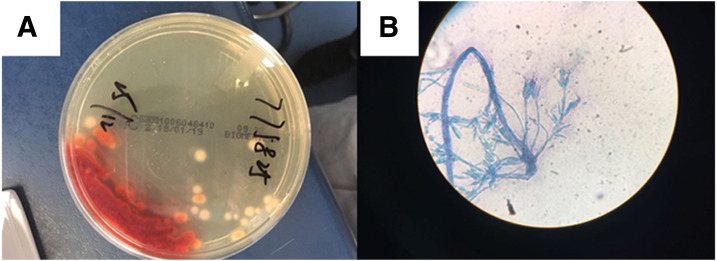
On the fifth day of admission, the NGS revealed that the most abundant pathogens in the BALF samples were T. marneffei (4,347 reads), Prevotella (671 reads), and Neisseria (150 reads), which raised the suspicion of T. marneffei infection. This was subsequently confirmed by fungal culture of BALF samples and blood culture, which demonstrated temperature-dependent mold-to-yeast conversion, typical colony, and microscopic morphology (Figure 2). Therefore, the patient was diagnosed with pulmonary T. marneffei infection. Amphotericin B was not administered because of the elevated SCr levels. Itraconazole and voriconazole were recommended, but they were rejected because the patient could not afford these drugs. Fortunately, oral posaconazole was covered in his medical insurance. Therefore, he was treated with oral posaconazole at the dose of 10 mL (400 mg) bid.
Figure 2.
Mycological findings of the patient. (A) Typical colony morphology with red pigment that diffuses into the medium. (B) Typical morphology of mold form, with branched septate hyphae and condiophores/phialoconidia typical of Penicillium species. This figure appears in color at www.ajtmh.org.
The patient was discharged on the seventh day of admission. One month later, his cough and fatigue had been resolved, but the serum SCr level increased to 323.2 umol/L. He denied having any symptom of infection or taking any self-medication that could cause renal injury except for immunosuppressants. Drug-level monitoring revealed that the serum drug level of tacrolimus increased to 20.5 ug/mL (reference range, 3–7 ug/mL). Considering the possibility of interaction between tacrolimus and posaconazole, it was speculated that posaconazole increased the serum level of tacrolimus by interfering with the metabolism of tacrolimus, resulting in renal injury. After adjusting the dose of immunosuppressants (tacrolimus 0.5 mg bid and mycophenolate mofetil 0.25 qd), the serum drug level of tacrolimus was decreased to normal level. Eight months since the initial admission, the serum SCr level decreased to 249 umol/L. The follow-up CT showed a considerable resolution in lung abnormalities without any evidence of relapse (Figure 1G and H). He received antifungal treatment for 6 months and recovered completely after stopping posaconazole. The long-term management plan was to continue with the immunosuppression regimen with careful follow-up for recrudescent infection.
DISCUSSION
Thanks to the widespread use of antiretroviral therapy, there is a significant decline in talaromycosis among HIV-infected patients.6 In recent years, an increasing number of T. marneffei infections have been reported in non–HIV-infected patients, especially in those with organ transplantation, hematological malignancies, and autoimmune diseases.7–9 Without timely diagnosis and treatment, the mortality rate can be as high as 50.6–97%.10,11 To our knowledge, this is the first case report describing efficacy of posaconazole in the treatment of localized pulmonary T. marneffei infection in a renal transplant recipient.
Talaromyces marneffei is a disease which is mostly prevalent in Southeast Asia and southern China.12 Some sporadic cases have been reported among Europeans, Americans, Africans, Singaporeans, and Koreans residing or traveling in the endemic areas.13 Although we still could not out the possibility of T. marneffei in live donor renal, we believed that the inhalation of conidiophores of T. marneffei was the primary mode of transmission in our case, as he developed symptoms after a trip to Laos. The most common reported symptoms of T. marneffei infection were fever, cough, and weight loss. Hemoptysis, although not a common symptom, had been reported in a previous study.13
The application of liposomal amphotericin B in T. marneffei infection was limited because of its nephrotoxicity.3 Itraconazole was usually given as maintenance therapy, after initial induction therapy with IV amphotericin B.14 It was only recommended as induction therapy in patients with mild disease. These two drugs were not suitable for our case. Although voriconazole was an alternative drug for the treatment of T. marneffei infection, it was associated with liver toxicity and neurological side effects including auditory and visual hallucinations.15 Posaconazole, a triazole with wide antifungal activities, may have additional advantages compared with itraconazole and voriconazole, especially in critically ill patients with organ dysfunction, as no renal or hepatic dosage adjustment was required.5 Previous study had reported the excellent in vitro activity of posaconazole against T. marneffei.4,16 In our case, the patient responded well to posaconazole, but his renal function declined. Considering the possibility of interaction between tacrolimus and posaconazole, it was speculated that posaconazole increased the serum level of tacrolimus by interfering with the metabolism of tacrolimus, resulting in renal injury.17 Our clinical strategy was to reduce the dose of tacrolimus by 50% when tacrolimus was coadministered with posaconazole. Seven months after adjusting the dose of immunosuppressants, the patient recovered completely.
CONCLUSION
Posaconazole may be a potentially promising therapy for T. marneffei infection that must be administered under close monitoring when used concurrently with immunosuppressors such as tacrolimus.
Acknowledgment:
We thank Ying-Na Zhao for help with photo editing.
REFERENCES
- 1.Chan JF, Lau SK, Yuen KY, Woo PC, 2016. Talaromyces (Penicillium) marneffei infection in non-HIV-infected patients. Emerg Microbes Infect 5: e19. [DOI] [PMC free article] [PubMed] [Google Scholar]
- 2.Shoham S, Dominguez EA; Practice ASTIDCo , 2019. Emerging fungal infections in solid organ transplant recipients: guidelines of the American society of transplantation infectious diseases community of Practice. Clin Transpl 33: e13525. [DOI] [PubMed] [Google Scholar]
- 3.Tollemar J, Klingspor L, Ringdén O, 2001. Liposomal amphotericin B (AmBisome) for fungal infections in immunocompromised adults and children. Clin Microbiol Infect 7 (Suppl 2): 68–79. [DOI] [PubMed] [Google Scholar]
- 4.Lei HL, Li LH, Chen WS, Song WN, He Y, Hu FY, Chen XJ, Cai WP, Tang XP, 2018. Susceptibility profile of echinocandins, azoles and amphotericin B against yeast phase of Talaromyces marneffei isolated from HIV-infected patients in Guangdong, China. Eur J Clin Microbiol Infect Dis 37: 1099–1102. [DOI] [PubMed] [Google Scholar]
- 5.Lau SK, et al. 2017. In vitro activity of posaconazole against Talaromyces marneffei by Broth microdilution and etest methods and comparison to itraconazole, voriconazole, and anidulafungin. Antimicrob Agents Chemother 61: e01480-16. [DOI] [PMC free article] [PubMed] [Google Scholar]
- 6.Le T, et al. 2011. Epidemiology, seasonality, and predictors of outcome of AIDS-associated Penicillium marneffei infection in Ho Chi Minh City, Viet Nam. Clin Infect Dis 52: 945–952. [DOI] [PMC free article] [PubMed] [Google Scholar]
- 7.Kawila R, Chaiwarith R, Supparatpinyo K, 2013. Clinical and laboratory characteristics of Penicilliosis marneffei among patients with and without HIV infection in Northern Thailand: a retrospective study. BMC Infect Dis 13: 464. [DOI] [PMC free article] [PubMed] [Google Scholar]
- 8.Xiao-Fen SU, Zhang NF, Liu CL, Dan-Hong SU, 2013. Disseminated Penicillium marneffei infection in immunocompetent patients: one case report and literature review. Chin J Respir Crit Care Med 3: 244–248. [Google Scholar]
- 9.Qiu Y, Liao H, Zhang J, Zhong X, Tan C, Lu D, 2015. Differences in clinical characteristics and prognosis of Penicilliosis among HIV-negative patients with or without underlying disease in southern China: a retrospective study. BMC Infect Dis 15: 525. [DOI] [PMC free article] [PubMed] [Google Scholar]
- 10.Hu Y, Zhang J, Li X, Yang Y, Zhang Y, Ma J, Xi L, 2013. Penicillium marneffei infection: an emerging disease in mainland China. Mycopathologia 175: 57–67. [DOI] [PubMed] [Google Scholar]
- 11.Ranjana KH, Priyokumar K, Singh TJ, Gupta ChC, Sharmila L, Singh PN, Chakrabarti A, 2002. Disseminated Penicillium marneffei infection among HIV-infected patients in Manipur state, India. J Infect 45: 268–271. [DOI] [PubMed] [Google Scholar]
- 12.Peng J, et al. 2017. Recovery from Talaromyces marneffei involving the kidney in a renal transplant recipient: a case report and literature review. Transpl Infect Dis 19: e12710. [DOI] [PubMed] [Google Scholar]
- 13.Wang P, Chen Y, Xu H, Ding L, Wu Z, Xu Z, Wang K, 2017. Acute disseminated Talaromyces marneffei in an immunocompetent patient. Mycopathologia 182: 751–754. [DOI] [PubMed] [Google Scholar]
- 14.Spanakis EK, Aperis G, Mylonakis E, 2006. New agents for the treatment of fungal infections: clinical efficacy and gaps in coverage. Clin Infect Dis 43: 1060–1068. [DOI] [PubMed] [Google Scholar]
- 15.Levine MT, Chandrasekar PH, 2016. Adverse effects of voriconazole: over a decade of use. Clin Transplant 30: 1377–1386. [DOI] [PubMed] [Google Scholar]
- 16.Filiotou A, Velegraki A, Giannaris M, Pirounaki M, Mitroussia A, Kaloterakis A, Archimandritis A, 2006. First case of Penicillium marneffei fungemia in Greece and strain susceptibility to five licensed systemic antifungal agents and posaconazole. Am J Med Sci 332: 43–45. [DOI] [PubMed] [Google Scholar]
- 17.Vanhove T, Bouwsma H, Hilbrands L, Swen JJ, Spriet I, Annaert P, Vanaudenaerde B, Verleden G, Vos R, Kuypers DRJ, 2017. Determinants of the magnitude of interaction between tacrolimus and voriconazole/posaconazole in solid organ recipients. Am J Transpl 17: 2372–2380. [DOI] [PubMed] [Google Scholar]


