Clinical vignettes
Case #1
An 81-year-old male with recent hospitalization for heart failure complicated with acute renal failure (RF). Workup demonstrated severe mitral regurgitation (MR) due to a flailed P2. The patient was discharged and referred to our center for further recommendations. The patient had persistent Class III NYHA symptoms and based on his age and comorbidities, including recent RF, he was offered MitraClip™ (Abbott, Illinois, USA) given his high risk for surgical intervention.
Case #2
A 54-year-old gentleman with a history of viral cardiomyopathy with left ventricular ejection fraction of 20% to 25%. His father has a history of non-ischemic cardiomyopathy and had a heart transplant at 55 years old. The patient has been managed by our heart failure team with goal-directed medical therapy including carvedilol, furosemide, sacubitril/valsartan, and Automatic Implantable Cardioverter Defibrillator implantation. Despite this management, he had persistent severe MR and Class III NYHA symptoms with recurrent heart failure hospitalizations. The patient was considered a high surgical risk and offered MitraClip™.
Surgical technique
Patient preparation and mitral valve (MV) assessment
Patients are brought to the cath lab or hybrid room and placed in the supine position. After induction of general anesthesia the patient is prepped and draped with access to both groins. We routinely place a foley catheter and arterial line, although some centers do not use invasive arterial pressure monitoring. Patients receive routine antibiotic prophylaxis. A complete transesophageal echocardiogram (TEE) is performed assessing the degree of MR, mitral valve area (MVA), MV gradients, and pulmonary vein flow pattern. We make sure to record the hemodynamic data (blood pressure and heart rate) at the time of TEE assessment and try to replicate the pre-procedure hemodynamic status after MitraClip™ implantation to assess improvement. The left atrial appendage is surveyed for thrombus. A significant subset of these patients have a history of atrial fibrillation and their anticoagulation is discontinued a few days prior to the procedure.
Procedural steps
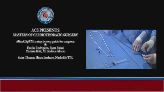
We divide this procedure into several key steps (Video 1).
Video 1.

MitraClipTM: a step by step guide for surgeons.
TEE evaluation and procedure planning
As mentioned above, we perform a complete TEE prior to starting the procedure. At this point, we review our final clip implantation strategy (i.e., clip location and number of clips) as well as clip size and width selection. There are four different MitraClips™ currently available. In general, we choose the XTR or XTW for big degenerative valves with central pathologies. In the case of commissural pathologies, it is sometimes difficult to maneuver the XTR or XTW clips as you usually have less leaflet length and higher risk for chord entanglement. In these cases, we prefer the NTR or NTW options. We avoid wide clips in small valves (i.e., MVA <4 cm2) at risk for mitral stenosis after clip implantation. Remember you need at least 6 and 9 mm of leaflet length to grasp for NTR/NTW and XTR/XTW MitraClips™, respectively, in order to minimize the risk of a single leaflet attachment complication.
Venous access
The right femoral head is located using a standard hemostat and fluoroscopy. Vascular ultrasound can also be a valuable tool to localize the right common femoral vein. We use the micropuncture technique for venous access and then place a 6F sheath to dilate the tract, followed by placement of two 6F Perclose Proglide Sutures (Abbott) off-set approximately 20 degrees rotation from each other. The patient is systemically heparinized to an ACT >250 seconds with a usual starting dose of 100 U/Kg.
Trans-septal access
We use an SL1 sheath (Abbott) and Baylis needle (Baylis Medical, Massachusetts, USA) for trans-septal puncture. Once we gain access into the left atrium (LA), we measure the initial LA pressure. We then place an Amplatzer extra-stiff wire (Cook Medical, Indiana, USA) with a 1-centimeter soft tip into the left superior pulmonary vein. The wire is advanced using TEE guidance to help avoid advancement into the left atrial appendage. Location of the trans-septal puncture is crucial for the success of the MitraClip™ procedure. The video illustrates the best access location.
Delivery guide and clip back-table preparation
We encourage the implanter to prepare their own device to increase facileness with the system (Video 2), however, many centers have their cath lab staff prepare the device on the back table.
Video 2.

Delivery guide & clip back-table preparation.
Insertion of delivery sheath
The delivery sheath is introduced after serial dilatation of the femoral vein and subcutaneous tract using a dilator set (LivaNova, London, UK) up to a 24F.
MitraClipTM positioning in LA
The MitraClip™ is then introduced and positioned above the MV in the desired location. Perpendicularity to the MV annular plane is crucial for symmetric leaflet grasping as well as clip rotation (Video 1).
MitraClipTM insertion into left ventricle (LV) and MV leaflet grasping
The MitraClip™ is then advanced into the LV making sure the clip does not dive anterior/posterior or medial/lateral. This helps prevent mal-location and potential chord entanglement. In addition, the rotation of the clip is assessed on TEE and fluoroscopy before and after crossing the MV. It is crucial to maintain clip rotational orientation so that minimal corrections are required following its descent into the LV. After the clip is positioned appropriately below the MV, it is then opened to 120°.
MV leaflet grasping assessment
Leaflet grasping is best achieved using the TEE LVOT view and is crucial to confirm good leaflet insertion. X-plane imaging (bicommissural and LVOT) can be helpful to confirm the position and grasp. After confirming the initial grasp and leaflet insertion, the clip is closed to 60°. Repeat imaging is performed to reconfirm leaflet insertion.
Clip closure and TEE assessment
Once leaflet insertion is confirmed the clip is then fully closed. We then perform a thorough assessment of residual MR, trans-MV gradient, pulmonary vein flow pattern, and LA pressure.
Delivery system removal
Once we are satisfied with the results, the final arm angle is tested. Once confirmed, the clip is deployed and delivery system removed. We again reassess the MitraClip™ result and quantitate the residual MR and discuss whether additional clips are required. The final LA pressure is also measured.
Atrial septal access determination for closure
The atrial septostomy is closed in <1% of cases. We consider closure in patients with severe right ventricular dysfunction, pulmonary hypertension, significant tricuspid regurgitation, or right-to-left shunting across the atrial septum.
Completion
Patients are extubated at the end of the procedure and transferred to the recovery room. Foley catheter and arterial monitoring are usually removed prior. Patients stay overnight in our telemetry unit and >85% are discharged home the next morning after completion of a transthoracic echocardiogram (TTE) demonstrating clip stability and unchanged or improved reduction of MR when compared to post-procedure TEE. All patients are discharged on ASA 81 mg/day. Plavix 75 mg daily for 30 days is added to patients that were not on an additional anticoagulant prior to this procedure.
Patients are reviewed at 30 days with a repeat TTE.
Comments
The MitraClip™ technology has been widely used over the last decade with outstanding reproducible results. It is the only FDA approved technology for transcatheter MV repair for patients with both primary and secondary moderate to severe MR. Several studies have demonstrated improved survival, improved symptoms, and quality of life, as well as decreased hospitalizations in patients treated with MitraClip™ technology (1-3). We have performed over 350 MitraClip™ procedures at our institution with reduction of MR to <2+ in over 95% of cases with <1% mortality. We believe that the success of our MV program is due to a comprehensive team including an imaging cardiologist, skilled sonographer, interventional cardiologist, and cardiac surgeon that work together during these cases.
Acknowledgments
Funding: None.
Open Access Statement: This is an Open Access article distributed in accordance with the Creative Commons Attribution-NonCommercial-NoDerivs 4.0 International License (CC BY-NC-ND 4.0), which permits the non-commercial replication and distribution of the article with the strict proviso that no changes or edits are made and the original work is properly cited (including links to both the formal publication through the relevant DOI and the license). See: https://creativecommons.org/licenses/by-nc-nd/4.0/.
Footnotes
Conflicts of Interest: ER and MAM are consultants, investigators, and train MitraClip™ implanters for Abbott. The other authors have no conflicts of interest.
References
- 1.Stone GW, Lindenfeld J, Abraham WT, et al. Transcatheter Mitral-Valve Repair in Patients with Heart Failure. N Engl J Med 2018;379:2307-18. 10.1056/NEJMoa1806640 [DOI] [PubMed] [Google Scholar]
- 2.Feldman T, Kar S, Elmariah S, et al. Randomized Comparison of Percutaneous Repair and Surgery for Mitral Regurgitation: 5-Year Results of EVEREST II. J Am Coll Cardiol 2015;66:2844-54. 10.1016/j.jacc.2015.10.018 [DOI] [PubMed] [Google Scholar]
- 3.Feldman T, Foster E, Glower DD, et al. Percutaneous repair or surgery for mitral regurgitation. N Engl J Med 2011;364:1395-406. 10.1056/NEJMoa1009355 [DOI] [PubMed] [Google Scholar]


