Abstract
The global concern of increasing number of children presenting with multisystem inflammatory syndrome in children (MIS-C) related to the coronavirus disease (COVID-19) has escalated the need for a case-oriented clinical approach that provides timely diagnosis and management. The aim of this study is to share our experience in managing 64 MIS-C patients of North African ethnicity guided by a risk-based algorithm. Sixty-four patients met the inclusion criteria, 19 (30%) patients were categorized as mild and moderate risk groups and cared for in an isolation ward and 45 patients who belonged to the high-risk group (70%) were admitted to the pediatric intensive care unit (PICU). Positive laboratory evidence of COVID-19 was found in 62 patients. Fever and dysfunction in 2 or more organs were confirmed in all cases (100%). Fifty patients (78%) presented with gastrointestinal symptoms, meanwhile only 10 patients (16%) had respiratory manifestations. Cardiac involvement was reported in 55 (86%) cases; hypotension and shock were found in 45 patients (70%) therein circulatory support and mechanical ventilations were needed for 45 and 13 patients respectively. Intravenous immunoglobulins (IVIG) were used for all cases and methylprednisolone was used in 60 patients (94%). Fifty-eight (91%) patients were discharged home after an average of 9 days of hospitalization. The mortality rate was 9% (6 patients). Conclusion. A single Egyptian center experience in the management of MIS-C patients guided by a proposed bed side algorithm is described. The algorithm proved to be a helpful tool for first-line responders, and helped initiate early treatment with IVIG.
Keywords: COVID-19, pediatric MIS-C, Kawasaki disease, IVIG, algorithm
Introduction
While diagnosis and management of MIS-C1 has been reported from epicenters in Europe2 and USA,3 little is published about MIS-C diagnosis and management in Egypt and North Africa.
The noticeable difference of the MIS-C presentation versus Kawasaki Disease (KD)4 resides in the older age of presentation, intense form of inflammation, more frequent gastrointestinal manifestations, different laboratory findings (eg, lymphopenia, thrombocytopenia, elevated troponin, N-terminal pro hormone B-type natriuretic peptide [NT-pro BNP], D-dimer, and ferritin), and higher propensity towards left ventricular dysfunction with shock.5
Since management is not universally established, it is reasonable to consider MIS-C with its spectrum as a unique syndrome with a different treatment plan than that for KD.2
Objective
We describe the spectrum of clinical presentation and management for a cohort of children with MIS-C from the epicenter of COVID-19 in Cairo, Egypt. Our “first golden hours” algorithm is based on classifying patients at presentation into risk criteria.
Methods
We carried out a retrospective observational study of pediatric patients admitted to the Children Hospital of Ain Shams University, the tertiary epicenter for COVID-19 in Central and Northern Metropolitan Cairo, Egypt between June 9th and August 18th, 2020. Patients were managed as guided by the proposed algorithm if they fulfilled the following criteria: (1) fever (≥38°C), (2) a history of prior infection or contact with a case of severe acute respiratory syndrome coronavirus-2 (SARS-CoV-2) or positive RT-PCR or serology, and (3) signs and symptoms of 2 or more organ system involvement. Since all cases presented during the pandemic with positive COVID-19 antigen and/or antibody with fever and positive inflammatory markers or lymphopenia, we determined that they represented the new MIS-C syndrome rather than KD or atypical KD.
Laboratory work-up included: SARS-CoV-2 infection status determination by reverse transcriptase–polymerase chain reaction (RT-PCR) of nasopharyngeal swabs, COVID-RT-PCR, IgG, and IgM, complete blood count (CBC), C-reactive protein (CRP), basic metabolic profile (BMP): (sodium [Na], potassium [k], alanine transaminase [ALT], aspartate aminotransferase [AST], blood urea nitrogen [BUN], and creatinine [Cr]), and Electrocardiogram (EKG) and Echocardiography (Echo) (using Philips EPIQ CVx Premium and Philips Portable Ultrasound CX50 MATRIX with Transesophageal Echocardiography).
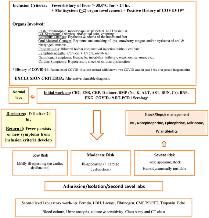
Patients were discharged if initial laboratory results were within normal limits, with a plan for a 24-hour follow up if fever persisted or other symptoms developed. In cases of positive laboratory results, patients were classified into low, moderate, or severe risk groups, and admitted into COVID-19 suspect/positive zones (Figure 1).
Figure 1.
MIS-C management based on risk criteria.
Abbreviations: IVF, intravenous fluids; IV, intravenous; LDH, lactate dehydrogenase; CMP, comprehensive metabolic profile; PT, prothrombin time; PTT, partial thromboplastin time; CT, computed tomography.
The Low-risk Group
Children looking mildly ill, presenting with fever, and symptoms of ≥ 2 organs involvement, with stable vital signs and no signs of cardiac dysfunction or hemodynamic instability.
The Moderate-risk Group
Ill-appearing children, with fever, symptoms of 2 or more organs involvement, and hemodynamically stable. Additional work-up includes a comprehensive metabolic profile (CMP), venous blood gas (VBG), lactate, ferritin, procalcitonin, lactic dehydrogenase (LDH), fibrinogen, PT/PTT, urine analysis (UA), urine culture and sensitivity (UCX), blood culture (Bl.cx.). As needed, workup includes abdominal imaging, cytokine panel, etc. Since troponin and B-type natriuretic peptide are useful markers for myocardial involvement, we selected to perform troponin levels only due to limited financial resources while trying to allocate the majority of the budget towards the costly therapeutics used.
The Severe-risk Group
Severely ill, toxic-appearing children, with evidence of shock or cardiac dysfunction and hemodynamic instability. The same moderate-risk group work-up, and workup based on specified clinical indications should be done. IL-6, IL-1 and TNF levels6 to be obtained if no response to therapy is noted. ICU admission is indicated for: persistent tachycardia, poor perfusion, hypotension, or shock.
MIS-C Therapeutics
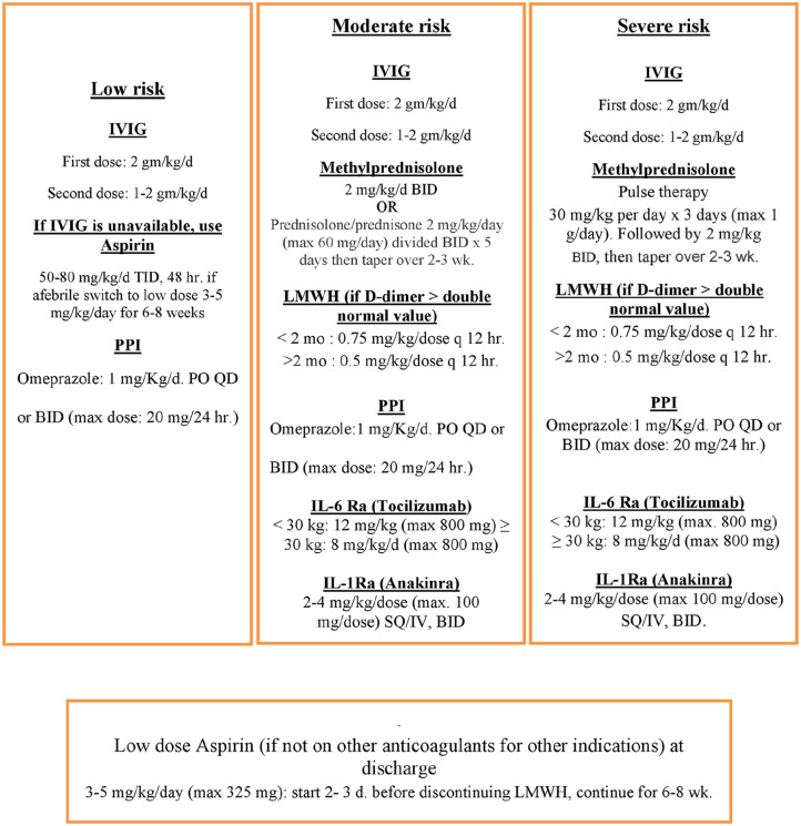
The cornerstone for our treatment of MIS-C was the early use of effective immunomodulatory therapy: Intravenous immunoglobulins (IVIG) and corticosteroids (Figure 2).7
Figure 2.
MIS-C therapeutics.
Discharge of MIS-C patients was considered if patients were (1) afebrile ≥ 48 hours, (2) oxygen saturation is ≥ 95% in room air, (3) tolerating oral intake (4) hemodynamically stable, with compensated cardiac function (with or without treatment) and (5) inflammatory marker levels were trending down.
It is prudent to ensure a short and long-term follow-up to these patients. Our patients were scheduled for follow up with hematology, rheumatology and cardiology services 2 weeks after discharge.
Ethical Approval and Informed Consent
The Ain Shams University ethics committee approved the study with a waiver of informed consent (FM-ASU P61/2020)
Results
Sixty-four patients (38 males; 26 females; median age of 7 years) met the inclusion criteria and were included in the study. Patients were classified into 1 of 3 categories according to the severity of clinical picture and the results of laboratory findings.
Nineteen (30%) patients were categorized as the mild and moderate risk groups and cared for in an isolation ward while 45 patients were classified into the high-risk group and admitted to the pediatric intensive care unit (PICU). With the exception of 2 patients (both of whom had a positive family history of positive COVID-19 diagnosis), 62 patients had a positive evidence of COVID-19 infection, as detected by different combinations of serological testing with a positive RT-PCR COVID-19. Fever was reported in 100% of the cases, followed by the presence of rash in 91% of the patients (58/64). Gastrointestinal manifestations, mainly abdominal pain, vomiting or diarrhea were noticeable in 78% (50/64) of the cases. A minority of patients, 4 cases (6%) presented with neurological symptoms (eg, seizures, headache or neck stiffness). Forty-five patients (70%) had symptoms and signs of shock. Thirteen patients (20%) required mechanical ventilation, with high frequency ventilation needed for 4 patients, while 9 patients were managed by conventional mechanical ventilation. Fifty-five patients had clinical and laboratory evidence of cardiac dysfunction, with some patients having more than one cardiac lesion on echocardiography. The variety of the echocardiographic findings including left ventricular dysfunction (LVD) in 22 patients, valvulitis in 35 patients, coronary artery changes in 20 patients and pericardial effusion in 7 patients. Echo was normal in 9 patients; all belonged to the mild group. Intravenous immunoglobulins were used for all cases and corticosteroids were used in 60 patients (94%); treatment with anti-IL-6 receptor antagonist was used for one patient. Prophylactic low molecular weight heparin was required for 52 patients (81%), while Aspirin was used for 35 (55%) cases. Fifty-eight (91%) patients were discharged home after a mean hospital stay of 9 days.
In our case series, 18 patients had underlying comorbid conditions. The co-morbidities included the following conditions: systemic lupus erythematosus, polyarteritis nodosa, hemophagocytic lympho-histiocytosis, juvenile rheumatoid arthritis, acute lymphoblastic leukemia, medulloblastoma, chronic kidney diseases, acute disseminated encephalomyelitis, Fehr’s syndrome, tetralogy of Fallot, gastrointestinal conditions (1 patient had ileostomy and 1 patient had intestinal tuberculosis), cystic fibrosis, diabetes mellitus, and Down syndrome. Six patients died (9%), 4 of them had severe underlying comorbidities. The 6 mortalities included: 1-month old neonate, a 3 months infant and 4 patients with severe comorbid conditions (2 children had acute lymphoblastic leukemia, aged 12 and 14, a 5-year old with medulloblastoma, and a 14-year-old patient with polyarteritis nodosa). Detailed demographic and clinical characteristics are outlined in Table 1.
Table 1.
Demographic, Clinical, and Selected Laboratory Characteristics.
| Characteristics | Patients no. (%) |
|---|---|
| Male sex—no. (%) | 38 (59%) |
| Median age (range) | 7 years (1 month-14 years) |
| Mild, moderate & severe risk groups—no. (%) | |
| High risk | 45 (70) |
| Mild & moderate risk—no (%) | 19 (30) |
| Level of medical care—no. (%) | |
| Isolation ward | 19 (30) |
| PICU | 45 (70) |
| Mechanical ventilation | 13 (20) |
| Conventional | 9 (14) |
| HFV | 4 (6) |
| Vasopressor and inotropic support | 45 (70) |
| Outcome—no. (%) | |
| Discharged alive | 58 (91) |
| Died | 6 (9) |
| Average length of hospital stay in days (range) | 9 (4-46) |
| Clinical symptoms and signs—no. (%) | |
| Fever | 64 (100) |
| Median fever duration in days (IQR) | 5 (3-10) |
| Rash | 58 (91) |
| Skin desquamation | 35 (55) |
| Conjunctivitis | 39 (61) |
| Gastrointestinal manifestations | 50 (78) |
| Neurologic manifestations | 4 (6) |
| Respiratory manifestations | 10 (15) |
| Cardiac manifestations | 55 (86) |
| Shock at presentation | 45 (70) |
| Underlying conditions—no. (%) | |
| Previously healthy | 46 (72) |
| Comorbidities—no. | 18 (28) |
| Rheumatological diseases | 5 |
| Renal disease | 1 |
| Cystic fibrosis | 1 |
| Neurological diseases | 2 |
| Malignancy | 3 |
| Gastrointestinal disorders | 2 |
| Cardiac diseases | 1 |
| Diabetes mellitus | 2 |
| Down syndrome | 1 |
| Diagnostic modality of SARS-COV-2—no. (%) | |
| Laboratory negative (+ve contacts) | 2 (3) |
| Laboratory positive patients | 62 (97) |
| Positive RT-PCR | 39/62 |
| Positive IgG | 50/62 |
| Positive IgM | 17/62 |
| Significant laboratory abnormalities—no. (%) | |
| High CRP | 64 (100%) |
| Hyperferritinemia | 60 (94) |
| Elevated D-dimer | 52 (81) |
| High troponin | 40 (63) |
| Lymphopenia | 45 (70) |
| Thrombocytopenia | 5 (8) |
| Thrombocytosis | 0 |
| Echocardiographic findings—no. | 64 |
| Normal | 9 (14%) |
| Abnormal | 55 (86%) |
| LVD | 22/55 |
| Valvulitis | 35/55 |
| Coronary artery changes | 20/55 |
| Pericardial effusion | 7/55 |
| Treatment modalities—no. (%) | |
| Intravenous immunoglobulins | 64 (100) |
| Methylprednisolone | 60 (94) |
| Anticoagulant therapya | 52 (81) |
| Aspirin | 35 (55) |
| IL-6 receptor antagonist | 1 |
Abbreviations: HFV, High frequency ventilation; IQR, Interquartile range; CRP, C-reactive protein; LVD, left ventricular dysfunction.
Considered for elevated D-dimer: more than double normal level.
We noticed a rapid progression of the clinical symptoms in some cases from the mild to the moderate/severe cases requiring the use of 2 doses of IVIG (we started with 2 gm/kg, then in the last 2 patients, we used only 1 gm/kg due to the limited supply during the pandemic). Slower IVIG administration was considered in patients with myocardial dysfunction to decrease the risk of fluid overload. The regimen and dosing of IVIG are summarized in Figure 2. Other management strategies used are detailed in Figure 2 and highlighted in the following points:
Decision about anticoagulation was based on the coagulation profile8 and clinical necessity. We speculated in our algorithm that a prophylactic dose of low molecular weight heparin (LMWH) should be enough for moderate and high-risk groups when their D-dimer is equal to or above 1000 mg/ml. LMWH was stopped 2 days before discharge while adding a low-dose aspirin that was continued for 4 to 6 weeks. The therapeutic dose of LMWH was used for only 1 patient with Systemic Lupus Erythematosus (SLE) with markedly elevated D-dimer (D-dimer was ≥ 3000 mg/ml).
Pediatric resuscitation guidelines were followed,9 and shock was managed with very careful intravenous fluid resuscitation. Epinephrine or norepinephrine were preferred for vasodilatory shock refractory to volume expansion (due to ventricular dysfunction). In children presenting with severe ventricular dysfunction, the addition of milrinone was helpful in some cases.10
Sepsis management was started pending culture results using empiric IV antibiotics (eg, beta-lactam agents or cephalosporins) followed by adding vancomycin for MIS-C presenting with shock syndrome and septic shock. Clindamycin was used if there were features consistent with toxin-mediated illness (eg, erythroderma). This usage was modified based on culture results and the patient’s clinical response to therapy. Antibiotics were discontinued once bacterial infection had been excluded and the child’s clinical status had stabilized.11
Anti-cytokine therapy was considered for the evidence of severe cytokine storm (eg, ≥3 times normal IL-6 values, markedly elevated ferritin, LDH and D-dimer levels), in the moderate and severe risk groups. This use was supported by most of the European studies.2,12 IL-6 receptor antagonists (IL-6Ra) were considered for use if symptoms persisted after the use of 2 doses of IVIG and pulse steroids. Only 1 patient required such use after demonstrating significantly elevated IL-6, ferritin, LDH, and D-dimer.
Interleukin-1 receptor antagonist (IL-1Ra) has a short half-life (4-6 hours) when compared to the long half-life of anti-IL-6 receptor antagonists (~16 days) and can be used if further cytokine storm control is needed13; our decision to use IL-6Ra was based on availability. Notably, however, treatment with more than 1 biologic is not recommended, due to lack of additional benefit and risk of increasing adverse effects.14
The use of antiviral therapies (eg, Remdesivir) in the management of children with MIS-C is uncertain15 and thus was not included in the algorithm, due to a lack of studies proving safety and efficacy at that time.
Discussion
Due to the lack of standardized well-established treatment protocols and for MIS-C and the relatively small number of reported cases from individual centers, we developed an algorithm for treating a series of 64 Egyptian children who met the criteria for MIS-C associated with SARS-CoV-2 infection.
Similar to the world’s reports of delayed presentation of MIS-C relative to the initial COVID-19 peak, the temporal relationship between the COVID-19 outbreak in Egypt and first case of MIS-C admitted to our center was in early June as Egypt experienced the surge in diagnosis of COVID-19 in early May 2020.16
The high number of cases reported from a single center spanning a 6-week period all belonged to low and middle-income families served in the university hospital setting, which fully covers the cost of admission and treatment. All patients belonged to the same ethnic Egyptian ancestry; essentially described as North Africans, a finding also in other registries,2,7 which seems to suggest either the presence of differences in the sequences of SARS-CoV-2 in various geographic locations17 or a possible genetic susceptibility as compared to KD.5
The median age of our patient registry is close to that of the Italian case registry reported by Verdoni et al,20 with a male predominance and no clear explanation other than a possible genetic predisposition, or increased exposure due to a male predominance in outdoor activities and chores for boys in lower income families.
In contrast to some studies from Europe and the USA that reported lower co-morbid conditions,7,18 in our patients, 47 (73%) were previously healthy, and 17 patients (27%) had significantly severe comorbid conditions.
The time from infection to onset of MIS-C symptoms varied widely, and many of our patients could not remember the exact timing of onset of symptoms, with most of the patients not recalling any antecedent symptoms, a common global finding.13,19 Regarding COVID-19 status, we reported a positive RT-PCR for SARS-CoV-2 in 39 (61%) of the patients, which is found to be slightly higher than most of the reports where SARS-CoV-2 positivity varied between 20% and 53% for patients admitted with MIS-C.7,20
Positive IgG antibodies were found in 78% (50/64) of patients, a figure similar to the 75% to 100% reported by the Italian study of Verdoni et al,20 which suggested a possible postinfectious immune response responsible for MIS-C.21
Similar to previous findings that gastrointestinal symptoms are prevalent in around 70% of patients,5 our patients showed a percentage of 78%. Our MIS-C patients had a relatively low incidence of respiratory system involvement of (10%) than most reported findings.22
A large cross-sectional study which included children positive for COVID-19 showed a requirement for invasive ventilation in 38% of their cases (18 out of 48 children)23 In our cases, we only resorted to mechanical ventilation in 13 cases out of 64 (20%). We speculate that this difference may be related to our early use of the immunomodulatory therapy.
Forty-five cases (70%) in our series presented with severe illness: shock requiring IV fluids and/or vasoactive drugs and ICU admission, a much higher figure than that reported from the early Canadian study of only 52% PICU admission.24 We suspect that this increased degree of severity was due to late presentation of patients, and likely contributed to the observed mortality.
To date, other multiple case series published in eastern US, Italy, UK, and France documented a comparatively lower cumulative percentage of IVIG use of 75% and steroid use in 59% of their case registries.5 We managed our patients according to the algorithm by early use of immunomodulatory therapy: IVIG and high dose steroids that were used in 100% & 94% of cases, respectively. Steroids were used most frequently in patients with severe clinical presentations, as reported by Elias et al,25 and 64% of respondents of the International Kawasaki Disease Registry (IKDR) across 38 institutions and 11 countries. Riphagen et al7 also stated that steroids play an essential role in controlling the inflammatory state related to the increased cytokines. Our MIS-C patients had signs and symptoms of an inflammatory state and we believe that steroids played an essential role for those who recovered.
Although most of the respondents of the International Kawasaki Disease Registry (IKDR) across 38 institutions and 11 countries reported a preference of IL-1Ra (58%) and TNF-a inhibitor (28%)25 use as compared to IL-6Ra (8%), in our case series we resorted to the use of IL-6Ra in one patient due to the availability of the drug as well as the markedly elevated level of IL-6.
We used the prophylactic dose of LMWH for the moderate and severe risk groups since children with MIS-C are at risk of thrombotic complications, endothelial damage, and coronary artery aneurysms. Low molecular weight heparin was used for 81% of the moderate and high-risk patients. Although only about one-third of the respondents in the survey of the IKDR25 supported its use, we anticipate that its use may positively influence the long-term coronary and myocardial consequences.
Cardiac involvement in our patients, as detected by EKG and Echocardiography, was present in 86% of the cases (55/64), which is higher than most reported studies,5 with considerable noticeable valvulitis.
Although mortality rate from other multicenter studies reported a lower percentage of 3%,26 we believe that the mortality in our study (9%) could have been lowered had we used ECMO which is not yet available at our facility. Additionally, we believe that the high rate of comorbid conditions may have contributed to our mortality rate. However, since all parents refused a postmortem examination, the contribution of the pre-existing conditions to the increased mortality rate could not be evaluated.
The limitations of this study reside in being a single-center, retrospective study, with a lack of access to some resources, such as ECMO.
Conclusion
This algorithm, used and developed at the Children’s Hospital of Ain Shams University, Cairo, Egypt for 64 MIS-C patients, provides healthcare workers with bed-side evaluation and management guide for children with COVID-19-associated MIS-C, however it should not supersede clinical judgement if other treatment/measures are deemed necessary, and should be used in conjunction with other developed algorithms and methodology.
Footnotes
Author Contributions: SM: author; drafted the article and final approval of the version to be published.
EMF: co-author; conception and design of the work.
AK: co-author; data collection; analysis and technical editing.
HMI: co-author; patients’ care; data collection and analyasis.
EAZ: co-author; data collection and provided care for study patients.
NHA: co-author; data collection and provided care for study patients.
MG: co-author; data collection; analysis and table drafting.
DHE: co-author; data collection and provided care for study patients.
AAAA: co-author; data collection and provided care for study patients.
YGE: co-author; data collection; analysis and patient’ care.
AO: scientific advisor.
M El-Meteini: scientific advisor.
M Elhodhod: scientific advisor.
Declaration of Conflicting Interests: The authors declared no potential conflicts of interest with respect to the research, authorship, and/or publication of this article.
Funding: The authors received no financial support for the research, authorship, and/or publication of this article.
ORCID iDs: Sanaa Mahmoud  https://orcid.org/0000-0002-9667-6763
Hanan Ibrahim  https://orcid.org/0000-0002-9813-2508
Asmaa AA Alsharkawy  https://orcid.org/0000-0001-5939-2844
Yasmin Gamal  https://orcid.org/0000-0002-4956-1031
References
- 1. Centers for Disease Control and Prevention Health Alert Network (HAN). Multisystem Inflammatory Syndrome in Children (MIS-C) Associated with Coronavirus Disease 2019 (COVID-19). Accessed July 9, 2020 https://emergency.cdc.gov/han/2020/han00432.asp
- 2. Whittaker E, Bamford A, Kenny J, et al. Clinical characteristics of 58 children with a pediatric inflammatory multisystem syndrome temporally associated with SARS-CoV-2. JAMA. 2020;324:259-269. [DOI] [PMC free article] [PubMed] [Google Scholar]
- 3. Cheung EW, Zachariah P, Gorelik M, et al. Multisystem inflammatory syndrome related to COVID-19 in previously healthy children and adolescents in New York City. JAMA. 2020;324:294-296. [DOI] [PMC free article] [PubMed] [Google Scholar]
- 4. McCrindle BW, Rowley AH, Newburger JW, et al. ; American Heart Association Rheumatic Fever, Endocarditis, and Kawasaki Disease Committee of the Council on Cardiovascular Disease in the Young; Council on Cardiovascular and Stroke Nursing; Council on Cardiovascular Surgery and Anesthesia; and Council on Epidemiology and Prevention. Diagnosis, treatment, and long-term management of Kawasaki disease: a scientific statement for health professionals from the American Heart Association. Circulation. 2017;135:e927-e999. [DOI] [PubMed] [Google Scholar]
- 5. Loke YH, Berul C, Harahsheh A. Multisystem inflammatory syndrome in children: is there a linkage to Kawasaki disease? Trends Cardiovasc Med. 2020;30:389-396. [DOI] [PMC free article] [PubMed] [Google Scholar]
- 6. Chang F-Y, Chen H-C, Chen P-J, et al. Immunologic aspects of characteristics, diagnosis, and treatment of coronavirus disease 2019 (COVID-19). J Biomed Sci. 2020;27:72. [DOI] [PMC free article] [PubMed] [Google Scholar]
- 7. Riphagen S, Gomez X, Gonzalez-Martinez C, et al. Hyperinflammatory shock in children during COVID-19 pandemic. Lancet. 2020;395:1607-1608. [DOI] [PMC free article] [PubMed] [Google Scholar]
- 8. Wright FL, Vogler TO, Moore EE, et al. Fibrinolysis shutdown correlates to thromboembolic events in severe COVID-19 infection. J Am Coll Surg. 2020;231:193-203.e1. [DOI] [PMC free article] [PubMed] [Google Scholar]
- 9. Edelson DP, Sasson C, Chan PS, et al. Interim guidance for basic and advanced life support in adults, children, and neonates with suspected or confirmed COVID-19: from the emergency cardiovascular care committee and get with the guidelines-resuscitation adult and pediatric task forces of the American Heart association. Circulation. 2020;141:e933-e943. [DOI] [PMC free article] [PubMed] [Google Scholar]
- 10. Davis AL, Carcillo JA, Aneja RK, et al. American College of Critical Care Medicine clinical practice parameters for hemodynamic support of pediatric and neonatal septic shock. Crit Care Med. 2017;45:1061-1093. [DOI] [PubMed] [Google Scholar]
- 11. Rhodes A, Evans LE, Alhazzani W, et al. Surviving sepsis campaign: international guidelines for management of sepsis and septic shock: 2016. Intensive Care Med. 2017;43:304-377. [DOI] [PubMed] [Google Scholar]
- 12. Belhadjer Z, Méot M, Bajolle F, Khraiche D, Legendre A, Abakka S. Acute heart failure in multisystem inflammatory syndrome in children (MIS-C) in the context of global SARS-CoV-2 pandemic. Circulation. 2020;142:429-436. [DOI] [PubMed] [Google Scholar]
- 13. Children’s Hospital of The King’s Daughters, Incorporated (CHKD), Children’s Medical Group, Inc., and CMG of North Carolina, Inc. (CMG), and Children’s Surgical Specialty Group, Inc. (CSSG). Version 2.6-July 17, 2020. [Google Scholar]
- 14. Dimopoulos G, DeMast Q, Markou N, et al. Favorable anakinra responses in severe Covid-19 patients with secondary hemophagocytic lymphohistiocytosis. Cell Host Microbe. 2020;28:117-123.e1. [DOI] [PMC free article] [PubMed] [Google Scholar]
- 15. Beigel JH, Tomashek KM, Dodd LE, et al. Remdesivir for the treatment of Covid-19 - preliminary report. N Engl J Med. 2020;383:993-994. [DOI] [PubMed] [Google Scholar]
- 16. Egypt statistics. https://data.europa.eu/euodp/en/data/dataset/covid-19-coronavirus-data/resource/260bbbde-2316-40eb-aec3-7cd7bfc2f590
- 17. Benvenuto D, Angeletti S, Giovanetti M, Bianchi M, Pascarella S, Cauda R. Evolutionary analysis of SARS-CoV-2: how mutation of Non-Structural Protein 6 (NSP6) could affect viral autophagy. J Infect. 2020;81:e24-e27. [DOI] [PMC free article] [PubMed] [Google Scholar]
- 18. Licciardi F, Pruccoli G, Denina M, Parodi E. SARS-CoV-2 – induced Kawasaki-like hyperinflammatory syndrome: a novel COVID phenotype in children. Pediatrics. 2020;146:e20201711. [DOI] [PubMed] [Google Scholar]
- 19. Toubiana J, Poirault C, Corsia A, et al. Kawasaki-like multisystem inflammatory syndrome in children during the covid-19 pandemic in Paris, France: prospective observational study. BMJ. 2020;369:m2094. [DOI] [PMC free article] [PubMed] [Google Scholar]
- 20. Verdoni L, Mazza A, Gervasoni A, et al. An outbreak of severe Kawasaki-like disease at the Italian epicentre of the SARS-CoV-2 epidemic: an observational cohort study. Lancet. 2020;395:1771-1778. [DOI] [PMC free article] [PubMed] [Google Scholar]
- 21. Mccrindle BW, Manlhiot C. SARS-CoV-2-related inflammatory multisystem syndrome in children: different or shared etiology and pathophysiology as Kawasaki disease? JAMA. 2020;324:246-248. [DOI] [PubMed] [Google Scholar]
- 22. Chiotos K, Bassiri H, Behrens EM, et al. Multisystem inflammatory syndrome in children during the COVID-19 pandemic: a case series. J Pediatric Infect Dis Soc. 2020;9:393-398. [DOI] [PMC free article] [PubMed] [Google Scholar]
- 23. Shekerdemian LS, Mahmood NR, Wolfe KK, et al. Characteristics and outcomes of children with coronavirus disease 2019 (COVID-19) infection admitted to US and Canadian pediatric intensive care units. JAMA Pediatr. 2020;174:868-873. [DOI] [PMC free article] [PubMed] [Google Scholar]
- 24. Sanna G, Serrau G, Bassareo PP, et al. Children’s heart and COVID-19: up-to-date evidence in the form of a systematic review. Eur J Pediatr. 2020;179:1079-1087. [DOI] [PMC free article] [PubMed] [Google Scholar]
- 25. Elias MD, McCrindle BW, Larios G, et al. Management of multisystem inflammatory syndrome in children associated with COVID-19: a survey from the International Kawasaki Disease Registry. CJC Open. 2020;2:632-640. [DOI] [PMC free article] [PubMed] [Google Scholar]
- 26. Kaushik S, Aydin SI, Derespina KR, et al. Multisystem inflammatory syndrome in children associated with severe acute respiratory syndrome coronavirus 2 infection (MIS-C): a multi-institutional study from New York City. J Pediatr. 2020;224:24-29. [DOI] [PMC free article] [PubMed] [Google Scholar]


