PLOS ONE logoLink to PLOS ONE
. 2021 Feb 8;16(2):e0246636. doi: 10.1371/journal.pone.0246636

Microvascular flow alterations in critically ill COVID-19 patients: A prospective study

Osama Abou-Arab 1,*, Christophe Beyls 1, Abdelilah Khalipha 1, Mathieu Guilbart 1, Pierre Huette 1, Stéphanie Malaquin 1, Benoit Lecat 1, Pierre-Yves Macq 1, Pierre Alexandre Roger 1, Guillaume Haye 1, Michaël Bernasinski 1, Patricia Besserve 1, Sandrine Soriot-Thomas 2, Vincent Jounieaux 3, Hervé Dupont 1, Yazine Mahjoub 1
Editor: Yu-Chang Yeh4
PMCID: PMC7870020  PMID: 33556081

Abstract

Background

Data on microcirculatory pattern of COVID-19 critically ill patients are scarce. The objective was to compare sublingual microcirculation parameters of critically ill patients according to the severity of the disease.

Methods

The study is a single-center prospective study with critically ill COVID-19 patients admitted in ICU. Sublingual microcirculation was assessed by IDF microscopy within 48 hours of ICU admission. Microcirculatory flow index (MFI), proportion of perfused vessel (PPV), total vessel density (TVD), De Backer score (DBS), perfused vessel density (PVD) and heterogeneity index (HI) were assessed. Patients were divided in 2 groups (severe and critical) according to the World health organization definition.

Findings

From 19th of March to 7th of April 2020, 43 patients were included. Fourteen patients (33%) were in the severe group and twenty-nine patients (67%) in the critical group. Patients in the critical group were all mechanically ventilated. The critical group had significantly higher values of MFI, DBS and PVD in comparison to severe group (respectively, PaCO2: 49 [44–45] vs 36 [33–37] mmHg; p<0,0001, MFI: 2.8 ± 0.2 vs 2.5 ± 0.3; p = 0.001, DBS: 12.7 ± 2.6 vs 10.8 ± 2.0 vessels mm-2; p = 0.033, PVD: 12.5 ± 3.0 vs 10.1 ± 2.4 mm.mm-2; p = 0.020). PPV, HI and TVD were similar between groups Correlation was found between microcirculatory parameters and PaCO2 levels.

Conclusion

Critical COVID-19 patients under mechanical ventilation seem to have higher red blood cell velocity than severe non-ventilated patients.

Introduction

The most severe Coronavirus disease 19 (COVID-19) patients require critical care support in dedicated intensive care units (ICUs) to deliver oxygen supplemental and mechanical ventilation [1]. During COVID-19 outbreak, the Surviving Sepsis Campaign emitted recommendations on the management of critically ill patients: oxygen supplemental is required when oxygen saturation (SpO2) is lower than 90% [2] and, if acute hypoxemic failure persists, tracheal intubation and mechanical ventilation are recommended. Nevertheless, clinical presentation of COVID-19 patients is uncommon and has been highlighted [3]. As the patient has a severe hypoxemia and compensated hyperventilation, no signs of fatigue or respiratory weakness are present probably due to the powerful hypocapnic ventilator inhibition [4, 5]. Timing for intubation and invasive ventilation in COVID-19 patients is a subject of debate [6]. In this new disease, microcirculatory impairment might explain disease progression and differences between invasively and non-invasively ventilated patients. [3]

Indeed, some reports have highlighted the increased lung perfusion in COVID-19 patients with probable intra-pulmonary shunt that is different from other causes of pneumonia [7, 8].

The aim of the study is to compare sublingual microcirculation of COVID-19 patients according the severity of the disease as defined by the World health organization [9].

Materials and methods

Ethics statement and population study

The study is an ancillary study of an ongoing single center observational study (CovidAmiens2020) conducted at Amiens Hospital University. According to French law on clinical research [10], the study was approved by our local institutional board (Comité de Protection des Personnes: chairman: Mr Salah Zerkly; registration identifier: PI2020_843_0026; approved on 15th of Mars 2020) and was registered on ClinicalTrial.gov on 21st of April 2020 (identifier: NCT04354558). The Agence nationale de security et du médicament notification was not required about our study. This study was conducted in accordance with the amended Declaration of Helsinski. Regarding French laws, our study was classified as a category 3 [11]. Thus, written consent was waved. The patient was orally informed in his right to oppose to the use of his data after delivering a written information on the study. A copy of the written information with the patient name was joined to the medical record.

All patients over 18 with COVID-19 disease confirmed by rt-PCR on a nasopharyngeal swab and admitted to our intensive care unit (ICU) for respiratory support were prospectively included.

Definition of severe and critical group

The severity was defined according the Worldwide Health Organization for COVID-19 case definition [9]. The patient was defined as severe (Severe group) when having a respiratory rate ≥ 30 breaths/min or oxygen saturation ≤ 90% on room air or signs of severe distress syndrome. The patient was defined as critical (Critical group) when having respiratory failure and requiring mechanical ventilation or shock or organ failure that requires ICU care.

Microcirculation assessment

Microcirculation was assessed 48 hours within ICU admission. An incident dark field imaging device (CytoCam®, Braedius Medical, Huizen, the Netherlands) was used to assess sublingual microcirculation. We measured the microvascular flow index (MFI), the total vessels density (TVD), the proportion of perfused vessels (PPV), the De Backer score (DBS) and the heterogeneity index (HI). As recommended by the European society task force of intensive care medicine in their second consensus, five video sequences of twenty seconds each, were recorded from 4 different sublingual areas for each patients [12]. The microcirculation image quality was assessed using the scoring system proposed by Massey et al [13].

Off record digital videos were analyzed in a semi-automated manner with a dedicated software (Automated Vascular Analysis 3.2, Microvision Medical, Amsterdam, the Netherlands) by an independent investigator blind to the patient characteristics. Only vessels with a diameter below 20 μm were assessed.

Data collection

For each patient, we collected demographic and biological data at ICU admission.

At the time of microcirculation assessment, blood gases, critical care severity scores, respiratory ventilation type, echocardiographic data (visual left ventricular ejection fraction and cardiac output), hemodynamics data (mean arterial pressure, heart rate, vasopressor use, capillary refill time and mottling score) were collected. All COVID-19 patients were under unfractioned heparin to achieve anti Xa activity between 0.5 and 0.8 UI/ml”

“Capillary refill time was performed as follows: a firm pressure was applied to the surface of the index finger distal phalanx. The pressure was increased until the skin became blank and maintained for 10s. The pressure was then released. The time for return to pre-existent skin color was measured using a chronometer [14].

Cardiac output was measured by Doppler echocardiography by the same operator (CB).”

Statistical analysis

No sample size was assessed for the study given the absence of data on Cytocam parameters in Covid-19 at the time of inclusion. We included the maximum of patients we could during the first wave of COVID-19 outbreak.

Data were presented as mean (standard deviation), median [interquartile range] or as numbers (percentage). Severe and critical groups were compared using a Mann-Whitney test, a chi-2 test or a Fischer exact test, as appropriate. Effect size on microcirculatory parameters was performed using Cohen D coefficient. Cohen D coefficient less than 0.2 was considered as small (less than 0.2), moderate (near 0.5), or large (more than 0.8) [14]. Correlations between microcirculatory parameters and blood gas were performed using a Spearman correlation test. A p value<0.05 for a statistical test was considered as significant. Statistical analysis was performed using SPSS software version 24 (IBM Corp, Armonk, NY).

Results

Demographic data (Table 1)

Table 1. Demographics between severe and critical patients.

Variables Severe group (n = 14) Critical group (n = 29) P value
Age; years 62 [50–68] 63 [57–68] 0.343
Male gender; n (%) 12 (86) 26 (90) 1.000
BMI; kg m-2 27.8 [24.3–34.7] 30.1 [29.2–33.2] 0.067
Medical history; n (%)
Hypertension 6 (42) 17 (59) 0.442
Diabetes 4 (29) 4 (14) 0.404
Dyslipidemia 4 (29) 5 (17) 0.442
Severe obesity 1 (7) 5 (17) 0.645
COPD/Asthma 0 1 (3) 1.000
Days from symptom onset to ICU admission; days 8 [5–10] 5 [2–8] 0.003
Days from ward to ICU admission; days 4 [2–5] 2 [1–2] 0.02
Time from hospital admission to intubation; hours - 2 [1–2] -
Biological investigations
WBC; mm-3 6400 [5800–8400] 8300 [7250–1050] 0.053
Lymphocyte count; mm-3 1000 [800–1300] 700 [600–1050] 0.015
Hemoglobin; g l-1 11.9 [11.0–12.7] 11.3 [10.2–12.5] 0.211
C reactive protein; mg l-1 152 [113–189] 217 [156–313] 0.049
Procalcitonin; ng ml-1 0.39 [0.08–0.59] 1.47 [0.42–2.66] 0.012
Platelet count; 103 mm-3 256 [235–313] 206 [150–321] 0.500
D-dimer; μg l-1 2.5 [1.5–5.06] 4.7 [1.0–7.9] 0.901
Fibrinogen; g l-1 6.2 [5.2–6.6] 6.9 [5.2–8.0] 0.124
Soluble fibrin complex; μg ml-1 4.0 [3.6–4.3] 4.3 [3.7–6.0] 0.554
Respiratory support; n (%)
Oxygen mask 11 (79) -
HFNC 3 (21) -
Mechanical ventilation - 29 (100)
CT scan at hospital admission
Ground glass opacities; n (%) 12 (86) 26 (90) 0.628
Crazy paving; n (%) 4 (29) 8 (27) 0.836
Consolidation; n (%) 7 (50) 15 (52) 0.882
Pulmonary embolism; n (%) 0 (0) 1 (3) 0.934
SOFA score at inclusion 3 [1–4] 10 [7–13] <0.0001
SAPS II score at inclusion 30 [23–32] 65 [55–71] <0.0001

COPD: chronic obstructive pulmonary disease; HFNC: high flow nasal oxygen cannula, SAPS II: simplified acute physiology score II, SOFA: sequential organ failure assessment. Data were expressed as median [interquartile range] or as number (percentage). Data were compared using Mann-Whitney, Chi-2 or a Fischer exact test.

From 19th of March to 7th of April 2020, 43 patients were included: 14 in the severe group (33%) and 29 in the critical group (67%).

Age, body mass index and comorbidities were similar between groups. Duration from the symptoms onset to ICU admission was similar between groups (8 [4–10] vs 7 [5–10] days; p = 0.894 for critical and severe groups, respectively). SOFA and SAPS II scores at inclusion were significantly higher in critical group when compared to severe group.

Biological investigations at ICU admission (Table 1)

The critical group showed a higher C reactive protein level (217 [156–313] vs 152 [113–189]; p = 0.049), a lower lymphocyte count (700 [600–1050] vs 1000 [800–1300] mm-3; p = 0.015) and a higher procalcitonin level (1.47 [0.42–2.66] vs 0.39 [0.08–0.59] ng ml-1) when compared to the severe group. White blood and platelet counts were similar in both groups. Both groups presented a similar coagulopathy with comparable high D-dimer and soluble complex levels.

Hemodynamics parameters, blood gas and ventilation at inclusion (Table 2)

Table 2. Respiratory, biological and microcirculatory data between severe and critical patients.

Variables Severe group (n = 14) Critical group (n = 29) P value
HR; bpm 96 [76–114] 87 [68–94] 0.468
MAP; mm Hg 95 [76–98] 81 [73–92] 0.117
SpO2; % 98 [96–100] 95 [94–96] 0.239
Cardiac output; l min-1 5.6 [4.0–6.6] 5.0 [4.0–6.6] 0.642
LVEF; % 60 [59–70] 55 [50–60] 0.085
Norepinephrine use; n (%) 0 (0) 13 (45) 0.003
Median dose; μg.kg.min-1 - 0.02 NA
CRT; sec 2 [1–3] 3 [2–4] 0.06
Mottling score
0 13 (93) 17 (59)
1 0 (0) 7 (24)
2 1 (7) 3 (10) 0.110
3 0 (0) 0 (0)
>3 0 (0) 2 (7)
Blood gases analysis
pH 7.46 [7.45–7.48] 7.36 [7.3–7.38] <0.0001
PaCO2; mmHg 36 [33–37] 49 [44–55] <0.0001
PaO2; mmHg 79 [71–110] 84 [74–100] 0.746
Lactate; mmol l-1 1.5 [1.1–1.7] 2.0 [1.7–2.5] 0.001
Ventilation settings
Compliance; ml H2O -1 - 41 [32–44] -
PEEP; cmH2O - 14 [12–15] -
Tidal volume; ml kg -1 - 5.5 [4.6–6.3] -
PaO2/FiO2 - 131 [91–145] -
Plateau pressure; cmH2O - 25 [23–26] -
Time from ICU admission to
inclusion; hours 20 [5–32] 21 [6–30] 0.682
HFNO before inclusion; hours 20 [5–32] - -
Microcirculatory parameters
MFI 2.5 ± 0.3 2.8 ± 0.2 0.001
PVD; mm.mm-2 10.1 ± 2.4 12.5 ± 3.0 0.020
PPV; % 98 ± 3 94 ± 9 0.090
TVD; mm.mm-2 13.8 ± 3.1 15.5 ± 3.3 0.224
DBS; n vessels.mm-2 10.8 ± 2.0 12.7 ± 2.6 0.033
Heterogeneity index 0.05 ± 0.06 0.1 ± 0.1 0.189
Went for mechanical ventilation within the 7 next days; n (%) 7 (50) - -
Duration of intubation; days 20 [15–29] 22 [16–32] 0.104
ICU mortality; n (%) 1 (8) 7 (24) <0.0001
Discharge from ICU; n (%) 13 (93) 21 (72) <0.0001
Discharge from hospital; (n%) 11 (79) 19 (66) <0.0001

HR: heart rate; MAP: mean arterial pressure; LVEF: left ventricular ejection fraction; CRT: capillary refill time; PEEP: positive end expiratory pressure; ICU: intensive care unit; MFI: microvascular flow index; PVD: perfused vessel density; TVD: total vessel density; PPV: proportion of perfused vessel; DBS: De Backer Score; NA: non-applicable. HFNO: High flow nasal oxygen. Data were expressed as median [interquartile range] or as number (percentage). Data were compared using Mann-Whitney, Chi-2 or a Fischer exact test.

MAP, HR, cardiac output and SpO2 were similar between groups. No significant difference in capillary refill time or mottling score was observed between groups.

PaO2 levels were similar between groups. The severe group presented a respiratory alkalosis compared to the critical group with a significant higher pH (respectively, 7.46 [7.45–7.48] vs 7.36 [7.3–7.38]; p<0.0001) and a significant lower PaCO2 (respectively, 36 [33–37] vs 49 [44–55] mmHg; <0.0001).

Microcirculation assessment (Table 2 and Fig 1)

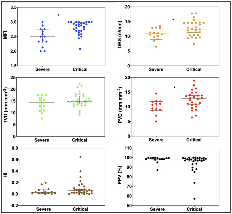
Fig 1. Comparisons of microcirculatory flow index (MFI), total vessel density (TVD), perfused vessel density (PVD), proportion of perfused vessel (PPV), De Backer score (DBS) and heterogeneity index (HI) in severe group and critical group.

Fig 1

*: P value <0.05 between groups comparisons using Mann-Whitney U test.

In the critical group, MFI, PVD and DBS were significantly higher in comparison to the severe group (respectively, for critical and severe groups, MFI: 2.8 ± 0.2 vs 2.5 ± 0.3, p = 0.001, D = 1.16; PVD: 12.5 ± 3.0 vs 10.1 ± 2.4 mm.mm-2, p = 0.02, D = 0.14 and DBS: 12.7 ± 2.6 vs 10.8 ± 2.0 vessels.mm-2, p = 0.033, D = 0.08).

PPV (p = 0.09, D = 0.05), TVD (p = 0.224, D = 0.55) and HI (p = .189, D = 1.15) were similar for both groups.

Correlation between PaO2, PaCO2 and microcirculation (Fig 2)

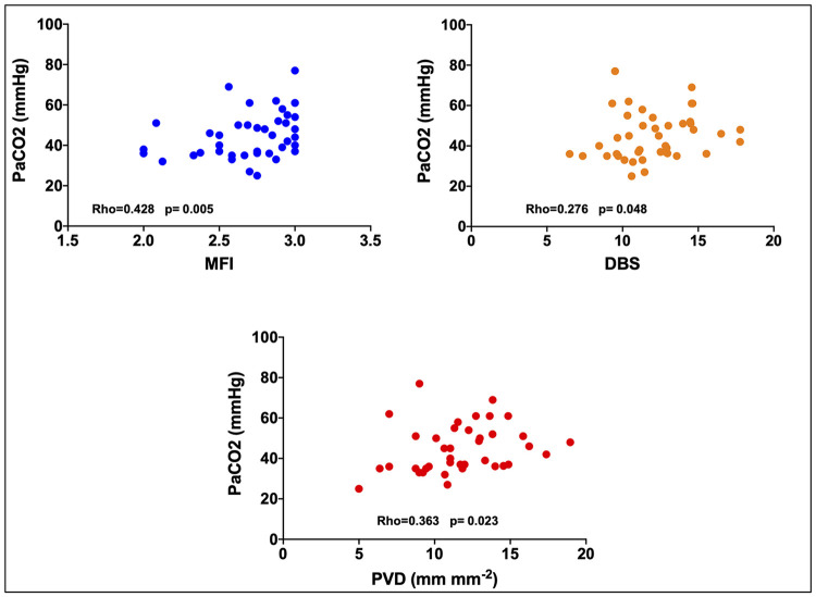
Fig 2. PaCO2 correlation with the microcirculatory flow index (MFI), the perfused vessel density (PVD) and the De Backer score (DBS).

Fig 2

PaCO2 appeared significantly correlated to MFI (Rho = 0.428; p = 0.005), PVD (Rho = 0.363; p = 0.023) and DBS (Rho = 0.276; p = 0.048) but not with PPV (Rho = 0.010; p = 0.948) TVD (Rho = 0.194; p = 0.224) and HI (Rho = -0.045; p = 0.777).

No significant correlations were found between PaO2 and MFI (Rho = 0.21; p = 0.893), PVD (Rho = -0.76; p = 0.644), DBS (Rho = -0.99; p = 0.534), PPV (Rho = -0.263; p = 0.092), TVD (Rho = -0.91; p = 0.573) and HI (Rho = 0.198; p = 0.209).

Discussion

Our findings suggest that the critical group had higher red blood cell velocity (higher MFI values) and a better vessel density (higher DBS and PVD). Capillary diffuse properties seem to be preserved given the values of PPV, TVD and HI.

Theses phenomenon do not appear related to PaO2 which is similar in both groups. PaCO2 levels seem to be correlated with microcirculation parameters.

We could have expected more altered microcirculatory parameters in the critical group than in the severe group with lower red blood cell velocities and a lower vessel density regarding the higher pro inflammatory state (higher C reactive protein and procalcitonin levels) and the higher severity scores (SAPS II and SOFA). Indeed, we learned from severe sepsis and septic shock that microcirculation in critically ill patients is usually impaired with an increase in heterogeneity, a decrease in vessel density and perfusions indices [15, 16]. It has been shown that vessel density and DBS are correlated with mortality in a large cohort of sepsis [17]. However, our critical group did not present the typical characteristics of septic shock (hyperkinetic cardiac state, high lactate level and high dose of norepinephrine. In our series, COVID-19 patients presented an isolated respiratory failure, sometimes associated with an acute kidney injury but with limited need for vasopressors and no clinical signs of hypo perfusion. These data are in accordance with previous reports on critically ill COVID-19 patients showing a low prevalence of septic shock or multi organ failure [18].

In a previous microcirculatory study on 9 COVID-19 patients under ECMO therapy, Carsetti et al. found that TVD and PVD were inversely correlated with D-dimer [19]. The same team found similar results on 12 COVID-19 patients under mechanical ventilation [20]. Values of microcirculatory parameters are similar to ours. However, we did not found any difference in D-dimer levels between our 2 groups.

Our results differ from other studies on severe virus’s infections. In a report on mechanically ventilated patients with Influenza infection, Salgado et al found a compromised microcirculation with a median MFI of 1.9 [21].

Hence, to explain our results, we focused on differences between respiratory parameters. Finding that MFI, PVD and DBS, the three most significant parameters, were correlated to PaCO2 levels (and hence to the type of ventilation) we hypothesized that hypocapnia might influence systemic microcirculation. In an experimental study on rabbits, Komori et al. have showed that systemic microcirculatory flow decreases with hypocapnia and is restored with the increase of PaCO2 (up to a certain threshold). Same findings are reported in a dog model for which hypercapnia improves vascular capacitance [22]. Moreover, hypocapnia is known to increase the hemoglobin affinity for oxygen, moving the hemoglobin-oxygen dissociation curve to the left and thus decreasing oxygen supply to organs.

In our series, there is no differences between groups concerning PaO2 and we did not find any correlation between microcirculatory parameters and PaO2. Several studies have shown that hypoxia enhances microcirculatory parameters trough a physiological adaptation to match oxygen delivery to demand [23]. So, microvascular blood flow increases to adapt to hypoxemic state.

Clinical studies on the effect of mechanical ventilation on microcirculation are scarce. In a recent physiologic paper, Ospina-Tascon et al. showed that sublingual microcirculation heterogeneity may decrease by reducing dead space ventilation in patient with ARDS [24]. The mechanism remains unclear but in a context of sepsis, heterogeneity in perfusion could alter both lung and systemic perfusions which seem intricately linked [25]. In our study, we did not find any difference between groups in heterogeneity index. Our hypothesis concerning the effect of PaCO2 on microcirculation needs further confirmation in clinical studies as our correlations (given Spearman Rho values) were moderate or weak and cofounders may not be excluded.

Another explanation is the higher dose of norepinephrine in the critical group with a potential capillary recruitment (Table 2). However, the effect of norepinephrine on microcirculation is still unclear. Jhanji et a. showed an increase in oxygen delivery with norepinephrine in patients with septic shock without improving MFI [26]. In a prospective randomized trial, Dubin et al. concluded to the lack of effect of norepinephrine on microcirculation [27]. However, once again, our pattern of microcirculation differed from septic shock and an absence of norepinephrine effect cannot be excluded even if parameters as cardiac index and mean arterial pressure were similar between groups. Moreover, the higher DBS, MFI and PVD values in most severe patients could reflect a state of loss in vascular tone with a more apparent hyperemic state. Pulmonary imaging and postmortem studies of COVID-19 patients have highlighted the hypothesis of an increased pulmonary blood flow with intrapulmonary vascular shunting responsible for hypoxemia [5, 7, 8]. Our data may reflect this hyperemic state.

Our study has several limitations. First, we recorded only one time-point measurement and, to confirm our results, different microcirculatory assessments, especially before and after tracheal intubation, would have been of great interest. A new tool called MicroTools software allowing an automated video analysis have been recently validated in a large international retrospective database [28]. This kind of automated analysis allowing resuscitation therapies targeted on microcirculatory parameters should be applied in further investigations. Second, we did not perform a sample size calculation. However, we applied the Cohen D test to assess the size effect of each parameter. Except for MFI, all parameters had a D value under 0.2 suggesting the absence of size effect making p value interpretation more reliable. Last, as patients in the sever group were spontaneously breathing, difficulty in sublingual assessment in awake patients may have influenced the results. However, the Massey score for image quality was comparable in the 2 groups. Finally, we did not evaluate central venous oxygen saturation which could be valuable to compare balance between oxygen consumption and oxygen delivery or you can say that we did not record central venous oxygen derived variables to compare oxygen consumption.

Conclusion

In this limited series of critically ill COVID-19 patients, microcirculation did not show heterogeneity as observed in septic shock. Moreover, patients under mechanical ventilation with higher severity scores but with higher PaCO2 levels, had higher red blood cell velocities as assessed by MFI. Further controlled clinical investigations are required to assess the effect of hypocapnia on microcirculation in this setting.

Acknowledgments

We thank Amiens university hospital staff for their support in the study process and their commitment in data collection during the pandemic.

Data Availability

All relevant data are within the paper. Raw data are available after notification and authorization of the competent authorities. In France, all computer data (including databases, in Cover Letter particular patient data) are protected by the National Commission on Informatics and Liberty (CNIL), the national data protection authority for France. CNIL is an independent French administrative regulatory body whose mission is to ensure that data privacy law is applied to the collection, storage, and use of personal data. As the database of this study was authorized by the CNIL, we cannot make available data without prior agreement of the CNIL. Requests may be sent to: picard.carl@chu-amiens.fr.

Funding Statement

The study was not funded.

References

  • 1.Richardson S, Hirsch JS, Narasimhan M, Crawford JM, McGinn T, Davidson KW, et al. Presenting Characteristics, Comorbidities, and Outcomes Among 5700 Patients Hospitalized With COVID-19 in the New York City Area. JAMA. 2020. 10.1001/jama.2020.6775 [DOI] [PMC free article] [PubMed] [Google Scholar]
  • 2.Alhazzani W, Møller MH, Arabi YM, Loeb M, Gong MN, Fan E, et al. Surviving Sepsis Campaign: Guidelines on the Management of Critically Ill Adults with Coronavirus Disease 2019 (COVID-19). Critical Care Medicine. 2020; 1 10.1097/CCM.0000000000004363 [DOI] [PMC free article] [PubMed] [Google Scholar]
  • 3.Gattinoni L, Chiumello D, Caironi P, Busana M, Romitti F, Brazzi L, et al. COVID-19 pneumonia: different respiratory treatments for different phenotypes? Intensive Care Medicine. 2020. [cited 20 Apr 2020]. 10.1007/s00134-020-06033-2 [DOI] [PMC free article] [PubMed] [Google Scholar]
  • 4.Ottestad W, Søvik S. COVID-19 patients with respiratory failure: what can we learn from aviation medicine? British Journal of Anaesthesia. 2020. [cited 20 Apr 2020]. 10.1016/j.bja.2020.04.012 [DOI] [PMC free article] [PubMed] [Google Scholar]
  • 5.Mahjoub Y, Rodenstein DO, Jounieaux V. Severe Covid-19 disease: rather AVDS than ARDS? Critical Care. 2020;24 10.1186/s13054-020-02972-w [DOI] [PMC free article] [PubMed] [Google Scholar]
  • 6.Arulkumaran N, Brealey D, Howell D, Singer M. Use of non-invasive ventilation for patients with COVID-19: a cause for concern? The Lancet Respiratory Medicine. 2020. [cited 25 Apr 2020]. 10.1016/S2213-2600(20)30181-8 [DOI] [PMC free article] [PubMed] [Google Scholar]
  • 7.Lang M, Som A, Mendoza DP, Flores EJ, Reid N, Carey D, et al. Hypoxaemia related to COVID-19: vascular and perfusion abnormalities on dual-energy CT. The Lancet Infectious Diseases. 2020. [cited 5 May 2020]. 10.1016/S1473-3099(20)30367-4 [DOI] [PMC free article] [PubMed] [Google Scholar]
  • 8.Ackermann M, Verleden SE, Kuehnel M, Haverich A, Welte T, Laenger F, et al. Pulmonary Vascular Endothelialitis, Thrombosis, and Angiogenesis in Covid-19. New England Journal of Medicine. 2020. [cited 15 Jun 2020]. 10.1056/NEJMoa2015432 [DOI] [PMC free article] [PubMed] [Google Scholar]
  • 9.Wu Z, McGoogan JM. Characteristics of and Important Lessons From the Coronavirus Disease 2019 (COVID-19) Outbreak in China: Summary of a Report of 72 314 Cases From the Chinese Center for Disease Control and Prevention. JAMA. 2020; 323: 1239: 10.1001/jama.2020.2648 [DOI] [PubMed] [Google Scholar]
  • 10.Toulouse E, Masseguin C, Lafont B, McGurk G, Harbonn A, Roberts JA, et al. French legal approach to clinical research. Anaesth Crit Care Pain Med. 2018;37: 607–614. 10.1016/j.accpm.2018.10.013 [DOI] [PubMed] [Google Scholar]
  • 11.Toulouse E, Lafont B, Granier S, Mcgurk G, Bazin J-E. French legal approach to patient consent in clinical research. Anaesthesia Critical Care & Pain Medicine. 2020;39: 883–885. 10.1016/j.accpm.2020.10.012 [DOI] [PubMed] [Google Scholar]
  • 12.Ince C, Boerma EC, Cecconi M, De Backer D, Shapiro NI, Duranteau J, et al. Second consensus on the assessment of sublingual microcirculation in critically ill patients: results from a task force of the European Society of Intensive Care Medicine. Intensive Care Med. 2018;44: 281–299. 10.1007/s00134-018-5070-7 [DOI] [PubMed] [Google Scholar]
  • 13.Massey MJ, Larochelle E, Najarro G, Karmacharla A, Arnold R, Trzeciak S, et al. The microcirculation image quality score: development and preliminary evaluation of a proposed approach to grading quality of image acquisition for bedside videomicroscopy. J Crit Care. 2013;28: 913–917. 10.1016/j.jcrc.2013.06.015 [DOI] [PubMed] [Google Scholar]
  • 14.Lakens D. Calculating and reporting effect sizes to facilitate cumulative science: a practical primer for t-tests and ANOVAs. Front Psychol. 2013; 4: 863: 10.3389/fpsyg.2013.00863 [DOI] [PMC free article] [PubMed] [Google Scholar]
  • 15.Farquhar I, Martin CM, Lam C, Potter R, Ellis CG, Sibbald WJ. Decreased capillary density in vivo in bowel mucosa of rats with normotensive sepsis. J Surg Res. 1996; 61: 190–196. 10.1006/jsre.1996.0103 [DOI] [PubMed] [Google Scholar]
  • 16.De Backer D, Creteur J, Preiser J-C, Dubois M-J, Vincent J-L. Microvascular blood flow is altered in patients with sepsis. Am J Respir Crit Care Med. 2002;166: 98–104. 10.1164/rccm.200109-016oc [DOI] [PubMed] [Google Scholar]
  • 17.for the ProCESS investigators, Massey MJ, Hou PC, Filbin M, Wang H, Ngo L, et al. Microcirculatory perfusion disturbances in septic shock: results from the ProCESS trial. Critical Care. 2018;22 10.1186/s13054-018-2240-5 [DOI] [PMC free article] [PubMed] [Google Scholar]
  • 18.Wang D, Hu B, Hu C, Zhu F, Liu X, Zhang J, et al. Clinical Characteristics of 138 Hospitalized Patients With 2019 Novel Coronavirus–Infected Pneumonia in Wuhan, China. JAMA. 2020; 323: 1061 10.1001/jama.2020.1585 [DOI] [PMC free article] [PubMed] [Google Scholar]
  • 19.Carsetti A, Damiani E, Casarotta E, Scorcella C, Domizi R, Montomoli J, et al. Sublingual microcirculation in patients with SARS-CoV-2 undergoing veno-venous extracorporeal membrane oxygenation. Microvascular Research. 2020;132: 104064 10.1016/j.mvr.2020.104064 [DOI] [PMC free article] [PubMed] [Google Scholar]
  • 20.Damiani E, Carsetti A, Casarotta E, Scorcella C, Domizi R, Adrario E, et al. Microvascular alterations in patients with SARS-COV-2 severe pneumonia. Annals of Intensive Care. 2020;10 10.1186/s13613-020-00680-w [DOI] [PMC free article] [PubMed] [Google Scholar]
  • 21.Salgado DR, Ortiz JA, Favory R, Creteur J, Vincent J-L, De Backer D. Microcirculatory abnormalities in patients with severe influenza A (H1N1) infection. Can J Anaesth. 2010;57: 940–946. 10.1007/s12630-010-9365-6 [DOI] [PMC free article] [PubMed] [Google Scholar]
  • 22.Rothe CF, Maass-Moreno R, Flanagan AD. Effects of hypercapnia and hypoxia on the cardiovascular system: vascular capacitance and aortic chemoreceptors. Am J Physiol. 1990;259: H932–939. 10.1152/ajpheart.1990.259.3.H932 [DOI] [PubMed] [Google Scholar]
  • 23.Casey DP, Joyner MJ. Compensatory vasodilatation during hypoxic exercise: mechanisms responsible for matching oxygen supply to demand: Hypoxic dependent vasodilatation during exercise. The Journal of Physiology. 2012;590: 6321–6326. 10.1113/jphysiol.2012.242396 [DOI] [PMC free article] [PubMed] [Google Scholar]
  • 24.Ospina-Tascón GA, Bautista DF, Madriñán HJ, Valencia JD, Bermúdez WF, Quiñones E, et al. Microcirculatory dysfunction and dead-space ventilation in early ARDS: a hypothesis-generating observational study. Annals of Intensive Care. 2020; 10.1186/s13613-020-00651-1 [DOI] [PMC free article] [PubMed] [Google Scholar]
  • 25.De Backer D, Ospina-Tascon G, Salgado D, Favory R, Creteur J, Vincent J-L. Monitoring the microcirculation in the critically ill patient: current methods and future approaches. Intensive Care Medicine. 2010;36: 1813–1825. 10.1007/s00134-010-2005-3 [DOI] [PubMed] [Google Scholar]
  • 26.Jhanji S, Stirling S, Patel N, Hinds CJ, Pearse RM. The effect of increasing doses of norepinephrine on tissue oxygenation and microvascular flow in patients with septic shock. Crit Care Med. 2009;37: 1961–1966. 10.1097/CCM.0b013e3181a00a1c [DOI] [PubMed] [Google Scholar]
  • 27.Dubin A, Pozo MO, Casabella CA, Pálizas F, Murias G, Moseinco MC, et al. Increasing arterial blood pressure with norepinephrine does not improve microcirculatory blood flow: a prospective study. Crit Care. 2009;13: R92 10.1186/cc7922 [DOI] [PMC free article] [PubMed] [Google Scholar]
  • 28.Hilty MP, Akin S, Boerma C, Donati A, Erdem Ö, Giaccaglia P, et al. Automated Algorithm Analysis of Sublingual Microcirculation in an International Multicentral Database Identifies Alterations Associated With Disease and Mechanism of Resuscitation. Crit Care Med. 2020;48: e864–e875. 10.1097/CCM.0000000000004491 [DOI] [PubMed] [Google Scholar]

Decision Letter 0

Yu-Chang Yeh

24 Nov 2020

PONE-D-20-32823

Systemic microcirculatory patterns of critically ill Covid-19 patients: a prospective study

PLOS ONE

Dear Dr. abou arab,

Thank you for submitting your manuscript to PLOS ONE. After careful consideration, we feel that it has merit but does not fully meet PLOS ONE’s publication criteria as it currently stands. Therefore, we invite you to submit a revised version of the manuscript that addresses the points raised during the review process.

Please submit your revised manuscript by Jan 08 2021 11:59PM. If you will need more time than this to complete your revisions, please reply to this message or contact the journal office at plosone@plos.org. When you're ready to submit your revision, log on to https://www.editorialmanager.com/pone/ and select the 'Submissions Needing Revision' folder to locate your manuscript file.

Please include the following items when submitting your revised manuscript:

  • A rebuttal letter that responds to each point raised by the academic editor and reviewer(s). You should upload this letter as a separate file labeled 'Response to Reviewers'.

  • A marked-up copy of your manuscript that highlights changes made to the original version. You should upload this as a separate file labeled 'Revised Manuscript with Track Changes'.

  • An unmarked version of your revised paper without tracked changes. You should upload this as a separate file labeled 'Manuscript'.

If you would like to make changes to your financial disclosure, please include your updated statement in your cover letter. Guidelines for resubmitting your figure files are available below the reviewer comments at the end of this letter.

If applicable, we recommend that you deposit your laboratory protocols in protocols.io to enhance the reproducibility of your results. Protocols.io assigns your protocol its own identifier (DOI) so that it can be cited independently in the future. For instructions see: http://journals.plos.org/plosone/s/submission-guidelines#loc-laboratory-protocols

We look forward to receiving your revised manuscript.

Kind regards,

Yu-Chang Yeh, M.D., Ph.D.

Academic Editor

PLOS ONE

Additional Editor Comments:

Please refer to the reviewers' comments and revise your manuscript as appropriate. Keep safe and all the best.

Journal Requirements:

When submitting your revision, we need you to address these additional requirements.

1. Please ensure that your manuscript meets PLOS ONE's style requirements, including those for file naming. The PLOS ONE style templates can be found at

https://journals.plos.org/plosone/s/file?id=wjVg/PLOSOne_formatting_sample_main_body.pdf and

https://journals.plos.org/plosone/s/file?id=ba62/PLOSOne_formatting_sample_title_authors_affiliations.pdf

2.Thank you for including your ethics statement:

"According to French law on clinical research [11], the study was declared and registered at the Commission Numérique Informatiques et Libertés (registration identifier:

I2020_843_0026) and was registered on ClinicalTrial.gov on 21st of April 2020 (identifier: NCT04354558). This study was conducted in accordance with the amended Declaration of Helsinski."   

a. Please amend your current ethics statement to include the full name of the ethics committee/institutional review board(s) that approved your specific study.

b. Once you have amended this/these statement(s) in the Methods section of the manuscript, please add the same text to the “Ethics Statement” field of the submission form (via “Edit Submission”).

For additional information about PLOS ONE ethical requirements for human subjects research, please refer to http://journals.plos.org/plosone/s/submission-guidelines#loc-human-subjects-research.

3. In the Methods, please state:

- Why written consent could not be obtained

- Whether the Institutional Review Board (IRB) approved use of oral consent

- How oral consent was documented

For more information, please see our guidelines for human subjects research: https://journals.plos.org/plosone/s/submission-guidelines#loc-human-subjects-research

4. Please provide a sample size and power calculation in the Methods, or discuss the reasons for not performing one before study initiation.

5. Thank you for stating the following financial disclosure:

"NO- The funders had no role in study design, data collection and analysis, decision to publish, or preparation of the manuscript."

At this time, please address the following queries:

  1. Please clarify the sources of funding (financial or material support) for your study. List the grants or organizations that supported your study, including funding received from your institution.

  2. State what role the funders took in the study. If the funders had no role in your study, please state: “The funders had no role in study design, data collection and analysis, decision to publish, or preparation of the manuscript.”

  3. If any authors received a salary from any of your funders, please state which authors and which funders.

  4. If you did not receive any funding for this study, please state: “The authors received no specific funding for this work.”

Please include your amended statements within your cover letter; we will change the online submission form on your behalf.

6. We note that you have indicated that data from this study are available upon request. PLOS only allows data to be available upon request if there are legal or ethical restrictions on sharing data publicly. For information on unacceptable data access restrictions, please see http://journals.plos.org/plosone/s/data-availability#loc-unacceptable-data-access-restrictions.

In your revised cover letter, please address the following prompts:

a) If there are ethical or legal restrictions on sharing a de-identified data set, please explain them in detail (e.g., data contain potentially identifying or sensitive patient information) and who has imposed them (e.g., an ethics committee). Please also provide contact information for a data access committee, ethics committee, or other institutional body to which data requests may be sent.

b) If there are no restrictions, please upload the minimal anonymized data set necessary to replicate your study findings as either Supporting Information files or to a stable, public repository and provide us with the relevant URLs, DOIs, or accession numbers. Please see http://www.bmj.com/content/340/bmj.c181.long for guidelines on how to de-identify and prepare clinical data for publication. For a list of acceptable repositories, please see http://journals.plos.org/plosone/s/data-availability#loc-recommended-repositories.

We will update your Data Availability statement on your behalf to reflect the information you provide.

7. Please ensure that you refer to Figure 1 and 2 in your text as, if accepted, production will need this reference to link the reader to the figure.

[Note: HTML markup is below. Please do not edit.]

Reviewers' comments:

Reviewer's Responses to Questions

Comments to the Author

1. Is the manuscript technically sound, and do the data support the conclusions?

The manuscript must describe a technically sound piece of scientific research with data that supports the conclusions. Experiments must have been conducted rigorously, with appropriate controls, replication, and sample sizes. The conclusions must be drawn appropriately based on the data presented.

Reviewer #1: Yes

Reviewer #2: Yes

**********

2. Has the statistical analysis been performed appropriately and rigorously?

Reviewer #1: Yes

Reviewer #2: Yes

**********

3. Have the authors made all data underlying the findings in their manuscript fully available?

The PLOS Data policy requires authors to make all data underlying the findings described in their manuscript fully available without restriction, with rare exception (please refer to the Data Availability Statement in the manuscript PDF file). The data should be provided as part of the manuscript or its supporting information, or deposited to a public repository. For example, in addition to summary statistics, the data points behind means, medians and variance measures should be available. If there are restrictions on publicly sharing data—e.g. participant privacy or use of data from a third party—those must be specified.

Reviewer #1: Yes

Reviewer #2: Yes

**********

4. Is the manuscript presented in an intelligible fashion and written in standard English?

PLOS ONE does not copyedit accepted manuscripts, so the language in submitted articles must be clear, correct, and unambiguous. Any typographical or grammatical errors should be corrected at revision, so please note any specific errors here.

Reviewer #1: Yes

Reviewer #2: Yes

**********

5. Review Comments to the Author

Please use the space provided to explain your answers to the questions above. You may also include additional comments for the author, including concerns about dual publication, research ethics, or publication ethics. (Please upload your review as an attachment if it exceeds 20,000 characters)

Reviewer #1: The authors showed interesting microcirculatory patterns in COVID-19 patients. I have some comments and questions :

1) The authors mentioned that sublingual microcirculation was assessed within 48 hours after ICU admission. What about the median inclusion time ? Did sublingual microcirculatory assessment was faster in critical group than severe group? (p.7)

2) For data collection for systemic hemodynamic variables, Was cardiac output measured by echocardiography in all patients ? if yes , was echocardiography performed by 1 persons due to operator dependent? For capillary refill time, could you mention how did you performed capillary refill time by eye ball or using chronometer ?

3) For demographic data, were SOFA and SAPS II scores calculated at ICU admission or time of inclusion ? It would be better if you mention severity scores at inclusion time. Was PaO2/FiO2 ratio in severe group different from patients in critical group. The authors mentions number of patients who required vasopressor, in these patients, were they shock ? Could you possibly mention numbers of patients with shock and without shock and did microcirculatory profiles differ between patients with and without shock.

4) Was the microcirculatory profiles related to lung recovery in critical group ?

5) Did microcirculatory alter in patients with COVID19 compare with healthy volunteers?

Reviewer #2: I would like to congratulate the authors for their courage and enthusiasm in performing such time consuming and labor intensive study protocol in the ICU.

I have just minor concerns:

1. The title could be more focussed beginning by deleting the word "systemic". Please rewrite the title specifically by adding more microcirculatory items like capillary, microvascular flow, alterations etc...

2. Please write COVID-19 with capital letters everywhere.

3. Abstract: Conclusion should be specified by replacing "better microcirculatory pattern" to more focussed description. What is better???? What is still wrong in the microcirculation???

4. Introduction: "Microcirculation pattern of Covid-19 might differ from other sepsis causes" could implicate that COVID-19 is (pre)sepsis. Please rewrite this sentence that doesn't confuse sepsis with this new disease.

5. Methods: MFI=microvascular flow index

6. Results: Please refer to figure 1 and 2 for the responsible data.

7. Discussion: First sentence is very superficial. Higher microcirculation parameters does mean nothing. Please redifine this e.g. hyperdynamic at least?? High density vasculary flows? high red blood cell velocity?

8. From limitations you could delete the third limitation hence there are thousands of studies which support the idea of sublingual microcirculation as the best paramter for follow-up of the end-organ perfusion ("systemic

(micro)circulation").

9. Finaly add in your discussion the more advanced novel automated microcirculation analysis which could be used for more detailed and accurate microcirculatory analysis published by Hilty et al. Crit Care Med 2020. Novel studies with novel and efficient microcirculation analysis programs could make the clinicians ICU life scientifically comfortable.

Tables: Please add more clinical data into the tables

- CT lungs for detecting pulmonary embolism; how many performed how many positive in each group?

- Day of illness?

- Day of intubation?

- Day of optiflow during the microcirculation measurement?

- What was the anticoagulation strategy? Profylaxes, therapeutic? which agent? UFH? LMWH? What was the follow-up/monitoring strategy for coagulation?

**********

6. PLOS authors have the option to publish the peer review history of their article (what does this mean?). If published, this will include your full peer review and any attached files.

If you choose “no”, your identity will remain anonymous but your review may still be made public.

Do you want your identity to be public for this peer review? For information about this choice, including consent withdrawal, please see our Privacy Policy.

Reviewer #1: No

Reviewer #2: No

[NOTE: If reviewer comments were submitted as an attachment file, they will be attached to this email and accessible via the submission site. Please log into your account, locate the manuscript record, and check for the action link "View Attachments". If this link does not appear, there are no attachment files.]

While revising your submission, please upload your figure files to the Preflight Analysis and Conversion Engine (PACE) digital diagnostic tool, https://pacev2.apexcovantage.com/. PACE helps ensure that figures meet PLOS requirements. To use PACE, you must first register as a user. Registration is free. Then, login and navigate to the UPLOAD tab, where you will find detailed instructions on how to use the tool. If you encounter any issues or have any questions when using PACE, please email PLOS at figures@plos.org. Please note that Supporting Information files do not need this step.

PLoS One. 2021 Feb 8;16(2):e0246636. doi: 10.1371/journal.pone.0246636.r002

Author response to Decision Letter 0


21 Dec 2020

Reviewer #1: The authors showed interesting microcirculatory patterns in COVID-19 patients. I have some comments and questions:

1) The authors mentioned that sublingual microcirculation was assessed within 48 hours after ICU admission. What about the median inclusion time?

Answer: We thank the reviewer for his comment. We checked for ICU exact admission time on our electronic health record software and match it with the Cytocam software record for each patient.

The median time inclusion was similar between group, around 20 hours. We added it in table 2 as follows:

Time from ICU admission to inclusion; hours 20 [5-32] 21 [6-30] 0.682

Comment: Did sublingual microcirculatory assessment was faster in critical group than severe group? (p.7)

Answer: Regarding the assessment, we proceeded with the same manner whatever the patient severity. We took the necessary time to record 5 videos of good quality as required by the semi-automatic software analysis. In cases of bad quality of videos, we tried as many attempts as necessary to obtain 5 videos of good quality. We did not specifically record the assessment duration. However, it seems to the operator that the duration of the assessment was faster in sedated patients compared to awake patients.

2) Comment For data collection for systemic hemodynamic variables, Was cardiac output measured by echocardiography in all patients ? if yes , was echocardiography performed by 1 persons due to operator dependent? For capillary refill time, could you mention how did you performed capillary refill time by eye ball or using chronometer ?

Answer: We thank the reviewer for his comment.

Cardiac output was measured by Doppler echocardiography. All measurements for all patients were performed by the same operator (CB). We added a sentence in the method section: “Cardiac output was measured by Doppler echocardiography by the same operator(CB).”

Regarding capillary refill time, measurements were standardized using a chronometer. We added details on the standardization of measurement as follows:

“Capillary refill time was performed as follows: a firm pressure was applied to the surface of the index finger distal phalanx. The pressure was increased until the skin became blank and maintained for 10s. The pressure was then released. The time for return to pre-existent skin color was measured using a chronometer [14].”

3) For demographic data, were SOFA and SAPS II scores calculated at ICU admission or time of inclusion ? It would be better if you mention severity scores at inclusion time.

Answer: We agree with reviewer. SAPS II and SOFA were calculated at inclusion time (time of microcirculation assessment) as mentioned in data collection section. We added “at inclusion” in the results section and in Table 1.

Comment: Was PaO2/FiO2 ratio in severe group different from patients in critical group.

Answer: We thank the reviewer for his relevant comment. However, in the severe group patients had a facial mask or a nasal canula which makes the exact estimation of FiO2 difficult. That is why we apologize for not being able to report this value for the severe group.

Comment: The authors mentions number of patients who required vasopressor, in these patients, were they shock? Could you possibly mention numbers of patients with shock and without shock and did microcirculatory profiles differ between patients with and without shock.

Answer: We thank the reviewer for his relevant comment. No patient had a shock state at inclusion. As reported in table 2, the dose of norepinephrine was quite low (0.02 μg.kg.min-1). Macro hemodynamics parameters were similar regarding cardiac output and heart rate with no septic shock pattern. Vasopressors use was probably related to sedation drugs (propofol) used to ensure mechanical ventilation.

4) Was the microcirculatory profiles related to lung recovery in critical group?

Answer: We thank the reviewer for his relevant comment. Unfortunately, only one time point measurement was performed. We agree that several a time point monitoring of microcirculation during the stay would had been very interesting. However, microcirculatory record was performed shortly after initiation of mechanical ventilation( i.e: worsening of lung function).

5) Did microcirculatory alter in patients with COVID19 compare with healthy volunteers?

Answer: We thank the reviewer for his relevant comment.

The guidelines on sublingual assessment (doi: 10.1007/s00134-018-5070-7) described the convective property according to the red blood cell velocities (explored by the MFI and the PVD), the diffuse capacity (explored by the TVD) and the heterogeneity in perfusion (HI)

No reference values were admitted but healthy subjects values are available from previous studies (doi: 10.1097/CCM.0b013e31823dae59).

In a healthy subject, a normal MFI is expected to be over 2.6 which was the case for our critical group but not in the severe group.

PVD in healthy subject was reported to be about 15 mm/mm2. PVD was less decreased in the severe group.

To summarize, the critical group tend to have higher red blood velocities when comparing the severe group (PVD and MFI higher)

In a healthy subject, TVD is around 15 mm/mm2 and HI around 0.03. In our population, these parameters were close to healthy subjects’ values. Hence, in our cohort COVID-19 patients did not seem to have convective impairment.

Reviewer #2: I would like to congratulate the authors for their courage and enthusiasm in performing such time consuming and labor intensive study protocol in the ICU.

Answer: we are very grateful to the reviewer for his consideration and encouragements.

I have just minor concerns:

1. The title could be more focussed beginning by deleting the word "systemic". Please rewrite the title specifically by adding more microcirculatory items like capillary, microvascular flow, alterations etc...

Answer: We thank the reviewer for his helpful comment. We changed the title as follows: “Microvascular flow alterations in critically ill COVID-19 patients: a prospective study.”

2. Please write COVID-19 with capital letters everywhere.

Answer: We made changes in the manuscript.

3. Abstract: Conclusion should be specified by replacing "better microcirculatory pattern" to more focussed description. What is better???? What is still wrong in the microcirculation???

Answer: We thank the reviewer for his comment and made changes to better describe the profiles. We made changes as follows:

Critical COVID-19 patients under mechanical ventilation seem to have higher red blood cell velocity than severe non-ventilated patients.

4. Introduction: "Microcirculation pattern of Covid-19 might differ from other sepsis causes" could implicate that COVID-19 is (pre)sepsis. Please rewrite this sentence that doesn't confuse sepsis with this new disease.

“Timing for intubation and invasive ventilation in COVID-19 patients is a subject of debate [6]. In this new disease, microcirculatory impairment might explain disease progression and differences between invasively and non-invasively ventilated patients. [3]

5. Methods: MFI=microvascular flow index

Answer: We apologize for this mistake and made changes

6. Results: Please refer to figure 1 and 2 for the responsible data.

Answer: We made changes and referred to figure 1 and 2 when required in the result section.

7. Discussion: First sentence is very superficial. Higher microcirculation parameters does mean nothing. Please redifine this e.g. hyperdynamic at least?? High density vasculary flows? high red blood cell velocity?

Answer: We thank the reviewer for his relevant comments and hope we better describe the profiles.

Here we made the following changes.

“Our findings suggest that the critical group had higher red blood cell velocity (higher MFI values) and a better vessel density (higher DBS and PVD). Capillary diffuse properties seem to be preserved given the values of PPV, TVD and HI.”

8. From limitations you could delete the third limitation hence there are thousands of studies which support the idea of sublingual microcirculation as the best paramter for follow-up of the end-organ perfusion ("systemic (micro)circulation").

Answer: We made changes and removed the sentence.

9. Finaly add in your discussion the more advanced novel automated microcirculation analysis which could be used for more detailed and accurate microcirculatory analysis published by Hilty et al. Crit Care Med 2020. Novel studies with novel and efficient microcirculation analysis programs could make the clinicians ICU life scientifically comfortable.

Answer: We thank the reviewer for his comment and added a sentence explaining the MicroTools.

“A new tool called MicroTools software allowing an automated video analysis have been recently validated in a large international retrospective database [29]. This kind of automated analysis allowing resuscitation therapies targeted on microcirculatory parameters should be applied in further investigations.”

Comments

Tables: Please add more clinical data into the tables

- CT lungs for detecting pulmonary embolism; how many performed how many positive in each group?

- Day of illness?

- Day of intubation?

- Day of optiflow during the microcirculation measurement?

Answer: We thank the reviewer for his comment and completed Table 1 and Table 2 as follows.

- What was the anticoagulation strategy? Profylaxes, therapeutic? which agent? UFH? LMWH? What was the follow-up/monitoring strategy for coagulation?

Answer: during the first wave of COVID-19 pandemic, our local institutional protocol included for all COVID-19 patients admitted to ICU, a therapeutic anticoagulation strategy with unfractioned heparin to achieve anti Xa activity between 0.5 and 0.8 UI/ml.

UFH was started at ICU admission. Anti Xa activity was checked daily and 4 hours after each dose change. We added a sentence in the method section “All COVID-19 patients were under unfractionned heparin to achieve anti Xa activity between 0.5 and 0.8 UI/ml”

Attachment

Submitted filename: reveiwer answer.docx

Decision Letter 1

Yu-Chang Yeh

15 Jan 2021

PONE-D-20-32823R1

Microvascular flow alterations  in critically ill COVID-19 patients: a prospective study

PLOS ONE

Dear Dr. abou arab,

Thank you for submitting your manuscript to PLOS ONE. Therefore, we invite you to submit a revised version of the manuscript that addresses the points raised during the review process.

Please submit your revised manuscript by Mar 01 2021 11:59PM. If you will need more time than this to complete your revisions, please reply to this message or contact the journal office at plosone@plos.org. When you're ready to submit your revision, log on to https://www.editorialmanager.com/pone/ and select the 'Submissions Needing Revision' folder to locate your manuscript file.

Please include the following items when submitting your revised manuscript:

  • A rebuttal letter that responds to each point raised by the academic editor and reviewer(s). You should upload this letter as a separate file labeled 'Response to Reviewers'.

  • A marked-up copy of your manuscript that highlights changes made to the original version. You should upload this as a separate file labeled 'Revised Manuscript with Track Changes'.

  • An unmarked version of your revised paper without tracked changes. You should upload this as a separate file labeled 'Manuscript'.

If you would like to make changes to your financial disclosure, please include your updated statement in your cover letter. Guidelines for resubmitting your figure files are available below the reviewer comments at the end of this letter.

If applicable, we recommend that you deposit your laboratory protocols in protocols.io to enhance the reproducibility of your results. Protocols.io assigns your protocol its own identifier (DOI) so that it can be cited independently in the future. For instructions see: http://journals.plos.org/plosone/s/submission-guidelines#loc-laboratory-protocols

We look forward to receiving your revised manuscript.

Kind regards,

Yu-Chang Yeh, M.D., Ph.D.

Academic Editor

PLOS ONE

Additional Editor Comments:

Thank you for carefully revising your manuscript. We have received the reply from all reviewers.

Please revise the sentence mentioned by the reviewer.

Please consider drawing a dot-boxplot to refine Figure 1.

[Note: HTML markup is below. Please do not edit.]

Reviewers' comments:

Reviewer's Responses to Questions

Comments to the Author

1. If the authors have adequately addressed your comments raised in a previous round of review and you feel that this manuscript is now acceptable for publication, you may indicate that here to bypass the “Comments to the Author” section, enter your conflict of interest statement in the “Confidential to Editor” section, and submit your "Accept" recommendation.

Reviewer #1: All comments have been addressed

Reviewer #2: All comments have been addressed

**********

2. Is the manuscript technically sound, and do the data support the conclusions?

The manuscript must describe a technically sound piece of scientific research with data that supports the conclusions. Experiments must have been conducted rigorously, with appropriate controls, replication, and sample sizes. The conclusions must be drawn appropriately based on the data presented.

Reviewer #1: Yes

Reviewer #2: Yes

**********

3. Has the statistical analysis been performed appropriately and rigorously?

Reviewer #1: Yes

Reviewer #2: Yes

**********

4. Have the authors made all data underlying the findings in their manuscript fully available?

The PLOS Data policy requires authors to make all data underlying the findings described in their manuscript fully available without restriction, with rare exception (please refer to the Data Availability Statement in the manuscript PDF file). The data should be provided as part of the manuscript or its supporting information, or deposited to a public repository. For example, in addition to summary statistics, the data points behind means, medians and variance measures should be available. If there are restrictions on publicly sharing data—e.g. participant privacy or use of data from a third party—those must be specified.

Reviewer #1: Yes

Reviewer #2: Yes

**********

5. Is the manuscript presented in an intelligible fashion and written in standard English?

PLOS ONE does not copyedit accepted manuscripts, so the language in submitted articles must be clear, correct, and unambiguous. Any typographical or grammatical errors should be corrected at revision, so please note any specific errors here.

Reviewer #1: Yes

Reviewer #2: Yes

**********

6. Review Comments to the Author

Please use the space provided to explain your answers to the questions above. You may also include additional comments for the author, including concerns about dual publication, research ethics, or publication ethics. (Please upload your review as an attachment if it exceeds 20,000 characters)

Reviewer #1: All comments are well addressed and it is much better than the previous one. However, I have few comments.

1 In discussion part "Finally, we did not record SvO2 which would have been valuable to compare oxygen consumption between groups". I think that this sentence should be change because we did not insert pulmonary arterial (PA) catheter in all patients and we will insert this PA catheter in case of pulmonary hypertension or in case that we would like to evaluate shunt effect in ARDS patients, May be you can write " we did not evaluate central venous oxygen saturation which was valuable to compare balance between oxygen consumption and oxygen delivery or you can say that we did not record central venous oxygen derived variables to compare oxygen consumption.

2 Please note Data was expressed in .... in Tables

Reviewer #2: Nice revision. Good luck! By adding the newest methods for analysing the microcirculation you make the follow-up for your study (and your patients ICU care) easier. "“A new tool called MicroTools software allowing an automated video analysis have been recently validated in a large international retrospective database [29]. This kind of automated analysis allowing resuscitation therapies targeted on microcirculatory parameters should be applied in further investigations.” Great sentence.

**********

7. PLOS authors have the option to publish the peer review history of their article (what does this mean?). If published, this will include your full peer review and any attached files.

If you choose “no”, your identity will remain anonymous but your review may still be made public.

Do you want your identity to be public for this peer review? For information about this choice, including consent withdrawal, please see our Privacy Policy.

Reviewer #1: No

Reviewer #2: No

[NOTE: If reviewer comments were submitted as an attachment file, they will be attached to this email and accessible via the submission site. Please log into your account, locate the manuscript record, and check for the action link "View Attachments". If this link does not appear, there are no attachment files.]

While revising your submission, please upload your figure files to the Preflight Analysis and Conversion Engine (PACE) digital diagnostic tool, https://pacev2.apexcovantage.com/. PACE helps ensure that figures meet PLOS requirements. To use PACE, you must first register as a user. Registration is free. Then, login and navigate to the UPLOAD tab, where you will find detailed instructions on how to use the tool. If you encounter any issues or have any questions when using PACE, please email PLOS at figures@plos.org. Please note that Supporting Information files do not need this step.

PLoS One. 2021 Feb 8;16(2):e0246636. doi: 10.1371/journal.pone.0246636.r004

Author response to Decision Letter 1


19 Jan 2021

Response to reviewer 1

Reviewer #1: All comments are well addressed and it is much better than the previous one.

Response: Thank you

However, I have few comments.

1 In discussion part "Finally, we did not record SvO2 which would have been valuable to compare oxygen consumption between groups". I think that this sentence should be change because we did not insert pulmonary arterial (PA) catheter in all patients and we will insert this PA catheter in case of pulmonary hypertension or in case that we would like to evaluate shunt effect in ARDS patients, May be you can write " we did not evaluate central venous oxygen saturation which was valuable to compare balance between oxygen consumption and oxygen delivery or you can say that we did not record central venous oxygen derived variables to compare oxygen consumption.

Response: Thank you. We changed the sentence in the manuscript.

2 Please note Data was expressed in .... in Tables

Response: Thank you. We made changes in both Tables as follows: “Data were expressed as median [interquartile range] or as number (percentage)”

Response to reviewer 2

Reviewer #2: Nice revision. Good luck! By adding the newest methods for analysing the microcirculation you make the follow-up for your study (and your patients ICU care) easier. "“A new tool called MicroTools software allowing an automated video analysis have been recently validated in a large international retrospective database [29]. This kind of automated analysis allowing resuscitation therapies targeted on microcirculatory parameters should be applied in further investigations.” Great sentence.

Response: thank you.

Decision Letter 2

Yu-Chang Yeh

25 Jan 2021

Microvascular flow alterations  in critically ill COVID-19 patients: a prospective study

PONE-D-20-32823R2

Dear Dr. abou arab,

We’re pleased to inform you that your manuscript has been judged scientifically suitable for publication and will be formally accepted for publication once it meets all outstanding technical requirements.

Within one week, you’ll receive an e-mail detailing the required amendments. When these have been addressed, you’ll receive a formal acceptance letter and your manuscript will be scheduled for publication.

An invoice for payment will follow shortly after the formal acceptance. To ensure an efficient process, please log into Editorial Manager at http://www.editorialmanager.com/pone/, click the 'Update My Information' link at the top of the page, and double check that your user information is up-to-date. If you have any billing related questions, please contact our Author Billing department directly at authorbilling@plos.org.

If your institution or institutions have a press office, please notify them about your upcoming paper to help maximize its impact. If they’ll be preparing press materials, please inform our press team as soon as possible -- no later than 48 hours after receiving the formal acceptance. Your manuscript will remain under strict press embargo until 2 pm Eastern Time on the date of publication. For more information, please contact onepress@plos.org.

Kind regards,

Yu-Chang Yeh, M.D., Ph.D.

Academic Editor

PLOS ONE

Additional Editor Comments (optional):

Reviewers' comments:

Acceptance letter

Yu-Chang Yeh

27 Jan 2021

PONE-D-20-32823R2

Microvascular flow alterations in critically ill COVID-19 patients: a prospective study

Dear Dr. Abou-Arab:

I'm pleased to inform you that your manuscript has been deemed suitable for publication in PLOS ONE. Congratulations! Your manuscript is now with our production department.

If your institution or institutions have a press office, please let them know about your upcoming paper now to help maximize its impact. If they'll be preparing press materials, please inform our press team within the next 48 hours. Your manuscript will remain under strict press embargo until 2 pm Eastern Time on the date of publication. For more information please contact onepress@plos.org.

If we can help with anything else, please email us at plosone@plos.org.

Thank you for submitting your work to PLOS ONE and supporting open access.

Kind regards,

PLOS ONE Editorial Office Staff

on behalf of

Dr. Yu-Chang Yeh

Academic Editor

PLOS ONE

Associated Data

    This section collects any data citations, data availability statements, or supplementary materials included in this article.

    Supplementary Materials

    Attachment

    Submitted filename: reveiwer answer.docx

    Data Availability Statement

    All relevant data are within the paper. Raw data are available after notification and authorization of the competent authorities. In France, all computer data (including databases, in Cover Letter particular patient data) are protected by the National Commission on Informatics and Liberty (CNIL), the national data protection authority for France. CNIL is an independent French administrative regulatory body whose mission is to ensure that data privacy law is applied to the collection, storage, and use of personal data. As the database of this study was authorized by the CNIL, we cannot make available data without prior agreement of the CNIL. Requests may be sent to: picard.carl@chu-amiens.fr.


    Articles from PLoS ONE are provided here courtesy of PLOS

    RESOURCES