



Abstract
Pancreatic cancer has the worst prognosis of all cancers due to disease aggressiveness and paucity of early detection platforms. We developed biomaterial scaffolds that recruit metastatic tumor cells and reflect the immune dysregulation of native metastatic sites. While this platform has shown promise in orthotopic breast cancer models, its potential in other models is untested. Herein, we demonstrate that scaffolds recruit disseminated pancreatic cells in the KPCY model of spontaneous pancreatic cancer prior to adenocarcinoma formation (3-fold increase in scaffold YFP+ cells). Furthermore, immune cells at the scaffolds differentiate early and late stage disease with greater accuracy (0.83) than the natural metastatic site (liver, 0.50). Early disease was identified by an approximately 2-fold increase in monocytes. Late stage disease was marked by a 1.5–2-fold increase in T cells and natural killer cells. The differential immune response indicated that the scaffolds could distinguish spontaneous pancreatic cancer from spontaneous breast cancer. Collectively, our findings demonstrate the utility of scaffolds to reflect immunomodulation in two spontaneous models of tumorigenesis, and their particular utility for identifying early disease stages in the aggressive KPCY pancreatic cancer model. Such scaffolds may serve as a platform for early detection of pancreatic cancer to improve treatment and prognosis.
Keywords: Metastasis, Biomaterials, Pancreatic Cancer, Immunomodulation
Introduction:
Pancreatic cancer has the worst prognosis of all cancers, with a 5-year survival rate of only 9% [1]. This poor outcome is due to the aggressive nature of the disease and its delayed detection. Over half of all cases go undiagnosed until tumor cells have already disseminated and formed metastases [1]. Early detection of pancreatic cancer presents many challenges, as it is often asymptomatic and the location and function of the pancreas make biopsy technically challenging with a relatively high risk for patients [2]. Even when the disease is deemed localized, pancreatic cancer cells can disseminate into circulation before the formation of macroscopic lesions at the pancreas [3, 4]. One study found circulating pancreatic cells in approximately 30% of patients with precancerous lesions [5]. The aggressive nature of the disease results in a survival rate of localized pancreatic cancer of about one in three – a stark contrast from other cancers (breast, kidney, prostate, etc.) where localized survival exceeds 90% [1].
Current screening technology limits detection of pancreatic cancer to lesions > 1 cm in diameter, yet the transition from pancreatic intraepithelial neoplasia (PanIN) to invasive cancer occurs at 5–8 mm [5, 6]. These delays mean that patients diagnosed at stage I disease are, on average, only 1.3 years younger than those diagnosed with stage IV [7]. However, the progression from initial mutations to stage I disease can take decades, which holds unrealized potential in early diagnostics. As a result of the rapid progression and overall low incidence of disease, screening has not been recommended in asymptomatic adults [8, 9]. Yet, high-risk populations that do undergo screening have increased odds of successful resection and improved life expectancy [6]. Radical new approaches and technologies for early detection could provide cost-effective alternatives that make screening more widely available, detect lesions prior to the development of malignancy, and improve therapeutic benefit.
An emerging technology for the early detection of metastasis is the implantation of biomaterial scaffolds that recruit metastatic tumor cells in vivo [10]. These studies, conducted in a diverse array of orthotopic and subcutaneous metastasis models, suggest that the immune cells recruited to the scaffold mimic dysregulation at native metastatic sites. We previously demonstrated success of such models using degradable microporous materials (e.g. poly(lactide-co-glycolide) and polycaprolactone (PCL)) in orthotopic syngeneic mouse and xenogeneic human models of breast cancer [11–18]. We have shown that changes in gene expression at the scaffold can accurately identify disease progression and predict resistance to tumor resection therapy [13]. Similar platforms have been employed in other models for the recruitment of leukemic [19], prostate [20], ovarian [21], and melanoma [22] cancer cells.
We investigated the ability of biomaterial scaffolds to recruit circulating pancreatic cells and identify tumor-dependent immunomodulation for the early detection of pancreatic cancer. We have previously shown that scaffolds successfully model the metastatic niche in models of orthotopic breast cancer [11–18]. However, in orthotopic models of metastasis, the cells have the intrinsic ability to metastasize and thus do not model the heterogeneity seen in humans where the majority of tumor cells are incapable of completing the metastatic cascade [23, 24]. Such orthotopic models are particularly ineffectual in studying early formation and dissemination of pancreatic cancer cells, as monitoring early mutations and lesions is critical to understanding disease onset. Spontaneous models provide a better platform for a more translational model of human tumor development and metastasis. Herein, we utilize the spontaneous KPCY (KrasG12D+/− p53R172H+/− Pdx1-Cre RosaYFP) model of pancreatic cancer, where lesions transition from PanIN to pancreatic ductal adenocarcinoma (PDAC) around 16 weeks of age [25]. In order to contextualize our results for detecting pancreatic cancer relative to our previous research, we also compare the ability of the scaffolds to detect disease progression in a spontaneous breast cancer model, PyMT (MMTV-PyMT+/− MMTV-Cre+/− RosaRFP) [26].
Materials and Methods:
Animal models
Animal studies were performed in accordance with institutional guidelines and protocols approved by the University of Michigan Institutional Animal Care and Use Committee (IACUC). KPCY (KrasG12D+/− p53R172H+/− Pdx1-Cre RosaYFP) mice were bred in house (Crawford and Shea labs) to model the spontaneous formation of metastatic pancreatic cancer, with CY (Pdx1-Cre RosaYFP) mice as the tumor free control [25, 27, 28]. In this model, the pancreatic epithelial cells exhibit YFP fluorescence. Scaffolds were implanted in 8-week old mice, prior to the formation of PDAC [25]. PyMT (MMTV-PyMT+/− MMTV-Cre+/− RosaRFP) mice (Figure 1A) were bred in house (Wicha lab) to model the spontaneous formation of metastatic breast cancer, with MMTV-Cre+/− RosaRFP mice as the tumor free control [26]. In this model, the breast epithelial cells exhibit RFP fluorescence. Scaffolds were implanted in 5- to 6-week old mice, when most mice have hyperplasia only [29]. Mice were monitored at least three times a week for the duration of the experiment for evidence of tumor-related morbidity. Mice were sacrificed at the designated time points or when mice appeared moribund (decreased body weight, limited physical activity).
Figure 1:

Late stage pancreatic cancer (22-week-old mice) is identified through cell and tissue infiltration at the scaffold. (A) Timeline of pancreatic cancer formation in the KPCY model. (B) YFP+ (green) disseminated pancreatic cells are more concentrated in scaffolds from KPCY mice than scaffolds from CY mice. Cells were identified with DAPI (blue). (C) Quantification of YFP intensity in the scaffold. Grey dotted line represents YFP- mice autofluorescence control, n = 30. (D) H&E staining of scaffolds explanted from CY and KPCY mice. (E) Quantification of cell and tissue infiltration in the scaffolds. KPCY scaffolds exhibited increased tissue infiltration (Eosin positive), increased cellular infiltration, higher cell density within infiltrated area, and decreased density variability (coefficient of variation; CV), n = 17–18. Black lines denote medians, *p < 0.05, **p < 0.01, ***p < 0.001, ****p < 0.0001.
Microsphere preparation
PCL microspheres were prepared as previously described [11–14, 18]. Briefly, a 6% (w/w) solution of PCL (Lactel Absorbable Polymers; inherent viscosity 0.65–0.85 dL/g) was emulsified in dichloromethane in 10% (w/v) poly(vinyl alcohol) and subsequently homogenized at 10,000 rpm for 1 min. The solution was stirred for 3 h to evaporate the dichloromethane solvent. Microspheres were collected by centrifugation at 2000 x g for 10 min and washed at least five times in deionized water. Finally, microspheres were lyophilized for 48 h and stored at room temperature under vacuum until use.
Scaffold fabrication
Microporous PCL scaffolds were prepared by mixing PCL microspheres and sodium chloride crystals (250–425 μm in diameter) at a 1:30 (w/w) ratio [11–14, 18]. This salt and polymer microsphere mixture was pressed in a steel die for 45 s at 1500 PSI. The resulting disks were heated at 60°C for 5 min per side to melt the polym er microparticles around the salt crystals to form a continuous structure. The salt was then leached from the scaffolds by immersion in deionized water for 1.5 h. Scaffolds were sterilized using 70% ethanol, rinsed with sterile water, dried on a sterile surface, and stored at −80°C unt il use.
Scaffold implantation
Scaffolds were implanted into the subcutaneous space of 8-week-old KPCY and CY mice, as previously described [11–14, 18]. During implantation, animals were anesthetized via isoflurane (2%, inhaled) and treated with carprofen analgesia (5 mg/kg, subcutaneous injection). The upper back was shaved and sterilized using Betadine and ethanol. A fenestrated sterile field was draped over the surgical area and a 1 cm incision was made in the upper back. Following incision, subcutaneous pockets were created perpendicular to the incision, into which sterilized scaffolds were inserted (2–8 scaffolds/mouse). The skin was then closed using sterile wound clips (Reflex 7 mm, Roboz Surgical Instrument Co). Mock surgery was conducted similarly, but without insertion of scaffolds.
Sectioning and staining of scaffolds
Scaffolds were flash frozen in liquid nitrogen and embedded in optimal cutting temperature (OCT) compound. Sectioning was conducted with the Cryostat Microm HM 525 (Thermo Scientific). Sections were fixed with 4% (w/v) paraformaldehyde for 10 min and subsequently washed with 1x phosphate buffered saline. Samples used to measure YFP intensity were adhered to cover slips with Fluoromount (with DAPI; Thermo Scientific). Fluorescent imaging was conducted using a Zeiss Axio Observer.Z1 inverted microscope. YFP intensity was quantified with the Zen software package (Zeiss).
For Hematoxylin and Eosin (H&E) staining, after fixation, slides were incubated with Hematoxylin (45 s), rinsed with deionized water, rinsed with 95% ethanol, incubated in Eosin (50 s), rinsed with deionized water, incubated in ethanol (60 s), incubated in xylene (3 × 30 s), and allowed to dry. Upon complete evaporation of xylene, a cover slip was attached using Cytoseal (Thermo Scientific). H&E imaging was conducted on a Leica DMIL inverted phase contrast microscope and analyzed with the ImageJ software.
Longitudinal scaffold explant
For the KPCY model, eight scaffolds were implanted in each mouse for longitudinal tracking of YFP+ cell infiltration and immune cell distribution in mice over time. Every two weeks, two scaffolds were explanted from each mouse (final explant at 16 weeks). For the PyMT model, two scaffolds were explanted from each mouse once a week and a fresh pair of scaffolds were implanted into the same location.
Mice were anesthetized as described above and shaved directly over the scaffold implant site. The surgical site was prepared as described and a 5–10 mm incision was made between the two implanted scaffolds. The incision was closed with #4–0 vicryl resorbable sutures (Ethicon). Explanted scaffolds from CY and KPCY mice were imaged (500Ex/540Em) with an IVIS Spectrum In Vivo Imaging System (PerkinElmer) to estimate the YFP+ cell infiltration. The scaffolds were then processed for flow cytometry.
Staging of pancreatic disease
Pancreata were explanted from mice between 12 and 16 weeks old. The tissues were fixed with formalin overnight and stored at 4°C in 70% ethanol. Samples were blinded and graded for acinar-to-ductal metaplasia (ADM), PanIN1, PanIN2, PanIN3, or PDAC.
Flow cytometry
Tissues were processed for flow cytometry as previously described [11, 12, 14, 18]. Scaffolds, livers, spleens, and pancreata were minced, digested (scaffolds and livers only) using Liberase (Roche) and strained through a 70 μm filter to produce a single cell suspension. Erythrocytes were lysed from blood samples using sequential 20 s exposure to 0.2% sodium chloride and 1.6% sodium chloride [30]. Cells were pelleted via centrifugation at 500 x g for 5 min. Samples were blocked using anti-CD16/32. All antibodies were obtained from Biolegend unless otherwise stated. Innate immune cells were identified with anti-mouse CD45 (AF700), CD11b (BV510; myeloid cells), F4/80 (PECy7; macrophages), Gr1 (PacBlue; neutrophils), Ly6C (PE; monocytes), and CD11c (APC; dendritic cells). Adaptive immune cells were identified with anti-mouse CD45 (AF700), CD4 (V500; CD4+ T cells; BD Biosciences), CD8 (PE; CD8+ T cells), CD19 (PacBlue; B cells), and CD49b (PECy7; natural killer (NK) cells). Pancreatic cells were identified as YFP+AF700-. Samples were run on either the MoFlo Astrios Flow Cytometer (Beckman Coulter; Figures 3, 5 and 6) or ZE5 Cell Analyzer (Bio-Rad; Figures 2 and 4) and data processed using FlowJo (TreeStar Inc.).
Figure 3:

Immune cell dynamics at the scaffold differentiate early and late stage pancreatic cancer. Heat maps were built using the fold change of immune cell populations in the KPCY mice relative to the CY mice for the (A) scaffold, (B) pancreas, and (C) liver. The scaffold can more successfully delineate disease stage then either the site of the primary tumor (pancreas) or the native metastatic site (liver). Cell types were identified as CD45+ (total immune cells), F4/80+CD11b+ (macrophages), Gr1+CD11b+ (neutrophils), Ly6C+CD11b+ (monocytes), CD11c+F4/80- (dendritic cells), CD19+ (B cells), CD4+ (CD4+ T cells), CD8+ (CD8+ T cells), and CD49b+ (NK cells).
Figure 5:
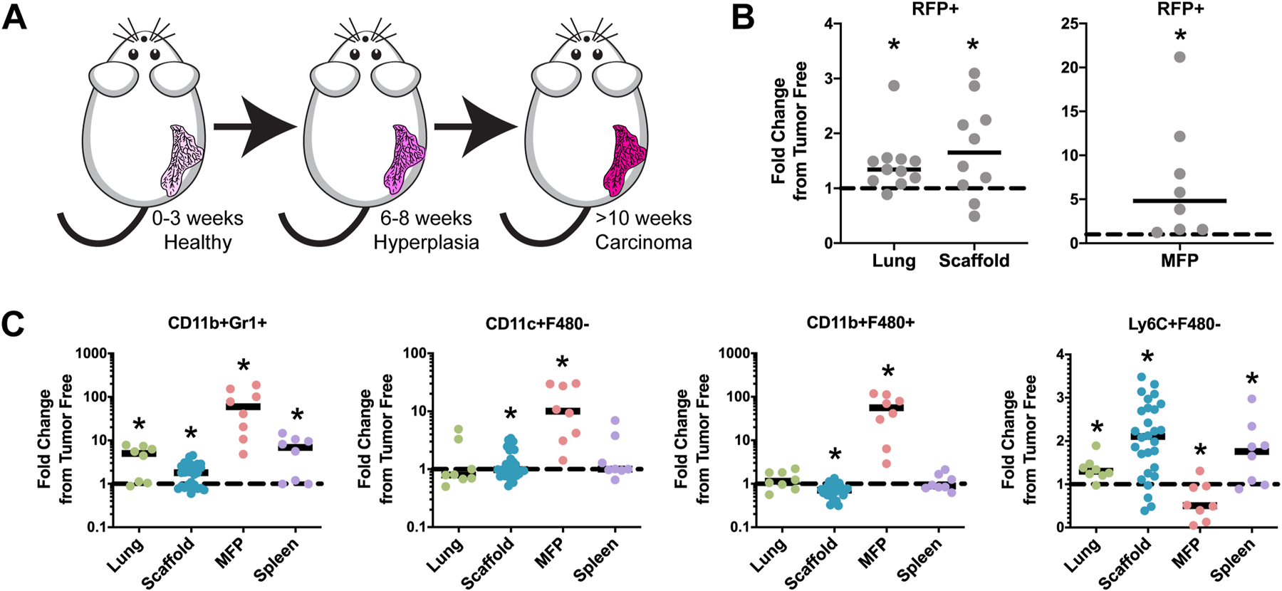
Scaffolds recruit tumor cells in the MMTV-PyMT model of spontaneous breast cancer and reflect tumor-dependent immunomodulation. (A) Timeline of breast cancer formation in the PyMT model. (B) The number of RFP+ cells increase in the scaffolds, lungs, and mammary fat pads (MFP) in PyMT+ mice relative to tumor free controls. (C) Innate immune cell types were identified as CD11b+Gr1+ (neutrophils), CD11c+F4/80- (dendritic cells), CD11b+F4/80+ (macrophages), and Ly6C+F480- (monocytes). The dotted lines represent the tumor free controls. *p < 0.05 relative to the tumor free control, n ≥ 8.
Figure 6:

Principal component analysis of KPCY pancreatic cancer (early disease and end stage disease) and PyMT breast cancer mice (end stage disease). Ellipses identify 90% confidence intervals. The two types of cancers diverge in the response of the immune cells as pancreatic cancer progresses.
Figure 2:

Disseminated pancreatic cells are identified in the scaffold prior to the formation of PDAC. (A) The percentage of YFP+ cells in the scaffold when mice are 12–16 weeks, as measured by flow cytometry. Black lines denote medians. Grey dotted lines represent YFP- mice autofluorescence control, n = 8–11. (B) YFP intensity at the scaffold measured by IVIS can distinguish KPCY and CY mice as early as 10 weeks of age in a longitudinal analysis. Grey dotted line represents YFP- mice autofluorescence control, n = 5. (C) Representative fluorescent images of the scaffolds under IVIS. *p < 0.05, **p < 0.01, ***p < 0.001, ****p < 0.0001.
Figure 4:

Temporal evolution of immune cells in the scaffold pre-metastatic niche prior to the formation of PDAC. The dotted lines represent the CY healthy controls. Cell types were identified as (A) CD45+ (total immune cells), (B) F4/80+CD11b+ (macrophages), (C) Gr1+CD11b+ (neutrophils), (D) Ly6C+CD11b+ (monocytes), (E) CD11c+F4/80- (dendritic cells), (F) CD19+ (B cells), (G) CD4+ (CD4+ T cells), (H) CD8+ (CD8+ T cells), and (I) CD49b+ (NK cells). * p < 0.05 relative to the CY control, # p < 0.05 over time, n = 5.
Hierarchical clustering and principal component analysis
Principal component analysis and unsupervised hierarchical clustering was performed in R, with data normalized across rows in the heat maps. Equation 1 was used to calculate accuracy of the separation of early and late stage disease by unsupervised hierarchical clustering.
| (1) |
Statistical analysis
All statistical tests were performed in GraphPad Prism 8. All graphs report mean ± standard error of the mean unless otherwise stated. Statistical tests were performed as a two-sided t-test with unequal variance unless otherwise stated (α = 0.05). Time-dependent comparisons were conducted using 2-way ANOVA (α = 0.05). Any outliers were identified and removed using the ROUT method with a Q-value of 1%.
Results:
Cellular infiltration of the scaffolds can identify late stage pancreatic cancer
We initially sought to verify that the scaffolds recruited disseminated pancreatic cells in late stage pancreatic cancer (22-week old mice). Accumulation of these cells was assessed through YFP fluorescence (Figure 1B-C). YFP intensity in KPCY scaffolds (179.5 ± 15.8 AU) was significantly higher (p < 0.0001) than in CY scaffolds (148.8 ± 7.3 AU) and scaffolds implanted in YFP-negative mice (126.2 ± 5.1 AU) which represent the off-target YFP expression driven by Pdx1 and the background autofluorescence level, respectively. These findings validate that the biomaterial scaffolds can successfully recruit pancreatic cells in the KPCY model.
Development of the metastatic niche is marked by an influx of both tumor and immune cells. Standard H&E staining (Figure 1D) demonstrated increased tissue infiltration and cellularity and a decrease in the coefficient of variation of density (measure of dispersion) (Figure 1E). Tissue infiltration (positively stained percentage of the scaffold section) was significantly higher (p < 0.01) in scaffolds from KPCY mice (89.3 ± 5.4 %) compared to CY mice (83.1 ± 6.3 %). Cellular density was 150% higher in scaffolds from KPCY mice. The coefficient of variation of cellular density was lower in scaffolds from KPCY mice (22.7 ± 6.9 %) compared to CY mice (33.6 ± 4.4 %). To account for the increased tissue infiltration, cellular density was normalized to tissue infiltration. Normalized cellular density was also significantly higher (p < 0.0001) in scaffolds from KPCY mice indicating increased cellular infiltration within the tissue. Thus, we concluded that histological changes at the biomaterial scaffolds can identify the metastatic niche in late stage disease.
Circulating YFP+ pancreatic cells are captured by scaffolds prior to PDAC formation
The concentration of YFP+ pancreatic cells in the scaffold was measured in 12- to 16-week old mice to investigate if early disseminated cells were captured before PDAC was detected (Figure S1). The relative number of YFP+ cells in each tissue from healthy CY mice were indistinguishable from background values (YFP- autofluorescence control), suggesting that pancreatic cells do not disseminate in the tumor-free control (Figure 2A). The percentage of YFP+ cells was significantly higher (p < 0.05) for scaffolds from KPCY mice (0.30 ± 0.22 %) relative to CY mice (0.09 ± 0.13 %) in early stage disease. In contrast, the percentage of YFP+ cells in the liver (0.64 ± 0.72 %) and blood (0.12 ± 0.14 %) did not increase relative to their healthy counterparts.
The temporal trends in YFP+ cells were captured through fluorescence imaging of scaffolds using IVIS. Scaffolds were explanted every 2 weeks from the mice and the YFP fluorescence was measured from each scaffold in KPCY versus CY littermates (Figure 2B). As early as 10 weeks of age, KPCY scaffolds demonstrated a 4.4-fold increase in YFP intensity over their CY controls. This relative increase in signal was maintained from 10–16 weeks of age (Figure 2C). These data indicate the significant accumulation of circulating YFP+ pancreatic cells at scaffolds as early as 10 weeks of age. Collectively, these findings demonstrate that the scaffolds can successfully capture disseminated pancreatic cells at disease stages consistent with the presence of circulating pancreatic cells in humans [3, 4].
Immune cells at the scaffolds can differentiate early and late stage pancreatic cancer
Cancer progresses, in part, through dysregulation of the immune system. We next investigated the immune cells dynamics at the scaffold relative to the pancreas and the liver, with the liver being the natural metastatic site (21–22 weeks). Immune composition was analyzed in early (14–16 weeks; PanIN) and late stages of disease (21–22 weeks; PDAC). Scaffolds collected from mice with early stage pancreatic cancer expressed 1.2- to 1.5-fold increases in Gr1+CD11b+ neutrophils, Ly6C+CD11b+ monocytes, and CD11c+F4/80- dendritic cells (Figure S2). Whereas, late stage disease caused a 1.4- to 1.9-fold change in CD45+ immune cells, F4/80+CD11b+ macrophages, CD4+ T cells, CD8+ T cells, CD19+ B cells, and CD49b+ NK cells in the scaffolds. Pancreata from early and late stage mice each exhibited 18-fold increases in CD45+ immune cells and 3.7-fold increases in Gr1+CD11b+ neutrophils (Figure S3). All immune populations at the liver exhibited high variability, with the only consistent trends being increases in CD45+ immune cells and decreases in CD49b+ NK cells (Figure S4).
The changes at the scaffold, pancreas, and liver were analyzed through unsupervised hierarchical clustering to assess the ability to delineate disease (primary tumor and metastatic sites, respectively). Despite the relatively small changes in the scaffold compared to the pancreas, the scaffold was able to classify early and late stage disease with an accuracy of 0.83 (Figure 3A). While the pancreas, unsurprisingly, was most sensitive to immune changes in early disease, it only separated early and late stage disease with an accuracy of 0.67 (Figure 3B). Analysis of the liver could not distinguish between early and late stage pancreatic cancer any better than random chance (accuracy = 0.50) (Figure 3C).
Immune cell dynamics at the scaffold in early pancreatic cancer
The immune cell dynamics throughout early pancreatic disease progression were next analyzed within the scaffolds every two weeks (Figure 4). CD45+ total immune cells, Ly6C+CD11b+ monocytes, and Gr1+CD11b+ neutrophils were consistently increased in scaffolds from KPCY mice relative to CY controls (Figure 4A, 4C, and 4D). F4/80+CD11b+ macrophages, CD11c+F4/80- dendritic cells, CD19+ B cells, and CD49b+ NK cell concentrations in the KPCY and CY mice were comparable and unchanged over time (Figure 4B, 4E, 4H, and 4I). CD4+ and CD8+ T cells were similar between KPCY and CY mice from 10–14 weeks, yet increased in KPCY mice at 16 weeks (3.17 ± 2.36 and 1.66 ± 0.86 fold from control, respectively), consistent with the observation that T cells were higher in late stage scaffolds than early stage (Figure 4F and 4G). These findings suggest that immune cells at the scaffolds can differentiate healthy mice from PanIN disease, in addition to separating PanIN disease from PDAC as shown in Figure 3.
The immune response at the scaffolds distinguishes divergent systemic immune alterations in spontaneous breast and pancreatic cancer
The immune dynamics observed at scaffolds in the KPCY model emphasize an adaptive immune response (T cells, B cells, and NK cells) relative to healthy controls and with disease progression. This observation contrasts with previous results for scaffolds implanted in orthotopic breast cancer models, which could result from either the model being orthotopic or being a different type of cancer. Thus, the KPCY spontaneous pancreatic cancer model was compared to the MMTV-PyMT spontaneous breast cancer model, as the PyMT model is metastatic and consistent with our previous use of scaffolds in orthotopic breast cancer (Figure 5A) [11–18]. We first analyzed the ability of the scaffolds to characterize spontaneous breast cancer and then assessed if immunomodulation at the scaffolds varied between breast and pancreatic cancer.
Scaffolds, lungs, and mammary fat pads were isolated from end stage PyMT mice (12–14 weeks of age), with the percentage of RFP+ mammary cells normalized to tumor free controls (Figure 5B). All tissues had significantly higher (p < 0.05) RFP+ content compared controls. We also tested the hypothesis that scaffold implantation may alter tumor progression in this model by measuring spleen mass (Figure S5A), tumor mass (Figure S5B), and liver (Figure S5C) and lung metastatic burden (Figure S5D). No significant alterations in disease progression were observed between PyMT mice implanted with scaffolds and those that received a mock surgery.
Similar to pancreatic cancer models, tumor dependent immune modulation was monitored in the MMTV-PyMT breast cancer model. Lungs, mammary fat pads, scaffolds, and spleens were analyzed at end point tumor size and normalized to controls (Figure 5C). CD11b+Gr1+ neutrophils increased in all diseased tissues. F4/80+CD11b+ macrophages increased in the mammary fat pad but decreased in the scaffold. Scaffolds further exhibited increases in CD11c+F4/80- dendritic cells and Ly6C+F4/80- monocytes in tumor bearing animals. Adaptive immune cell populations were also analyzed (Figure S6) and did not demonstrate significant changes from tumor-free control in scaffolds.
We next measured longitudinal tumor-dependent immunomodulation of biomaterial scaffolds (Figure S7). Mice were followed from the early onset of disease (5 weeks) until end stage morbidity (11 weeks). In the PyMT model CD45+ cells, Ly6C+CD11b+ monocytes, CD11c+F4/80- dendritic cells, and CD19+ B cells decreased over time. F480+CD11b+ macrophages and CD49b+ NK cells increased over time. These data demonstrate significant tumor-dependent immunomodulation at the scaffold in spontaneous breast cancer.
Finally, we compared the divergent immune response to biomaterial scaffolds in breast and pancreatic cancer. We implanted scaffolds in both models prior to the formation of invasive disease and explanted scaffolds at end stage tumor size (breast and pancreatic cancer) or early disease (pancreatic cancer) (Figure 6). Both models were analyzed by the same flow cytometry panels and normalized to the tumor free control to remove variation associated with different background mouse strains (C57/BL6 for KPCY and FVB/N for PyMT). Principal component analysis showed considerable overlap of PyMT scaffolds (blue circle) with early stage KPCY scaffolds (red circle). Yet, there were strong deviations between the PyMT scaffolds and those from late stage KPCY mice (green circle). This clustering suggests that the initial changes at the scaffold may have low dependence on the type of cancer. In contrast, immunomodulation at the scaffold in late stage breast and pancreatic cancer is largely driven by the origin of disease. Spontaneous breast cancer exhibited larger increases in CD11c+ and Ly6C+ cells (Figure 5) while spontaneous pancreatic cancer was marked by increases in CD49b+, CD4+, CD19+, and CD8+ cells (Figure 3). These findings demonstrate the potential of the scaffolds to not only detect early metastatic disease, but also identify unique dysregulation of the immune system with cancer development in different organs.
Discussion:
Improving survival and quality of life for pancreatic cancer is dependent on early detection, and therefore treatment, of the disease. Currently, the rates of pancreatic cancer diagnosis and mortality are nearly identical, and these numbers have remained relatively stagnant over the last 30 years [31, 32]. Scaffolds may serve as a site that can be readily accessed and monitored and thus provide an opportunity for detection [10]. In this report, we investigated the use of biomaterial scaffolds for early detection of spontaneous pancreatic cancer. The scaffolds successfully identified pancreatic disease, prior to the formation of PDAC, through immune cell evolution and circulating pancreatic cell recruitment.
Rhim et al. have identified circulating pancreatic cells in the blood and livers of KPCY mice as early as 8–10 weeks [33]. We reliably detected YFP+ pancreatic cells in the scaffolds of KPCY mice at increased levels relative to CY mice at this early time point, while the liver and blood could not detect these increases (Figure 2). The median percentage of YFP+ cells in the scaffolds of KPCY mice was nearly identical to the livers, the natural metastatic site. This result validates the scaffolds as a source of pancreatic cells that can monitor disease progression. While detecting circulating pancreatic cells is possible due to the genetically engineered mouse model, cancer cells in human disease are not fluorescently labeled. This study provides a proof of principal for detection of pancreatic cells at an engineered site. Detection would be more challenging in humans without a genetic fluorescent label, yet potentially still feasible based on evaluation of cytokeratin and pancreatic tissue specific marker (e.g. PDX1) expression and morphological changes (Figure 1).
No biomarkers have been confirmed for pancreatic cancer diagnostics. Imaging and endoscopic ultrasound of the pancreas are often unable to distinguish PDAC from pancreatitis lesions, which can result in overdiagnosis and overtreatment [32, 34]. Only highly invasive biopsy can conclusively identify pancreatic cancer, which is only conducted once the lesions are identified through screening, often once the disease has already become metastatic. Moreover, cancer antigen 19–9 (CA-19–9) is the lone biomarker used to monitor the progression of the disease [5, 6]. CA-19–9 has proven clinical value but two major limitations – it is not pancreatic cancer-specific and 5–10% of people do not express this antigen. Transgenic mouse models have been used to identify other biomarkers (i.e. Kras mutations, circulating pancreatic cells, Mucin-1, VEGFR2) for pancreatic cancer [32, 35]. Yet, none of these markers outperform CA-19–9.
Emerging clinical evidence indicates circulating pancreatic cells are present not only in PDAC patients, but also in patients with benign cystic lesions [36]. Furthermore, this same study found that these cell counts were not correlated with cyst size, tumor stage, or CA-19–9. Thus, there is a critical need for peripheral biomarkers for early detection that are independent of circulating pancreatic cells in the blood. These findings highlight the opportunity of the scaffolds’ ability to delineate disease through analysis of a metastatic niche that can contain tumor cells and also various immune cell populations.
The immune cells at the scaffold provided a signature that classified early and late stage pancreatic cancer with an accuracy of 0.83 (Figure 3). The accuracy of the immune signature is particularly promising as it was derived from non-optimized data. Further analyses into gene and protein expression of the scaffolds could produce a multi-variate signature that accurately recapitulates the progression of pancreatic cancer [13].
We observed a decrease in CD4+ cell numbers in KPCY mice during precancerous stages (10–12 weeks), but an escalation during PDAC (>16 weeks) (Figures 3–4). The depletion of CD4+ cells in KPC mice has been shown to completely abrogate the formation of PanIN lesions [37], while higher levels of CD4+ infiltration were found to correlate with more advanced pancreatic carcinomas [38]. Other reports have found that CD4+ cells are decreased in PDAC [39], but the outcome of this loss varies with the specific subset. Th1 CD4+ cells are reported to suppress pancreatic cancers [40], but Th2 and Th17 CD4+ cells promote PDAC progression and correlate with poor clinical outcomes [41–43]. These findings are consistent with the pro-metastatic role of CD4+ cells in this model. Phenotypic and genomic analyses of these cells have the potential to derive a multi-variate signature that can delineate early and late stage pancreatic cancer from healthy controls. Our findings pose an interesting contrast with work reported by others that CD4+ and CD8+ T cells are suppressed in pancreatic cancer [44]. Analysis of the unique scaffold T cell response could provide an avenue to better investigate the role of T cells in pancreatic cancer therapies.
Although immune cells are critical in the metastatic tumor microenvironment, many other cells have a role in niche development [45]. The work presented herein focused primarily on immune cell profiling due to observed population dynamics during tumor progression in orthotopic breast cancer [18]. In this model, the deposited extracellular matrix at the scaffolds was minimally changed, but the percentage of endothelial cells increased in tumor-bearing mice relative to healthy controls [14]. Herein, we observe changes in tissue infiltration in the KPCY model (Figure 1D-E) that indicate the potential for other cell populations to be altered with disease progression. This evidence suggests that while immune dynamics can be used for the detection of pancreatic cancer progression, a number of other cell populations and factors may exhibit similar dynamics and contribute to pancreatic tumor cell recruitment.
Tumor cells are recruited to scaffolds in both spontaneous breast and pancreatic cancer, yet we observe a divergent immune response to scaffolds in these two models. In KPCY mice, we found an increase in T cells that is organ of origin-specific, as no such change occurred in the scaffolds in the spontaneous breast cancer model (Figure 6, S6-S7) nor has this finding been observed in scaffolds implanted in orthotopic breast cancer models [11, 13, 15, 17, 18]. Changes in lymphocytes largely drive the differential response between pancreatic cancer and breast cancer. Early fluctuations in the PyMT models at the scaffolds primarily occur in the myeloid cells. These divergent immune responses in the same biomaterial scaffold indicate that tumors of different origin modulate the systemic immune response in distinct ways that are then represented at the implant.
Biomaterial implants induce a classical foreign body response beginning with an acute phase (characterized by neutrophils, monocytes, and leukocytes) and a subsequent chronic phase (characterized by macrophages, foreign body giant cells, and fibroblasts) [46]. The immune response to microporous PCL scaffolds is well studied and reaches steady state two weeks after implantation in tumor free mice [18]; however, the presence of a tumor leads to dynamic changes in the immune response at the implant [13]. Herein, all scaffolds were implanted for at least two weeks prior to explant to rule out dynamics associated with the acute response and isolate effects of disease progression. Time-matched scaffolds were implanted in healthy mice to serve as a control that accounts for effects of scaffold implantation. Furthermore, in the PyMT model, tissue mass and metastatic burden were compared between mock and scaffold-implanted mice with no significant difference between groups (Figure S5). Taken together, these findings indicate that biomaterial implantation does not significantly alter disease progression, yet is able to capture the associated immune dynamics.
In this report, the recruitment of circulating pancreatic cells or breast cancer cells and changes in the immune cell composition can identify signs of cancer progression prior to the development of overt carcinoma. Interestingly, the pancreatic cancer and breast cancer models primarily metastasize to the liver and lung respectively, yet both types of cancer have tumor cell metastasis to the scaffold. The ability of the subcutaneous scaffolds to successfully detect disease evolution for both pancreatic and breast cancer may provide novel opportunity for development of early detection strategies for cancer progression.
Supplementary Material
Footnotes
Publisher's Disclaimer: This is a PDF file of an unedited manuscript that has been accepted for publication. As a service to our customers we are providing this early version of the manuscript. The manuscript will undergo copyediting, typesetting, and review of the resulting proof before it is published in its final form. Please note that during the production process errors may be discovered which could affect the content, and all legal disclaimers that apply to the journal pertain.
Data Availability:
All data required to evaluate the conclusions of this manuscript are present in the main text and supplemental data. Additional data will be available from the authors upon request.
References:
- [1].Siegel RL, Miller KD, Jemal A, Cancer statistics, 2020, CA Cancer J Clin 70(1) (2020) 7–30. [DOI] [PubMed] [Google Scholar]
- [2].Lee CJ, Scheiman J, Anderson MA, Hines OJ, Reber HA, Farrell J, Kochman ML, Foley PJ, Drebin J, Oh YS, Ginsberg G, Ahmad N, Merchant NB, Isbell J, Parikh AA, Stokes JB, Bauer T, Adams RB, Simeone DM, Risk of malignancy in resected cystic tumors of the pancreas < or =3 cm in size: is it safe to observe asymptomatic patients? A multi-institutional report, J Gastrointest Surg 12(2) (2008) 234–42. [DOI] [PubMed] [Google Scholar]
- [3].Klein CA, Parallel progression of primary tumours and metastases, Nat Rev Cancer 9(4) (2009) 302–12. [DOI] [PubMed] [Google Scholar]
- [4].Haeno H, Gonen M, Davis MB, Herman JM, Iacobuzio-Donahue CA, Michor F, Computational modeling of pancreatic cancer reveals kinetics of metastasis suggesting optimum treatment strategies, Cell 148(1–2) (2012) 362–75. [DOI] [PMC free article] [PubMed] [Google Scholar]
- [5].Chari ST, Kelly K, Hollingsworth MA, Thayer SP, Ahlquist DA, Andersen DK, Batra SK, Brentnall TA, Canto M, Cleeter DF, Firpo MA, Gambhir SS, Go VL, Hines OJ, Kenner BJ, Klimstra DS, Lerch MM, Levy MJ, Maitra A, Mulvihill SJ, Petersen GM, Rhim AD, Simeone DM, Srivastava S, Tanaka M, Vinik AI, Wong D, Early detection of sporadic pancreatic cancer: summative review, Pancreas 44(5) (2015) 693–712. [DOI] [PMC free article] [PubMed] [Google Scholar]
- [6].McGuigan A, Kelly P, Turkington RC, Jones C, Coleman HG, McCain RS, Pancreatic cancer: A review of clinical diagnosis, epidemiology, treatment and outcomes, World J Gastroenterol 24(43) (2018) 4846–4861. [DOI] [PMC free article] [PubMed] [Google Scholar]
- [7].Yu J, Blackford AL, Dal Molin M, Wolfgang CL, Goggins M, Time to progression of pancreatic ductal adenocarcinoma from low-to-high tumour stages, Gut 64(11) (2015) 1783–9. [DOI] [PMC free article] [PubMed] [Google Scholar]
- [8].Owens DK, Davidson KW, Krist AH, Barry MJ, Cabana M, Caughey AB, Curry SJ, Doubeni CA, Epling JW Jr., Kubik M, Landefeld CS, Mangione CM, Pbert L, Silverstein M, Simon MA, Tseng CW, Wong JB, Screening for Pancreatic Cancer: US Preventive Services Task Force Reaffirmation Recommendation Statement, JAMA 322(5) (2019) 438–444. [DOI] [PubMed] [Google Scholar]
- [9].Henrikson NB, Aiello Bowles EJ, Blasi PR, Morrison CC, Nguyen M, Pillarisetty VG, Lin JS, Screening for Pancreatic Cancer: Updated Evidence Report and Systematic Review for the US Preventive Services Task Force, JAMA 322(5) (2019) 445–454. [DOI] [PMC free article] [PubMed] [Google Scholar]
- [10].Morris AH, Orbach SM, Bushnell GG, Oakes RS, Jeruss JS, Shea LD, Engineered Niches to Analyze Mechanisms of Metastasis and Guide Precision Medicine, Cancer Res (2020). [DOI] [PMC free article] [PubMed]
- [11].Bushnell GG, Rao SS, Hartfield RM, Zhang Y, Oakes RS, Jeruss JS, Shea LD, Microporous scaffolds loaded with immunomodulatory lentivirus to study the contribution of immune cell populations to tumor cell recruitment in vivo, Biotechnol Bioeng 117(1) (2020) 210–222. [DOI] [PMC free article] [PubMed] [Google Scholar]
- [12].Bushnell GG, Hardas TP, Hartfield RM, Zhang Y, Oakes RS, Ronquist S, Chen H, Rajapakse I, Wicha MS, Jeruss JS, Shea LD, Biomaterial Scaffolds Recruit an Aggressive Population of Metastatic Tumor Cells In Vivo, Cancer Res 79(8) (2019) 2042–2053. [DOI] [PMC free article] [PubMed] [Google Scholar]
- [13].Oakes RS, Bushnell GG, Orbach SM, Kandagatla P, Zhang Y, Morris AH, Hall MS, LaFaire P, Decker JT, Hartfield RM, Brooks MD, Wicha MS, Jeruss JS, Shea LD, Metastatic Conditioning of Myeloid Cells at a Subcutaneous Synthetic Niche Reflects Disease Progression and Predicts Therapeutic Outcomes, Cancer Res 80(3) (2020) 602–612. [DOI] [PMC free article] [PubMed] [Google Scholar]
- [14].Bushnell GG, Hong X, Hartfield RM, Zhang Y, Oakes RS, Rao SS, Jeruss JS, Stegemann JP, Deng CX, Shea LD, High Frequency Spectral Ultrasound Imaging to Detect Metastasis in Implanted Biomaterial Scaffolds, Ann Biomed Eng 48(1) (2020) 477–489. [DOI] [PMC free article] [PubMed] [Google Scholar]
- [15].Aguado BA, Caffe JR, Nanavati D, Rao SS, Bushnell GG, Azarin SM, Shea LD, Extracellular matrix mediators of metastatic cell colonization characterized using scaffold mimics of the pre-metastatic niche, Acta Biomater 33 (2016) 13–24. [DOI] [PMC free article] [PubMed] [Google Scholar]
- [16].Aguado BA, Hartfield RM, Bushnell GG, Decker JT, Azarin SM, Nanavati D, Schipma MJ, Rao SS, Oakes RS, Zhang Y, Jeruss JS, Shea LD, Biomaterial Scaffolds as Pre-metastatic Niche Mimics Systemically Alter the Primary Tumor and Tumor Microenvironment, Adv Healthc Mater 7(10) (2018) e1700903. [DOI] [PMC free article] [PubMed] [Google Scholar]
- [17].Azarin SM, Yi J, Gower RM, Aguado BA, Sullivan ME, Goodman AG, Jiang EJ, Rao SS, Ren Y, Tucker SL, Backman V, Jeruss JS, Shea LD, In vivo capture and label-free detection of early metastatic cells, Nat Commun 6 (2015) 8094. [DOI] [PMC free article] [PubMed] [Google Scholar]
- [18].Rao SS, Bushnell GG, Azarin SM, Spicer G, Aguado BA, Stoehr JR, Jiang EJ, Backman V, Shea LD, Jeruss JS, Enhanced Survival with Implantable Scaffolds That Capture Metastatic Breast Cancer Cells In Vivo, Cancer Res 76(18) (2016) 5209–18. [DOI] [PMC free article] [PubMed] [Google Scholar]
- [19].Lee J, Li M, Milwid J, Dunham J, Vinegoni C, Gorbatov R, Iwamoto Y, Wang F, Shen K, Hatfield K, Enger M, Shafiee S, McCormack E, Ebert BL, Weissleder R, Yarmush ML, Parekkadan B, Implantable microenvironments to attract hematopoietic stem/cancer cells, Proc Natl Acad Sci U S A 109(48) (2012) 19638–43. [DOI] [PMC free article] [PubMed] [Google Scholar]
- [20].Bersani F, Lee J, Yu M, Morris R, Desai R, Ramaswamy S, Toner M, Haber DA, Parekkadan B, Bioengineered implantable scaffolds as a tool to study stromal-derived factors in metastatic cancer models, Cancer Res 74(24) (2014) 7229–38. [DOI] [PMC free article] [PubMed] [Google Scholar]
- [21].de la Fuente A, Alonso-Alconada L, Costa C, Cueva J, Garcia-Caballero T, Lopez-Lopez R, Abal M, M-Trap: Exosome-Based Capture of Tumor Cells as a New Technology in Peritoneal Metastasis, J Natl Cancer Inst 107(9) (2015). [DOI] [PMC free article] [PubMed] [Google Scholar]
- [22].Ko CY, Wu L, Nair AM, Tsai YT, Lin VK, Tang L, The use of chemokine-releasing tissue engineering scaffolds in a model of inflammatory response-mediated melanoma cancer metastasis, Biomaterials 33(3) (2012) 876–85. [DOI] [PMC free article] [PubMed] [Google Scholar]
- [23].Talmadge JE, Singh RK, Fidler IJ, Raz A, Murine models to evaluate novel and conventional therapeutic strategies for cancer, Am J Pathol 170(3) (2007) 793–804. [DOI] [PMC free article] [PubMed] [Google Scholar]
- [24].Hanahan D, Weinberg RA, Hallmarks of cancer: the next generation, Cell 144(5) (2011) 646–74. [DOI] [PubMed] [Google Scholar]
- [25].Lee JW, Komar CA, Bengsch F, Graham K, Beatty GL, Genetically Engineered Mouse Models of Pancreatic Cancer: The KPC Model (LSL-Kras(G12D/+); LSL-Trp53(R172H/+); Pdx-1-Cre), Its Variants, and Their Application in Immuno-oncology Drug Discovery, Curr Protoc Pharmacol 73 (2016) 14 39 1–14 39 20. [DOI] [PMC free article] [PubMed] [Google Scholar]
- [26].Feinberg TY, Zheng H, Liu R, Wicha MS, Yu SM, Weiss SJ, Divergent Matrix-Remodeling Strategies Distinguish Developmental from Neoplastic Mammary Epithelial Cell Invasion Programs, Dev Cell 47(2) (2018) 145–160 e6. [DOI] [PMC free article] [PubMed] [Google Scholar]
- [27].Olive KP, Jacobetz MA, Davidson CJ, Gopinathan A, McIntyre D, Honess D, Madhu B, Goldgraben MA, Caldwell ME, Allard D, Frese KK, Denicola G, Feig C, Combs C, Winter SP, Ireland-Zecchini H, Reichelt S, Howat WJ, Chang A, Dhara M, Wang L, Ruckert F, Grutzmann R, Pilarsky C, Izeradjene K, Hingorani SR, Huang P, Davies SE, Plunkett W, Egorin M, Hruban RH, Whitebread N, McGovern K, Adams J, Iacobuzio-Donahue C, Griffiths J, Tuveson DA, Inhibition of Hedgehog signaling enhances delivery of chemotherapy in a mouse model of pancreatic cancer, Science 324(5933) (2009) 1457–61. [DOI] [PMC free article] [PubMed] [Google Scholar]
- [28].Hingorani SR, Wang L, Multani AS, Combs C, Deramaudt TB, Hruban RH, Rustgi AK, Chang S, Tuveson DA, Trp53R172H and KrasG12D cooperate to promote chromosomal instability and widely metastatic pancreatic ductal adenocarcinoma in mice, Cancer Cell 7(5) (2005) 469–83. [DOI] [PubMed] [Google Scholar]
- [29].Lin EY, Jones JG, Li P, Zhu L, Whitney KD, Muller WJ, Pollard JW, Progression to malignancy in the polyoma middle T oncoprotein mouse breast cancer model provides a reliable model for human diseases, Am J Pathol 163(5) (2003) 2113–26. [DOI] [PMC free article] [PubMed] [Google Scholar]
- [30].Akuthota P, Shamri R, Weller PF, Isolation of human eosinophils, Curr Protoc Immunol Chapter 7 (2012) Unit 7 31. [DOI] [PMC free article] [PubMed] [Google Scholar]
- [31].SEER, Cancer Stat Facts: Pancreatic Cancer, 2019. https://seer.cancer.gov/statfacts/html/pancreas.html.
- [32].Tummers WS, Willmann JK, Bonsing BA, Vahrmeijer AL, Gambhir SS, Swijnenburg RJ, Advances in Diagnostic and Intraoperative Molecular Imaging of Pancreatic Cancer, Pancreas 47(6) (2018) 675–689. [DOI] [PMC free article] [PubMed] [Google Scholar]
- [33].Rhim AD, Mirek ET, Aiello NM, Maitra A, Bailey JM, McAllister F, Reichert M, Beatty GL, Rustgi AK, Vonderheide RH, Leach SD, Stanger BZ, EMT and dissemination precede pancreatic tumor formation, Cell 148(1–2) (2012) 349–61. [DOI] [PMC free article] [PubMed] [Google Scholar]
- [34].Lennon AM, Wolfgang CL, Canto MI, Klein AP, Herman JM, Goggins M, Fishman EK, Kamel I, Weiss MJ, Diaz LA, Papadopoulos N, Kinzler KW, Vogelstein B, Hruban RH, The early detection of pancreatic cancer: what will it take to diagnose and treat curable pancreatic neoplasia?, Cancer Res 74(13) (2014) 3381–9. [DOI] [PMC free article] [PubMed] [Google Scholar]
- [35].Herreros-Villanueva M, Bujanda L, Non-invasive biomarkers in pancreatic cancer diagnosis: what we need versus what we have, Ann Transl Med 4(7) (2016) 134. [DOI] [PMC free article] [PubMed] [Google Scholar]
- [36].Rhim AD, Thege FI, Santana SM, Lannin TB, Saha TN, Tsai S, Maggs LR, Kochman ML, Ginsberg GG, Lieb JG, Chandrasekhara V, Drebin JA, Ahmad N, Yang YX, Kirby BJ, Stanger BZ, Detection of circulating pancreas epithelial cells in patients with pancreatic cystic lesions, Gastroenterology 146(3) (2014) 647–51. [DOI] [PMC free article] [PubMed] [Google Scholar]
- [37].Zhang Y, Yan W, Mathew E, Bednar F, Wan S, Collins MA, Evans RA, Welling TH, Vonderheide RH, di Magliano MP, CD4+ T lymphocyte ablation prevents pancreatic carcinogenesis in mice, Cancer immunology research 2(5) (2014) 423–435. [DOI] [PMC free article] [PubMed] [Google Scholar]
- [38].Clark CE, Hingorani SR, Mick R, Combs C, Tuveson DA, Vonderheide RH, Dynamics of the immune reaction to pancreatic cancer from inception to invasion, Cancer research 67(19) (2007) 9518–9527. [DOI] [PubMed] [Google Scholar]
- [39].Chang JH, Jiang Y, Pillarisetty VG, Role of immune cells in pancreatic cancer from bench to clinical application: an updated review, Medicine 95(49) (2016). [DOI] [PMC free article] [PubMed] [Google Scholar]
- [40].Pushalkar S, Hundeyin M, Daley D, Zambirinis CP, Kurz E, Mishra A, Mohan N, Aykut B, Usyk M, Torres LE, The pancreatic cancer microbiome promotes oncogenesis by induction of innate and adaptive immune suppression, Cancer discovery 8(4) (2018) 403–416. [DOI] [PMC free article] [PubMed] [Google Scholar]
- [41].Zhang Y, Lazarus J, Steele NG, Yan W, Lee H-J, Nwosu ZC, Halbrook CJ, Menjivar RE, Kemp SB, Sirihorachai VR, Regulatory T-cell depletion alters the tumor microenvironment and accelerates pancreatic carcinogenesis, Cancer Discovery 10(3) (2020) 422–439. [DOI] [PMC free article] [PubMed] [Google Scholar]
- [42].McAllister F, Bailey JM, Alsina J, Nirschl CJ, Sharma R, Fan H, Rattigan Y, Roeser JC, Lankapalli RH, Zhang H, Oncogenic Kras activates a hematopoietic-to-epithelial IL-17 signaling axis in preinvasive pancreatic neoplasia, Cancer cell 25(5) (2014) 621–637. [DOI] [PMC free article] [PubMed] [Google Scholar]
- [43].De Monte L, Reni M, Tassi E, Clavenna D, Papa I, Recalde H, Braga M, Di Carlo V, Doglioni C, Protti MP, Intratumor T helper type 2 cell infiltrate correlates with cancer-associated fibroblast thymic stromal lymphopoietin production and reduced survival in pancreatic cancer, Journal of Experimental Medicine 208(3) (2011) 469–478. [DOI] [PMC free article] [PubMed] [Google Scholar]
- [44].Chang JH, Jiang Y, Pillarisetty VG, Role of immune cells in pancreatic cancer from bench to clinical application: An updated review, Medicine (Baltimore) 95(49) (2016) e5541. [DOI] [PMC free article] [PubMed] [Google Scholar]
- [45].Aguado BA, Bushnell GG, Rao SS, Jeruss JS, Shea LD, Engineering the pre-metastatic niche, Nat Biomed Eng 1 (2017). [DOI] [PMC free article] [PubMed] [Google Scholar]
- [46].Chandorkar Y, R. K, Basu B, The Foreign Body Response Demystified, ACS Biomaterials Science & Engineering 5(1) (2019) 19–44. [DOI] [PubMed] [Google Scholar]
Associated Data
This section collects any data citations, data availability statements, or supplementary materials included in this article.
Supplementary Materials
Data Availability Statement
All data required to evaluate the conclusions of this manuscript are present in the main text and supplemental data. Additional data will be available from the authors upon request.