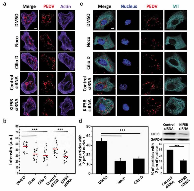
Figure 2.

Microtubule, dynein and kinesin-1 affect PEDV fusion and accumulation in the perinuclear region. (a) Fluorescence images of Vero cells first respectively treated with control DMSO, Noco, Cilio D, control siRNA and KIF5B siRNA, then infected with DiD-labeled PEDV particles for 1 h and finally fixed. Actin was stained with AbFluor 488-conjugated phalloidin. Scale bar, 5 μm. (b) Statistical analysis on the DiD fluorescence intensity from (a) (n = 20 cells). a.u.: arbitrary units. (c) Fluorescence images of Vero cells first transfected with EGFP-MT and respectively treated with control DMSO, Noco, Cilio D, control siRNA and KIF5B siRNA and then infected with DiD-labeled PEDV particles. Nuclei were stained with DAPI. Scale bar, 5 μm. (d) Quantification on the percentage virions within 2 μm of the nuclei in 15 infected cells respectively treated with control DMSO, Cilio D, Noco, control siRNA and KIF5B siRNA. The KIF5B level was determined using Western blot with the KIF5B antibody, and equal loading was verified using the anti-GAPDH antibody. Each data point represents mean ± standard deviation from three independent experiments. Statistical analysis of all data was performed using one-way ANOVA (***, P < 0.001)
