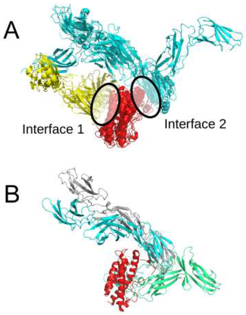
Figure 1:

A) Two classes of interaction between IL23R and IL23A homologs observed among the structures in the PDB. The IL23A homologs are shown in red, IL23R homologs corresponding to two different interaction types are shown in yellow and cyan. B) The best model among the top 10 predictions in our human submission. The model is in gray, the structure of the native complex (PDB ID 5MZV) is in red (IL23A), green (IL12B) and cyan (IL23R).
