

The Journal has been informed of an error in the article by Song and colleagues (1), published in the November 2017 issue. In Figure 7, panel C (HIF-1α) and panel D (Negative Ctrl) in the bottom row (normal human nasal epithelial cells) incorrectly show views of the same sample. The authors inadvertently selected these panels when they were assembling the figure. A revised version of Figure 7 with the correct panels C and D is included here.
Figure 7.
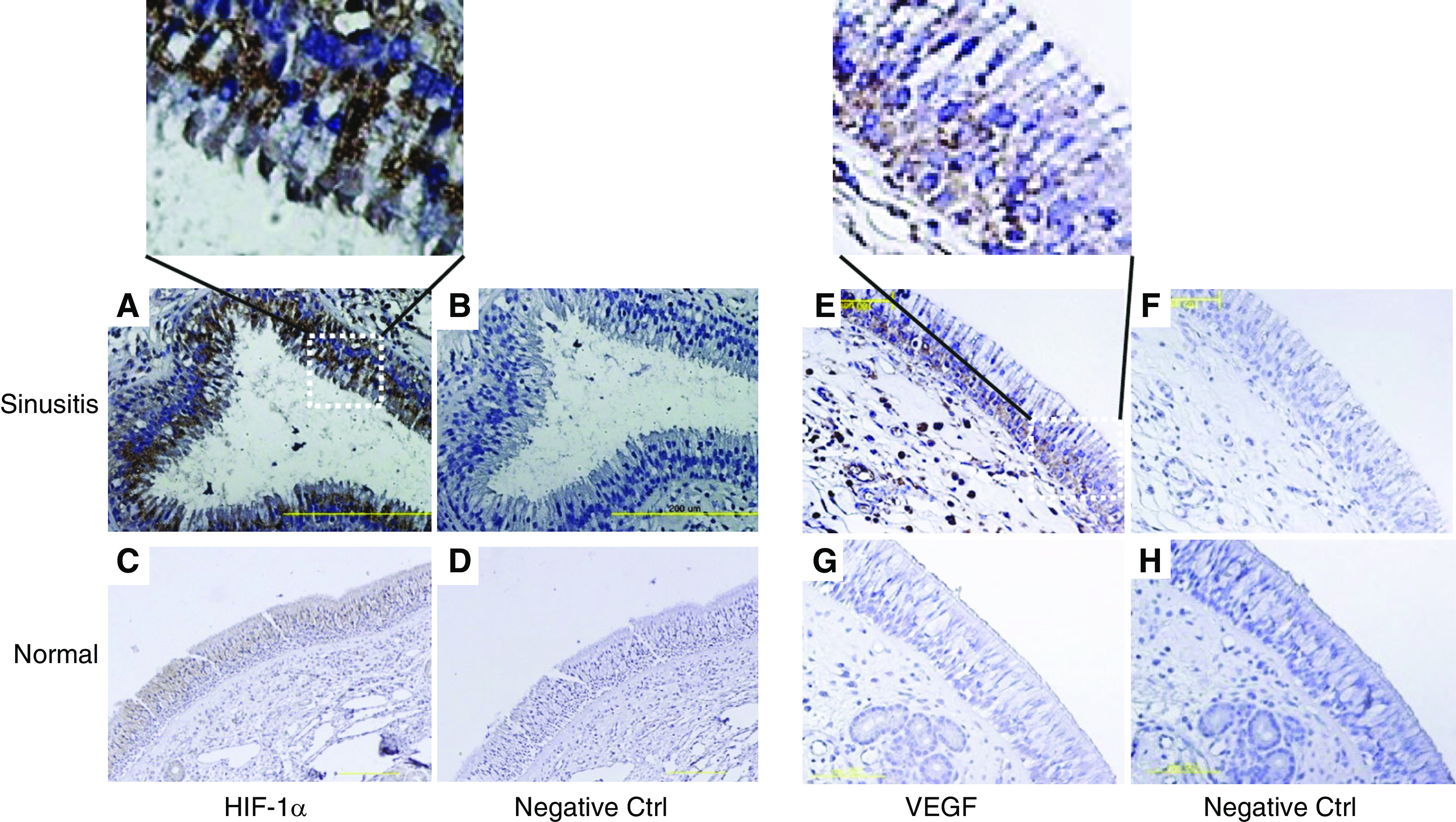
[revised image]. Immunohistochemical staining with anti–HIF-1α antibody and anti-VEGF antibody in the sinus mucosa. (A–D) High HIF-1α expression was indicated by strong reactivity in sinus epithelium from a patient with sinusitis. The left inset figure is the magnification of the epithelial layer of figure 7A. (E–H) Pronounced expression of VEGF was observed in sinus epithelium from a patient with sinusitis. The right inset figure is the magnification of the epithelial layer of Figure 7E. Scale bars: 200 μm in panels A and B, 50 μm in panels C and D, and 100 μm in panels E–H.
The authors apologize to the readers and the Journal for any inconvenience this may have caused.
Reference
- 1.Song HA, Kim YS, Cho HJ, Kim SI, Kang MJ, Kim JH, Min HJ, Kang JW, Yoon JH, Kim CH. Hypoxia modulates epithelial permeability via regulation of vascular endothelial growth factor in airway epithelia. Am J Respir Cell Mol Biol. 2017;57:527–535. doi: 10.1165/rcmb.2016-0080OC. [DOI] [PubMed] [Google Scholar]