PLOS ONE logoLink to PLOS ONE
. 2021 Feb 11;16(2):e0245725. doi: 10.1371/journal.pone.0245725

Presence of myxoid stromal change and fibrotic focus in pathological examination are prognostic factors of triple-negative breast cancer: Results from a retrospective single-center study

Hirotsugu Yanai 1, Katsuhiro Yoshikawa 1,2, Mitsuaki Ishida 2,*, Koji Tsuta 2, Mitsugu Sekimoto 1, Tomoharu Sugie 1
Editor: Elda Tagliabue3
PMCID: PMC7877644  PMID: 33571189

Abstract

Background

Stromal reaction is an important prognostic factor in several cancers, and the presence of myxoid change was assessed as a poor prognostic factor in colorectal cancer. However, the prognostic significance of myxoid change in triple-negative breast cancer (TNBC) remains unknown. This study aimed to determine the prognostic significance of myxoid change and fibrotic focus (FF), which is a fibrotic area within the tumor and considered a poor prognostic indicator in patients with TNBC.

Methods

We enrolled 62 patients with TNBC and reviewed the surgically resected specimens to evaluate myxoid change and FF in the tumor using previously outlined criteria. We evaluated tumor-infiltrating lymphocytes (TILs) using hematoxylin and eosin slides. Overall survival (OS) and relapse-free survival (RFS) were compared based on the presence of myxoid change and/or FF, and the risk factors for RFS were analyzed.

Results

Myxoid change and FF were observed in 25.8% and 33.9% of specimens, respectively. Based on stromal lymphocyte infiltration, 19 patients (30.6%) had high TILs, while the remaining 43 patients (69.4%) had low/intermediate TILs. Presence of myxoid change was significantly correlated with poor OS and RFS (p = 0.040 and 0.031, respectively). FF was also significantly correlated with poor OS and RFS (p = 0.012 and 0.028, respectively). The combination of myxoid change and FF was an independent and poor prognostic factor according to the multivariate analysis (HR 11.61; 95% CI 1.027–131.2; p = 0.048). Presence of myxoid change and FF were significantly associated with low/intermediate TILs in the stroma (p = 0.013).

Conclusions

Histopathological assessment of myxoid change and FF in TNBC may be a useful, practical, and easily assessable method for predicting prognosis in patients with TNBC, which should be confirmed in larger prospective studies. Diagnostic criteria for the establishment of myxoid change and FF in TNBC must be established, and their underlying molecular events must be clarified.

Introduction

Recent studies have highlighted the important role of the tumor microenvironment in growth, invasion, and metastasis of cancer [1]. The tumor microenvironment consists of a variety of cells, including inflammatory cells, fibroblasts, myofibroblasts, and endothelial cells, and the extracellular matrix [1]. It has been recognized that cancer-associated fibroblasts are one of the main components of the stromal reaction of carcinoma, and they play an important role in constituting the tumor microenvironment through the secretion of cytokines and extracellular matrix [1, 2]. Moreover, cancer-associated fibroblasts have been reported to exhibit prognostic implications in various types of carcinomas, including breast cancer [24].

More or less, stromal reaction is frequently observed in the invasive carcinoma tissues [5]. Recent studies have revealed that the patterns of stromal reaction (desmoplastic reaction) due to carcinoma invasion play an important role in the prognosis of cancer patients [5]. The prognostic significance of the desmoplastic reaction has been characterized in colorectal cancer [6, 7]. The patterns of desmoplastic reaction were classified as mature, intermediate, or immature, depending on the presence of myxoid stroma and hyalinized collagen in the invasive front of the tumor. These patterns were identified as independent prognostic factors for recurrence-free survival in patients with colorectal cancer. While mature group had the highest recurrence-free survival rates, the immature group (presence of myxoid change) had the worst prognosis [6, 7]. Furthermore, stromal reaction routinely occurs in breast cancer tissues. However, the prognostic significance of the different stromal reaction patterns in breast cancer tissues remains inadequately evaluated [4, 8]; nevertheless, it has been reported that the presence of fibrotic focus (FF), characterized by a fibrotic area containing fibroblasts and collagen, which is surrounded by a cellular zone of infiltrating carcinoma, is a poor prognostic factor in patients with breast cancer [9, 10]. Triple-negative breast cancer (TNBC) is characterized by the lack of estrogen and progesterone receptors and human epidermal growth factor receptor 2 (HER2). It is known to be one of the most aggressive subtypes of breast cancer. However, the significance of stromal reaction patterns, particularly in TNBC, remains unknown. Thus, the aim of this study was to determine the prognostic significance of the presence of myxoid change and FF in patients with TNBC.

Materials and methods

Patient selection

We selected 165 consecutive patients with TNBC who underwent surgical resection at the Department of Surgery of Kansai Medical University Hospital between January 2006 and December 2018. Patients who received neoadjuvant chemotherapy or those who had a special type of invasive carcinoma were excluded; 62 patients with TNBC were included.

This study was conducted in accordance with the principles of the Declaration of Helsinki and was approved by the Institutional Review Board of the Kansai Medical University Hospital (Approval #2019234). Informed consent was obtained from patients through the opt-out methodology, owing to the retrospective design of the study, with no new risk to the participants. Information regarding this study, such as the inclusion criteria, and the opportunity to opt out was provided through the hospital’s website.

Histopathological analysis

Surgically resected specimens were fixed with formalin, sectioned, and stained with hematoxylin and eosin. More than 2 experienced diagnostic pathologists independently evaluated all histopathological diagnoses. Myxoid change was histopathologically characterized by the presence of amorphous extracellular substances, which comprise an amphophilic or slightly basophilic material within the fibrous stroma, similar to the criteria reported for colorectal cancer [6, 7]. The presence of myxoid change was evaluated in the invasive front and within the tumor. Moreover, the presence of FF in breast cancer was also evaluated according to the previously reported criteria, which was diagnosed when there was a fibrotic area within the tumor containing fibroblasts and collagen, surrounded by a highly cellular zone of infiltrating carcinoma and measuring more than 1 mm [10]. In addition, we also evaluated the presence of myxoid change and FF using the matched pre-operative biopsy specimens.

Furthermore, we evaluated tumor-infiltrating lymphocytes (TILs) using hematoxylin and eosin slides according to the recommendation of the TIL Working Group [11]. We classified ≥60% stromal TILs as high TILs, and <60% stromal TILs as low/intermediate TILs [12]. Assessments of myxoid change, FF, and TILs were independently performed by more than 2 experienced researchers, after reviewing all surgically resected slides.

We used the American Joint Committee on Cancer (8th edition)/Union for International Cancer Control (9th edition) TNM classification and stage groupings. Histopathological grading was based on the Nottingham histological grade [13]. According to a meta-analysis on patients with TNBC, a Ki-67 labeling index (LI) of 40% or more was considered high [14].

Statistical analyses

All analyses were performed using SPSS Statistics 25.0 (IBM, Armonk, NY, USA). The association between groups was evaluated using Fisher’s exact test for categorical variables and Mann–Whitney U test was used for continuous variables. The overall survival (OS) and relapse-free survival (RFS) rates were evaluated using a Kaplan–Meier analysis; log-rank tests were used to compare between groups. The Cox proportional-hazards model was used to examine the relationship between clinicopathological parameters and survival. Agreement between two groups was analyzed using the Kappa test. P < 0.05 was considered to indicate statistical significance.

Results

Patient characteristics

This study comprised 62 female patients. The patients’ clinical characteristics are summarized in Table 1. The median age at initial diagnosis was 68 years (range: 31–93 years). All patients were diagnosed with TNBC according to histopathological findings, and all samples were diagnosed as “invasive carcinoma, no special type”. The median tumor diameter was 20 mm (range: 2–55 mm). Patients were staged as follows: I (26 patients), IIA (23 patients), IIB (5 patients), IIIA (4 patients), IIIB (3 patients), and IIIC (1 patient). Based on the histopathology, 2, 28, and 32 patients were graded 1, 2, and 3, respectively. The Ki-67 LI was high, low, and not-tested among 37, 21, and 4 patients, respectively. Based on stromal lymphocyte infiltration, 19 patients (30.6%) were classified as high TILs, while the remaining 43 patients (69.4%) were classified as low/intermediate TILs. The observation period was up to 60 months in all the patients, during which 9 (14.5%) patients experienced relapse (all had distant metastasis, and none experienced local recurrence), 7 (11.3%) patients died due to the disease, and 3 (4.8%) patients died of unrelated causes.

Table 1. Clinical characteristics of patients with triple-negative breast cancer.

Factors N %
Total 62  
Age (years)  
Median (range) 68 (31–93)  
Menopausal status
Premenopausal 9 14.5
Postmenopausal 52 83.9
Unknown 1 1.6
Body mass index
Median (range) 23.2 (16.2–32.2)  
Tumor size (mm)
Median (range) 20 (2–55)  
Pathological stage
I 26 41.9
IIA 23 37.1
IIB 5 8.1
IIIA 4 6.5
IIIB 3 4.8
IIIC 1 1.6
Lymph node status
Positive 14 22.6
Negative 33 53.2
Not tested 15 24.2
Lymphatic invasion
Positive 53 85.5
Negative 9 14.5
Venous invasion
Positive 37 59.7
Negative 25 40.3
Nottingham histological grade
1 2 3.2
2 28 45.2
3 32 51.6
Ki-67 labeling index (LI)
High 37 59.7
Low 21 33.9
Not tested 4 6.5
Stromal tumor-infiltrating lymphocytes
High 19 30.6
Low/intermediate 43 69.4
Adjuvant chemotherapy
Performed 34 54.8
Not performed 25 40.3
Undetermined 3 4.8

Association between myxoid change and survival

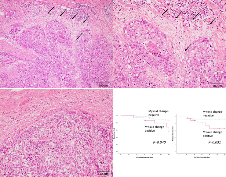
Myxoid change was observed in 16 patients (25.8%) in this cohort (Fig 1A and 1B). Fig 1C shows that there was only fibrotic stroma without myxoid change around the tumour nests. Table 2 summarizes the clinicopathological factors in myxoid change-positive and–negative patients. Myxoid change was significantly associated with low/intermediate TILs in the stroma (p = 0.001). However, there were no significant differences in pathological stage, lymph node status, and Nottingham histological grades between the groups. Fig 1D shows the OS and RFS curves of myxoid change-positive and -negative patients. Presence of myxoid change was significantly correlated with both poor OS (p = 0.040) and RFS (p = 0.031) in patients with TNBC.

Fig 1. Myxoid change as an independent predictor of survival in patients with triple-negative breast cancer.

Fig 1

(a) (b) Representative image of myxoid change in triple-negative breast cancer. Accumulation of amorphous stromal substances around the tumor nests (arrows) (x 100 (a) and x 200 (b)). (c) Only fibrous stroma is present, and no myxoid change is observed around the tumor nests (x 200). (d) Kaplan–Meier curves for the overall survival (OS) and relapse-free survival (RFS) of patients with triple-negative breast cancer. OS (left) and RFS (right) curves among myxoid change-positive (red line) and myxoid change-negative (blue line) patients.

Table 2. Association of myxoid change with clinicopathological factors.

Factors Myxoid change -positive (n = 16) Myxoid change -negative (n = 46) P-value
Age (years; median ± standard deviation) 67±17 68±14 0.910
Menopausal status  
Premenopausal 3 6 0.686
Postmenopausal 13 39  
Unknown 0 1  
Body mass index  
  21.1±3.3 23.9±3.6 0.017
Tumor size (mm)  
≦20 5 27 0.083
>20 11 19  
Pathological stage  
I + II 12 42 0.187
III 4 4  
Lymph node status  
Positive 4 10 0.456
Negative 6 27  
Not tested 6 9  
Lymphatic invasion  
Positive 16 37 0.096
Negative 0 9  
Venous invasion  
Positive 13 24 0.074
Negative 3 22  
Nottingham histological grade  
1+2 5 21 0.386
3 11 25  
Ki-67 labeling index (LI)
High 9 27 1.00
Low 6 16  
Not tested 1 3  
Stromal tumor-infiltrating lymphocytes  
High 0 19 0.001
Low/intermediate 16 27  
Adjuvant chemotherapy  
Performed 7 27 0.549
Not performed 7 18  
Undetermined 2 1  

Association between FF and survival

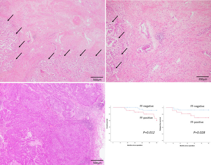
FF was noted in 21 patients (33.9%) in this cohort (Fig 2A and 2B). Fig 2C shows tumor nests without FF. Table 3 summarizes the clinicopathological factors in FF-positive and -negative patients. FF was significantly associated with venous invasion and a high Nottingham histological grade (p = 0.003 and 0.033, respectively). However, pathological stage, lymph node status, and lymphatic invasion were not significantly different across both groups. Presence of FF was significantly associated with low/intermediate TILs in the stroma (p = 0.001). Fig 2D shows the OS and RFS curves of FF-positive and -negative patients. Presence of FF was significantly correlated with poor OS (p = 0.012) and RFS (p = 0.028) in patients with TNBC.

Fig 2. Fibrotic focus as an independent predictor of survival in patients with triple-negative breast cancer.

Fig 2

(a) (b) Representative image of fibrotic focus (FF) in triple-negative breast cancer. Fibrotic area within the tumor containing collagen surrounded by a highly cellular zone of infiltrating carcinoma (arrows) (x 40 (a) and x 100 (b)). (c) No FF is observed within the tumor (x 40). (d) Kaplan-Meier curves for the overall survival (OS) and relapse-free survival (RFS) of triple-negative patients with breast cancer. OS (left) and RFS (right) curves among FF-positive (red line) and FF-negative (blue line) patients.

Table 3. Association of fibrotic focus with clinicopathological factors.

Factors Fibrotic focus -positive (n = 21) Fibrotic focus -negative (n = 41) P-value
Age (years; median ± standard deviation) 74±16 67±14 0.067
Menopausal status  
Premenopausal 3 6 1.00
Postmenopausal 18 34  
Unknown 0 1  
Body mass index  
  23.4±3.6 23.0±3.7 0.800
Tumor size (mm)  
≦20 7 25 0.060
>20 14 16  
Pathological stage  
I + II 17 37 0.426
III 4 4  
Lymph node status  
Positive 7 7 0.080
Negative 7 26  
Not tested 7 8  
Lymphatic invasion  
Positive 20 33 0.150
Negative 1 8  
Venous invasion  
Positive 18 19 0.003
Negative 3 22  
Nottingham histological grade  
1+2 6 24 0.033
3 15 17  
Ki-67 labeling index (LI)
High 13 23 0.783
Low 7 15  
Not tested 1 3  
Stromal tumor-infiltrating lymphocytes  
High 0 19 0.001
Low/intermediate 21 22  
Adjuvant chemotherapy  
Performed 8 26 0.158
Not performed 11 14  
Undetermined 2 1  

Association of combined myxoid change and FF with clinicopathological parameters

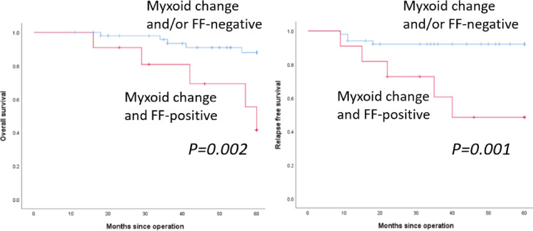
Both myxoid change and FF were observed simultaneously in 11 patients (17.7%), and only myxoid change or FF were noted in 5 and 10 patients, respectively. Fig 3 shows the OS and RFS curves of myxoid change- and FF-positive vs. myxoid change- and/or FF-negative patients. Presence of both myxoid change and FF was significantly correlated with poor OS (p = 0.002) and RFS (p = 0.001) in patients with TNBC.

Fig 3. Survival for myxoid change- and fibrotic focus-positive patients vs. myxoid change- and/or fibrotic focus-negative patients.

Fig 3

Overall survival (OS) (left) and relapse-free survival (RFS) (right) curves in myxoid change and FF-positive (red line) and myxoid change- and/or FF-negative (blue line) patients.

Table 4 summarizes the association of both myxoid change- and FF-positive patients vs. myxoid change- and/or FF-negative patients with clinicopathological factors. Myxoid change- and FF-positive patients were significantly associated with the presence of venous invasion and a high Nottingham histological grade (p = 0.002 and 0.044, respectively) (Table 4). However, pathological stage, lymph node status, lymphatic invasion, and Ki-67 LI were not significantly different between both groups. Presence of myxoid change and FF were significantly associated with low/intermediate TILs in the stroma (p = 0.013).

Table 4. Association of myxoid change and fibrotic focus with clinicopathological factors.

Factors Myxoid change and fibrotic focus-positive (n = 11) Myxoid change and/or fibrotic focus-negative (n = 51) P-value
Age (years old; median ± standard deviation) 64±19 68±14 0.658
Menopausal status  
Premenopausal 2 7 0.660
Postmenopausal 9 43  
Unknown 0 1  
Body mass index  
  21.9±3.7 23.3±3.6 0.246
Tumor size (mm)  
≦20 3 28 0.182
>20 8 23  
Pathological stage  
I + II 8 46 0.142
III 3 5  
Lymph node status  
Positive 3 11 0.344
Negative 3 30  
Not tested 5 10  
Lymphatic invasion  
Positive 11 42 0.348
Negative 0 9  
Venous invasion  
Positive 11 26 0.002
Negative 0 25  
Nottingham histological grade  
1+2 2 28 0.044
3 9 23  
Ki-67 labeling index (LI)
High 4 25 0.730
Low 6 23  
Not tested 1 3  
Stromal tumor-infiltrating lymphocytes  
High 0 19 0.013
Low/intermediate 11 32  
Adjuvant chemotherapy  
Performed 4 30 0.297
Not performed 6 19  
Undetermined 1 2  

Moreover, a multivariate analysis of RFS clearly demonstrated that the simultaneous presence of myxoid change and FF was an independent poor prognostic factor (hazard ratio [HR] 11.61; 95% confidence interval [CI] 1.027–131.2; p = 0.048) (Table 5). In addition, a multivariate analysis of RFS, performed as a separate factor of myxoid change or FF showed that both myxoid change (HR 1.78; 95% CI 0.13–23.7; p = 0.66) and FF (HR 5.18; 95% CI 0.34–79.7; p = 0.24) were not independent poor prognostic factors.

Table 5. Multivariate analysis of relapse-free survival.

Factor Multivariate analysis
  HR 95% CI P-value
Tumor size (mm)  
20 < vs. ≦ 20 0.297 0.021–4.093 0.364
Lymph node status  
Positive vs. negative 11.82 0.647–216.0 0.096
Lymphatic invasion  
Positive vs. negative 4.846×10⁵ 0 0.980
Venous invasion  
Positive vs. negative 0.560 0.016–19.50 0.749
Nottingham histological grade  
3 vs. 1+2 1.721 0.109–27.16 0.700
Ki-67 labelling index (LI)  
High vs. low/intermediate 2.240 0.163–30.86 0.547
Stromal tumor-infiltrating lymphocytes  
High vs. low 2.775 0.186–41.43 0.459
Adjuvant chemotherapy  
Not performed vs. performed 5.462 0.519–57.52 0.158
Myxoid change and fibrotic focus  
Positive vs. negative 11.61 1.027–131.2 0.048

Association between myxoid change, FF, and a combination of myxoid change and FF and survival in patients with adjuvant chemotherapy

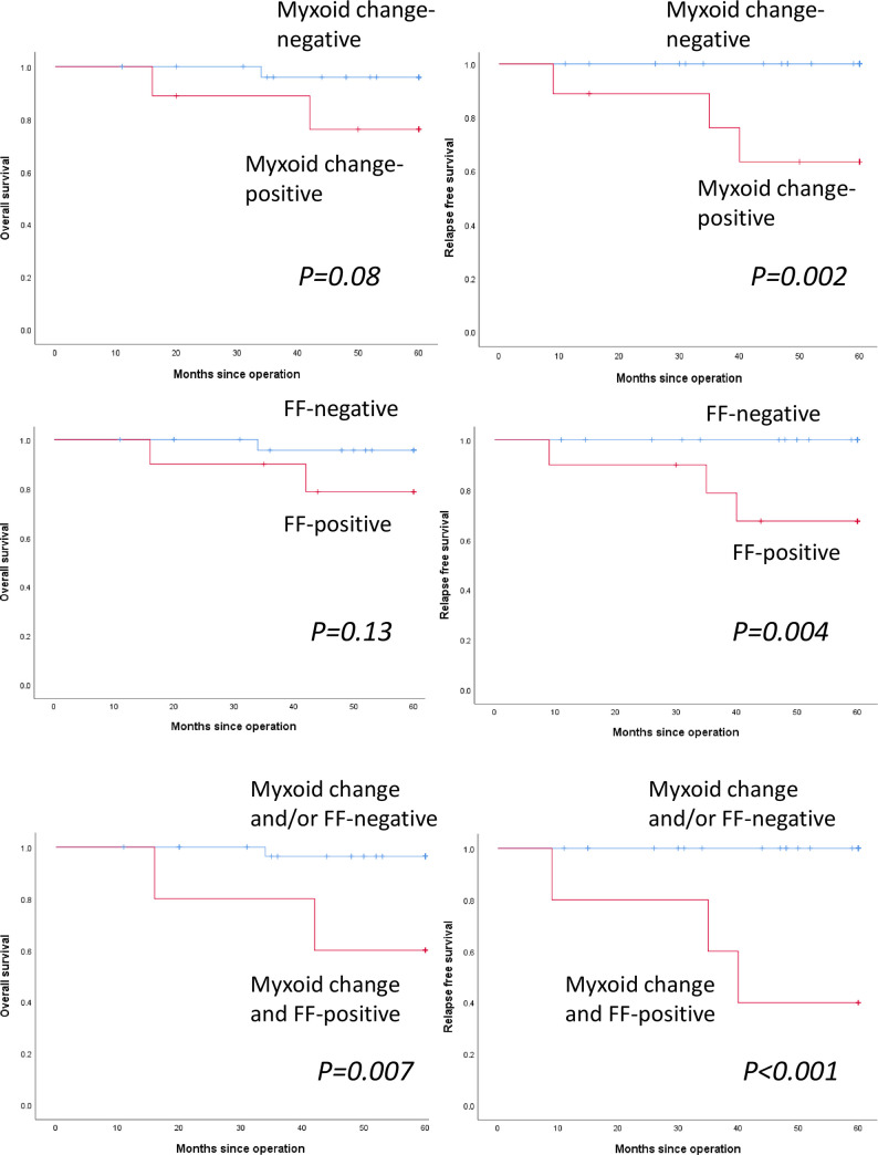
Consequently, we performed survival analyses in patients with or without adjuvant chemotherapy. Myxoid change was observed in 7 of 34 patients (20.6%) with adjuvant chemotherapy. Fig 4A shows the OS and RFS curves of myxoid change-positive and -negative patients with adjuvant chemotherapy. Presence of myxoid change was significantly correlated with poor RFS (p = 0.002), but not with OS (p = 0.080). FF was observed in 8 of 34 patients (23.5%) with adjuvant chemotherapy. Fig 4B shows the OS and RFS curves of FF-positive and -negative patients with adjuvant chemotherapy. Presence of FF was significantly correlated with poor RFS (p = 0.004), but not with OS (p = 0.13). Both myxoid change and FF were observed in 4 of 34 patients (11.8%) with adjuvant chemotherapy. Fig 4C shows the OS and RFS curves of myxoid change- and FF-positive vs myxoid change- and/or FF-negative patients with adjuvant chemotherapy. The presence of a combination of myxoid change and FF was significantly correlated with poor OS (p = 0.007) and RFS (p<0.001).

Fig 4. Survival analyses for myxoid change, fibrotic focus (FF), and a combination of myxoid change and FF in patients with adjuvant chemotherapy.

Fig 4

(a) Kaplan–Meier curves for the overall survival (OS) and relapse-free survival (RFS) of patients with adjuvant chemotherapy. OS (left) and RFS (right) curves among myxoid change-positive (red line) and myxoid change-negative (blue line) patients. (b) OS (left) and RFS (right) curves among FF-positive (red line) and FF-negative (blue line) patients. (c) Overall survival (OS) (left) and relapse-free survival (RFS) (right) curves in myxoid change and FF-positive (red line) and myxoid change- and/or FF-negative (blue line) patients.

Association between myxoid change, FF, and a combination of myxoid change and FF and survival in patients without adjuvant chemotherapy

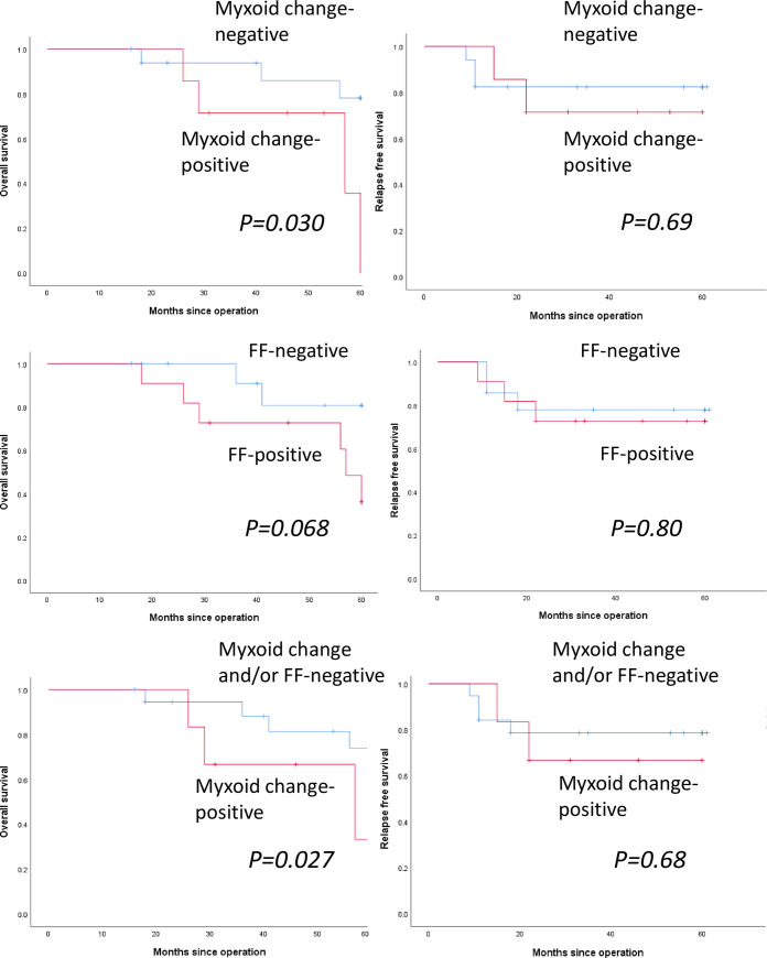
Myxoid change was observed in 7 of 25 patients (28.0%) without adjuvant chemotherapy. Fig 5A shows the OS and RFS curves of myxoid change-positive and -negative patients without adjuvant chemotherapy. Presence of myxoid change was significantly correlated with poor OS (p = 0.030), but not with poor RFS (p = 0.069). FF was observed in 11 of 25 patients (44.0%) without adjuvant chemotherapy. Fig 5B shows the OS and RFS curves of FF-positive and -negative patients without chemotherapy. Presence of FF was not significantly correlated with OS (p = 0.068) and RFS (p = 0.080). Both myxoid change and FF were observed in 6 of 25 patients (24.0%) without adjuvant chemotherapy. Fig 5C shows the OS and RFS curves of myxoid change- and FF-positive vs myxoid change- and/or FF-negative patients without adjuvant chemotherapy. The presence of a combination of myxoid change and FF was significantly correlated with poor OS (p = 0.027), but not with RFS (p = 0.68).

Fig 5. Survival analyses for myxoid change, fibrotic focus (FF), and a combination of myxoid change and fibrotic focus in patients without adjuvant chemotherapy.

Fig 5

(a) Kaplan–Meier curves for the overall survival (OS) and relapse-free survival (RFS) of patients without adjuvant chemotherapy. OS (left) and RFS (right) curves among myxoid change-positive (red line) and myxoid change-negative (blue line) patients. (b) OS (left) and RFS (right) curves among FF-positive (red line) and FF-negative (blue line) patients. (c) Overall survival (OS) (left) and relapse-free survival (RFS) (right) curves in myxoid change and FF-positive (red line) and myxoid change- and/or FF-negative (blue line) patients.

Comparison of the presence of myxoid change between pre-operative biopsy specimens and surgically resected specimens

Pre-operative biopsy specimens were available in 44 patients (71%) in this cohort. Of the 11 myxoid change-positive patients diagnosed via surgically resected specimens, 9 patients were positive for myxoid change via biopsy specimens. Besides, of the 33 myxoid change-negative patients diagnosed via surgically resected specimens, 25 patients were negative for myxoid change via the biopsy specimens. Regarding the presence of myxoid change, there was a moderate correlation between biopsy and surgically resected specimens (Kappa statistics: 0.487, p = 0.001). FF was noted only in 5 biopsy specimens; thus, statistical analysis was not performed.

Discussion

TNBC is one of the most challenging molecular subtypes of invasive breast cancer because of its aggressive clinical behavior and the scarcity of targeted therapies; however, recently, an anti-programmed cell death 1 (anti-PD-L1) targeted therapy has emerged as a promising treatment for TNBC [15]. There are no established prognostic factors in patients with TNBC, although some histopathological prognostic indicators have been proposed. FF is a scar-like area in the center of a carcinoma, representing hypoxia [10]. One report demonstrated that FF was associated with higher histological grades, higher T stage, frequent lymph node metastasis, and poor prognosis [16]. In contrast, another study concluded that FF was found in as many as 18% of patients with breast cancer (including all molecular subtypes), and hence was not a significant prognostic factor [4]. In the present cohort, the presence of FF was a significant poor prognostic indicator for both OS and RFS, which is consistent with the results of the first study [10, 16].

Stromal reaction has also attracted attention in various types of carcinomas [6, 7] because the tumor microenvironment plays important roles in cancer growth, invasion, and metastasis. Further, stromal reaction is one of the main components of the tumor microenvironment [1]. However, stromal reaction in breast cancer has not received enough attention, although the tumor–stroma ratio (or percentage) has been shown to have a prognostic value [17, 18]. In addition, it has been reported that stromal myxoid change represented hyaluronan accumulation and was significantly associated with lymph node metastasis and a higher histological tumor grade; however, it was not an independent prognostic indicator in patients with breast cancer (molecular subtypes were not evaluated) [19]. Moreover, a recent report showed that 26.9% of the breast cancer specimens, which included all molecular subtypes, had myxoid (immature) stroma, and patients with myxoid stroma showed a significantly poorer survival [8]. Myxoid stroma in breast cancer was linked to a higher histological grade and positive lymph nodes [20]. Therefore, the presence of myxoid stroma may be a poor prognostic indicator in breast cancer, consistent with the findings in colorectal cancer [6, 7]. However, the prognostic significance of the presence of myxoid stroma in patients with TNBC had not been explored. In the present study, we demonstrated that myxoid change was a significant poor prognostic indicator for OS and RFS in patients with TNBC. Interestingly, we further revealed that the simultaneous presence of myxoid change and FF was significantly correlated with poorer OS and RFS, compared with the presence of either myxoid change or FF. To the best of our knowledge, this is the first study, through multivariate analysis, to show that a combination of myxoid change and FF was an independent poor prognostic indicator in patients with TNBC. The results of the present study indicate that histopathological assessment of myxoid change and FF in breast cancer tissues of patients with TNBC may be a useful, practical, and easily assessable method for predicting such patients’ prognosis. Although the present study demonstrated a moderate correlation between presence of myxoid change and both pre-operative biopsy and surgically resected specimens, the prognostic significance of presence of myxoid change in the pre-operative biopsy specimens must be clarified in a larger cohort.

Moreover, for the first time, the present study analyzed the survival significance of the presence of myxoid change and/or FF in patients with TNBC receiving adjuvant chemotherapy or not. According to the results of the present study, the presence of myxoid change, FF, and the combination of myxoid change and FF were significant poor prognostic factors for RFS in patients with adjuvant chemotherapy. However, this trend was not noted in patients without adjuvant chemotherapy. Besides, myxoid change-, FF-, and myxoid change and FF-positive patients showed similar survival curves regardless of the presence of adjuvant chemotherapy; however, myxoid change-, FF-, and myxoid change and/or FF-negative patients without adjuvant chemotherapy showed poorer survival curves compared to those of patients with adjuvant chemotherapy. Accordingly, adjuvant chemotherapy may not be efficient in improving outcomes in myxoid change- and/or FF-positive patients with TNBC.

However, the molecular events explaining the association of myxoid change with poor prognosis remain unknown, and this must be revealed to establish novel targeted therapies to treat patients with myxoid stroma who show a poor survival. Notably, there have been reports that the possible hypothesis explaining molecular events in myxoid change in colorectal cancer was associated with inhibition of angiogenesis, which hinders access of the cancer cells by immune cells, leading to altered effects of cancer-associated fibroblasts on the local tumor microenvironment [6].Notably, in the present cohort, the combination of myxoid change and FF was significantly associated with low/intermediate TILs. Moreover, the association between TILs and desmoplasia in metastatic breast cancer has also been reported [21]. Therefore, the presence of myxoid change and FF in TNBC stroma may be a barrier to immune-cell infiltration. Furthermore, the detailed mechanism of myxoid change in the tumor stroma, which represents hyaluronan accumulation, is unclear, although the patterns of stromal reaction are recognized to be associated with tumor microenvironment, including immune cells and cancer-associated fibroblasts [6]. Neoadjuvant chemotherapy may affect the tumor microenvironment, which may lead to a change of the stromal reaction of TNBC. Thus, additional studies are needed to clarify the molecular events underlying the formation of myxoid change in TNBC stroma.

There are some limitations in the present study. First, the present retrospective study included a relatively small number of patients with TNBC. Therefore, additional validation studies with better designs are needed to clarify the prognostic significance of myxoid change and FF in patients with TNBC. Second, definitive assessable and reproductive criteria for myxoid change and FF are needed. Therefore, we performed this study using the previously described criteria of myxoid change that were used in colorectal cancer [6, 7] and FF [10]. Diagnostic criteria of myxoid change and FF for breast cancer specimens must be established for routine use to predict the prognosis.

Conclusions

This study clearly suggests that myxoid change and FF are independent poor prognostic indicators in patients with TNBC of the present cohort. Additionally, the combination of myxoid change and FF is also an independent poor prognostic indicator in patients with TNBC, as seen in the multivariate analysis in this study. Besides, our findings suggest that the presence of myxoid change and FF in TNBC stroma may be a barrier to immune-cell infiltration. The results of the present study indicate that histopathological assessment of myxoid change and FF in breast cancer tissues of patients with TNBC may be a useful, practical, and easily assessable method for predicting the prognosis of patients with TNBC, which should be confirmed in prospective studies with larger samples. In addition, diagnostic criteria for the establishment of myxoid change and FF in TNBC must be established. Additional studies are also required to clarify the molecular events underlying the myxoid change in TNBC.

Supporting information

S1 Dataset

(PDF)

Data Availability

All relevant data are in the paper and the Supporting Information files.

Funding Statement

The authors received no specific funding for this work.

References

  • 1.Liu T, Zhou L, Li D, Andl T, Zhang Y. Cancer-associated fibroblasts build and secure the tumor microenvironment. Front Cell Dev Biol. 2019; 7: 60 10.3389/fcell.2019.00060 [DOI] [PMC free article] [PubMed] [Google Scholar]
  • 2.Sahai E, Astsaturov I, Cukierman E, DeNardo DG, Egeblad M, Evans RM, et al. A framework for advancing our understanding of cancer-associated fibroblasts. Nat Rev Cancer. 2020; 20: 174–186. 10.1038/s41568-019-0238-1 [DOI] [PMC free article] [PubMed] [Google Scholar]
  • 3.Eiro N, Gonzalez LO, Fraile M, Cid S, Schneider J, Vizoso FJ. Breast cancer tumor stroma: cellular components, phenotypic heterogeneity, intercellular communication, prognostic implications and therapeutic opportunities. Cancers (Basel). 2019; 11: E664 10.3390/cancers11050664 [DOI] [PMC free article] [PubMed] [Google Scholar]
  • 4.Ahn S, Cho J, Sung J, Lee JE, Nam SJ, Kim KM, et al. The prognostic significance of tumor-associated stroma in invasive breast carcinoma. Tumour Biol. 2012; 33: 1573–1580. 10.1007/s13277-012-0411-6 [DOI] [PubMed] [Google Scholar]
  • 5.Kalluri R. The biology and function of fibroblasts in cancer. Nat Rev Cancer. 2016; 16: 582–598. 10.1038/nrc.2016.73 [DOI] [PubMed] [Google Scholar]
  • 6.Ueno H, Kanemitsu Y, Sekine S, Ishiguro M, Ito E, Hashiguchi Y, et al. A multicenter study of the prognostic value of desmoplastic reaction categorization in stage II colorectal cancer. Am J Surg Pathol. 2019; 43: 1015–1022. 10.1097/PAS.0000000000001272 [DOI] [PubMed] [Google Scholar]
  • 7.Ueno H, Shinto E, Shimazaki H, Kajiwara Y, Sueyama T, Yamamoto J, et al. Histologic categorization of desmoplastic reaction: its relevance to the colorectal cancer microenvironment and prognosis. Ann Surg Oncol. 2015; 22: 1504–1512. 10.1245/s10434-014-4149-9 [DOI] [PubMed] [Google Scholar]
  • 8.Zhai Q, Fan J, Lin Q, Liu X, Li J, Hong R, et al. Tumor stromal type is associated with stromal PD-L1 expression and predicts outcomes in breast cancer. PLoS One. 2019; 14: e0223325 10.1371/journal.pone.0223325 [DOI] [PMC free article] [PubMed] [Google Scholar]
  • 9.Mujtaba SS, Ni YB, Tsang JY, Chan SK, Yamaguchi R, Tanaka M, et al. Fibrotic focus in breast carcinomas: relationship with prognostic parameters and biomarkers. Ann Surg Oncol. 2013; 20: 2842–2849. 10.1245/s10434-013-2955-0 [DOI] [PubMed] [Google Scholar]
  • 10.Van den Eynden GG, Colpaert CG, Couvelard A, Pezzella F, Dirix LY, Vermeulen PB, et al. A fibrotic focus is a prognostic factor and a surrogate marker for hypoxia and (lymph)angiogenesis in breast cancer: review of the literature and proposal on the criteria of evaluation. Histopathology. 2007; 51: 440–451. 10.1111/j.1365-2559.2007.02761.x [DOI] [PubMed] [Google Scholar]
  • 11.Salgado R, Denkert C, Demaria S, Sirtaine N, Klauschen F, Pruneri G, et al. The evaluation of tumor-infiltrating lymphocytes (TILs) in breast cancer: recommendations by an International TILs Working Group 2014. Ann Oncol. 2015; 26: 259–271. 10.1093/annonc/mdu450 [DOI] [PMC free article] [PubMed] [Google Scholar]
  • 12.Denkert C, von Minckwitz G, Darb-Esfahani S, Lederer B, Heppner BI, Weber KE, et al. Tumour-infiltrating lymphocytes and prognosis in different subtypes of breast cancer: a pooled analysis of 3771 patients treated with neoadjuvant therapy. Lancet Oncol. 2018; 19: 40–50. 10.1016/S1470-2045(17)30904-X [DOI] [PubMed] [Google Scholar]
  • 13.Elston CW, Ellis IO. Pathological prognostic factors in breast cancer. I. The value of histological grade in breast cancer: experience from a large study with long-term follow-up. Histopathology. 1991; 19: 403–410. 10.1111/j.1365-2559.1991.tb00229.x [DOI] [PubMed] [Google Scholar]
  • 14.Wu Q, Ma G, Deng Y, Luo W, Zhao Y, Li W, et al. Prognostic value of Ki-67 in patients with resected triple-negative breast cancer: A meta-analysis. Front. Oncol. 2019; 9: 1068 10.3389/fonc.2019.01068 [DOI] [PMC free article] [PubMed] [Google Scholar]
  • 15.Schmid P, Adams S, Rugo HS, Schneeweiss A, Barrios CH, Iwata H, et al. Atezolizumab and nab-paclitaxel in advanced triple-negative breast cancer. N Engl J Med. 2018; 379: 2108–2121. 10.1056/NEJMoa1809615 [DOI] [PubMed] [Google Scholar]
  • 16.Hasebe T, Sasaki S, Imoto S, Mukai K, Yokose T, Ochiai A. Prognostic significance of fibrotic focus in invasive ductal carcinoma of the breast: a prospective observational study. Mod Pathol. 2002; 15: 502–516. 10.1038/modpathol.3880555 [DOI] [PubMed] [Google Scholar]
  • 17.Gujam FJ, Edwards J, Mohammed ZM, Going JJ, McMillan DC. The relationship between the tumour stroma percentage, clinicopathological characteristics and outcome in patients with operable ductal breast cancer. Br J Cancer. 2014; 111: 157–165. 10.1038/bjc.2014.279 [DOI] [PMC free article] [PubMed] [Google Scholar]
  • 18.Moorman AM, Vink R, Heijmans HJ, van der Palen J, Kouwenhoven EA. The prognostic value of tumour-stroma ratio in triple-negative breast cancer. Eur J Surg Oncol. 2012; 38: 307–313. 10.1016/j.ejso.2012.01.002 [DOI] [PubMed] [Google Scholar]
  • 19.Wernicke M, Piñeiro LC, Caramutti D, Dorn VG, Raffo MM, Guixa HG, et al. Breast cancer stromal myxoid changes are associated with tumor invasion and metastasis: a central role for hyaluronan. Mod Pathol. 2003; 16: 99–107. 10.1097/01.MP.0000051582.75890.2D [DOI] [PubMed] [Google Scholar]
  • 20.Walker RA. The complexities of breast cancer desmoplasia. Breast Cancer Res. 2001; 3: 143–145. 10.1186/bcr287 [DOI] [PMC free article] [PubMed] [Google Scholar]
  • 21.Chen I X, Chauhan VP, Posada J, Ng MR, Wu MW, Adstamongkonkul P, et al. Blocking CXCR4 alleviates desmoplasia, increases T-lymphocyte infiltration, and improves immunotherapy in metastatic breast cancer. Proc Natl Acad Sci U S A. 2019; 116: 4558–4566. 10.1073/pnas.1815515116 [DOI] [PMC free article] [PubMed] [Google Scholar]

Decision Letter 0

Elda Tagliabue

23 Sep 2020

PONE-D-20-23411

Presence of myxoid stromal change and fibrotic focus in pathological examination are prognostic factors of triple-negative breast cancer: Results from a retrospective single-center study

PLOS ONE

Dear Dr. Ishida,

Thank you for submitting your manuscript to PLOS ONE. After careful consideration, we feel that it has merit but does not fully meet PLOS ONE’s publication criteria as it currently stands. Therefore, we invite you to submit a revised version of the manuscript that addresses the points raised during the review process.

Please ensure that your decision is justified on PLOS ONE’s publication criteria and not, for example, on novelty or perceived impact.

Please submit your revised manuscript by December 1st. If you will need more time than this to complete your revisions, please reply to this message or contact the journal office at plosone@plos.org. When you're ready to submit your revision, log on to https://www.editorialmanager.com/pone/ and select the 'Submissions Needing Revision' folder to locate your manuscript file.

Please include the following items when submitting your revised manuscript:

  • A rebuttal letter that responds to each point raised by the academic editor and reviewer(s). You should upload this letter as a separate file labeled 'Response to Reviewers'.

  • A marked-up copy of your manuscript that highlights changes made to the original version. You should upload this as a separate file labeled 'Revised Manuscript with Track Changes'.

  • An unmarked version of your revised paper without tracked changes. You should upload this as a separate file labeled 'Manuscript'.

If you would like to make changes to your financial disclosure, please include your updated statement in your cover letter. Guidelines for resubmitting your figure files are available below the reviewer comments at the end of this letter.

If applicable, we recommend that you deposit your laboratory protocols in protocols.io to enhance the reproducibility of your results. Protocols.io assigns your protocol its own identifier (DOI) so that it can be cited independently in the future. For instructions see: http://journals.plos.org/plosone/s/submission-guidelines#loc-laboratory-protocols

We look forward to receiving your revised manuscript.

Kind regards,

Elda Tagliabue

Academic Editor

PLOS ONE

Journal Requirements:

When submitting your revision, we need you to address these additional requirements.

1. Please ensure that your manuscript meets PLOS ONE's style requirements, including those for file naming. The PLOS ONE style templates can be found at

https://journals.plos.org/plosone/s/file?id=wjVg/PLOSOne_formatting_sample_main_body.pdf and

https://journals.plos.org/plosone/s/file?id=ba62/PLOSOne_formatting_sample_title_authors_affiliations.pdf

2.In your Data Availability statement, you have not specified where the minimal data set underlying the results described in your manuscript can be found. PLOS defines a study's minimal data set as the underlying data used to reach the conclusions drawn in the manuscript and any additional data required to replicate the reported study findings in their entirety. All PLOS journals require that the minimal data set be made fully available. For more information about our data policy, please see http://journals.plos.org/plosone/s/data-availability.

Upon re-submitting your revised manuscript, please upload your study’s minimal underlying data set as either Supporting Information files or to a stable, public repository and include the relevant URLs, DOIs, or accession numbers within your revised cover letter. For a list of acceptable repositories, please see http://journals.plos.org/plosone/s/data-availability#loc-recommended-repositories. Any potentially identifying patient information must be fully anonymized.

Important: If there are ethical or legal restrictions to sharing your data publicly, please explain these restrictions in detail. Please see our guidelines for more information on what we consider unacceptable restrictions to publicly sharing data: http://journals.plos.org/plosone/s/data-availability#loc-unacceptable-data-access-restrictions. Note that it is not acceptable for the authors to be the sole named individuals responsible for ensuring data access.

We will update your Data Availability statement to reflect the information you provide in your cover letter.

3.Your ethics statement should only appear in the Methods section of your manuscript. If your ethics statement is written in any section besides the Methods, please delete it from any other section.

[Note: HTML markup is below. Please do not edit.]

Reviewers' comments:

Reviewer's Responses to Questions

Comments to the Author

1. Is the manuscript technically sound, and do the data support the conclusions?

The manuscript must describe a technically sound piece of scientific research with data that supports the conclusions. Experiments must have been conducted rigorously, with appropriate controls, replication, and sample sizes. The conclusions must be drawn appropriately based on the data presented.

Reviewer #1: Partly

Reviewer #2: Partly

**********

2. Has the statistical analysis been performed appropriately and rigorously?

Reviewer #1: Yes

Reviewer #2: No

**********

3. Have the authors made all data underlying the findings in their manuscript fully available?

The PLOS Data policy requires authors to make all data underlying the findings described in their manuscript fully available without restriction, with rare exception (please refer to the Data Availability Statement in the manuscript PDF file). The data should be provided as part of the manuscript or its supporting information, or deposited to a public repository. For example, in addition to summary statistics, the data points behind means, medians and variance measures should be available. If there are restrictions on publicly sharing data—e.g. participant privacy or use of data from a third party—those must be specified.

Reviewer #1: Yes

Reviewer #2: Yes

**********

4. Is the manuscript presented in an intelligible fashion and written in standard English?

PLOS ONE does not copyedit accepted manuscripts, so the language in submitted articles must be clear, correct, and unambiguous. Any typographical or grammatical errors should be corrected at revision, so please note any specific errors here.

Reviewer #1: Yes

Reviewer #2: Yes

**********

5. Review Comments to the Author

Please use the space provided to explain your answers to the questions above. You may also include additional comments for the author, including concerns about dual publication, research ethics, or publication ethics. (Please upload your review as an attachment if it exceeds 20,000 characters)

Reviewer #1: In this paper the authors studied stromal reaction in TNBC tumors as prognostic factors. Specifically they assessed that the presence of myxoid change and fibrotic focus are poor prognostic factors in TNBC patients in term of overall survival and disease free survival and that the combination of both parameters was an independent poor prognostic factor.

The major limitation of this study lies on the difficulty to generalize the clinical value of the prognostic factors described due to the fact that the authors analyzed only a cohort of 62 TNBC patients without a validation cohort.

The paper is potentially interesting anyway a number of compulsory aspects need to be clarified and the following comments should be addressed before the manuscript can be considered for publication:

Major points:

• The authors analyzed only a cohort of 62 TNBC patients. Please add a validation cohort.

• As reported in Table 1, 56.5% of patients received adjuvant chemotherapy and 38.7% did not received chemotherapy. The analysis of OS and RFS for all stromal prognostic factors considered should be performed considering separately patients who received/not received treatment. Please add these analyses.

• In figures 1a and 2a the authors report only a representative image at large magnification for myxoid change and fibrotic focus respectively. Please add also images at lower magnification and images of tumor sample negative for myxoid change and fibrotic focus.

• Correlation analysis with clinicopathological factors, reported in Table 2 was done only considering patients with both mixoid change and fibrotic focus together. Please add correlation analysis considering separately the two stromal characteristics.

Minor points:

• In the introduction section the authors should better support sentences at lines 62-65 with references from literature

• Table 1: tumor size is considered as a continuous variable. Could the authors indicate tumor size as they do in Table 2 and 3 for correlation and multivariate analyzes?

• As regards the analysis of TILs, the paper the authors refer to, considers also “intermediate TILs as 10%<tils≤59%”. analysis="">

Overall the manuscript can be interesting but it cannot be accepted in the present form. A major revision is mandatory before publication.</tils≤59%”.>

Reviewer #2: Hirotsugu Yanai and collaborators examined on 62 triple negative breast cancers the clinical meaning of the histopathological assessment of myxoid change and FF fibrotic focus. Although their results seem to be novel and promising there are important issues that need to be addressing.

Major issues

Methods

1. Although there are two cited papers regarding the criteria to detect myxoid change it would be useful to clearly explain the characteristics taking into account for the analysis.

2. There is any score to classify positive and negative tissues (%, number of cells, area)?, what was to cutoff to consider a positive tissue?

Results

1. Please include at least two more representative pictures of myxoid changes and FF, and clearly indicate by arrows the area and changes of interest. Please also include images from patients in which the myxoid changes or FF were not observed.

2. On table 2 why positive patients to FF or Myxoid change were grouped with negative patients?. I would group the tumors as following to get a better idea of the independent and coordinate changes: 1) Myxoid change and fibrotic focus-positive (N=11) 2) Myxoid change positive-only (N=5) 3) fibrotic focus-positive (N=10) and 4) Myxoid change and fibrotic focus-positive negative (N=36).

3. As previously mentioned on table 3 it is necessary to show multivariate analysis of stroma changes occurring independently and in co-occurrence, to get an idea of how this phenomena impact tumor biology alone or in combination.

4. Although this is one of the first efforts to associate the presence myxoid changes and FF with clinical outcomes and patients features on TNBC, unfortunately by the limited number of included tumors there is not an optimal statistical strategy that can be robustly applied and the conclusion are not strongly supported, thus I encourage the authors to include some independent cohorts to confirm some of their findings. Since hematoxylin and eosin is the only necessary test to evaluate stroma reactions, I will suggest including some triple negative TCGA tumors that can be evaluated from https://cancer.digitalslidearchive.org/#!/CDSA/brca and clinical and TILS information is available.

5. Since most of the triple negative tumors are now being treated in neoadjuvant therapy, it is possible to examine this stroma reactions from biopsy tissues?. The authors described that they discard an important set of tumoral samples from their original cohort due to neoadjuvant treatment. So 1) It is possible to compare the histopathological assessment of the surgical analyzed tissues, included in this study, with their matched diagnosis biopsies to define how much a biopsy tissue can recapitulate what is detected in a larger tissue (surgical sample) 2) it is possible to include the analysis of this excluded patients based on their biopsy tissue? Please discuss this point.

Minor

Since a Fisher test was computed to examine the significance of the association between two kinds of classification the most accurate term for table 2 and methods section is association instead of correlation.

**********

6. PLOS authors have the option to publish the peer review history of their article (what does this mean?). If published, this will include your full peer review and any attached files.

If you choose “no”, your identity will remain anonymous but your review may still be made public.

Do you want your identity to be public for this peer review? For information about this choice, including consent withdrawal, please see our Privacy Policy.

Reviewer #1: No

Reviewer #2: Yes: Sandra Romero-Cordoba

[NOTE: If reviewer comments were submitted as an attachment file, they will be attached to this email and accessible via the submission site. Please log into your account, locate the manuscript record, and check for the action link "View Attachments". If this link does not appear, there are no attachment files.]

While revising your submission, please upload your figure files to the Preflight Analysis and Conversion Engine (PACE) digital diagnostic tool, https://pacev2.apexcovantage.com/. PACE helps ensure that figures meet PLOS requirements. To use PACE, you must first register as a user. Registration is free. Then, login and navigate to the UPLOAD tab, where you will find detailed instructions on how to use the tool. If you encounter any issues or have any questions when using PACE, please email PLOS at figures@plos.org. Please note that Supporting Information files do not need this step.

PLoS One. 2021 Feb 11;16(2):e0245725. doi: 10.1371/journal.pone.0245725.r002

Author response to Decision Letter 0


3 Dec 2020

November 30, 2020

Dr. Joerg Heber

Editor-in-Chief

PLoS ONE

Resubmission - Manuscript ID: PONE-D-20-23411

Dear Dr. Heber:

I would like to resubmit an article for publication in PLoS ONE, titled “Presence of myxoid stromal change and fibrotic focus in pathological examination are prognostic factors of triple-negative breast cancer: Results from a retrospective single-center study.”

Your comments, as well as those of the reviewers, were highly insightful and enabled us to greatly improve the quality of our manuscript. In the following pages, I have provided our point-by-point response to all comments and have highlighted the revised portions of the manuscript.

I hope that you will find our revised manuscript suitable for publication in PLoS ONE. I look forward to hearing from you at your earliest convenience.

Sincerely,

Mitsuaki Ishida

Kansai Medical University

2-5-1, Shinmachi, Hirakata City

Osaka, 573-1010, Japan

Tel: +81-72-804-2794

Fax: +81-72-804-2794

E-mail: ishidamt@hirakata.kmu.ac.jp

Response to the comments of Reviewer #1

Thank you very much for reviewing our manuscript. We appreciate your constructive comments. We have made the following revisions in response to the issues raised by you.

1. The authors analyzed only a cohort of 62 TNBC patients. Please add a validation cohort.

Response: This study cohort included a relatively small number of patients with triple-negative breast cancer (TNBC), as mentioned in the Discussion section. As you have mentioned, a validation cohort is required to clarify the results of the present study. However, at this point, we are unable to validate the results, and we aim to undertake an international validation cohort study to clarify the usefulness of myxoid change and fibrotic focus in TNBC in the near feature.

2. As reported in Table 1, 56.5% of patients received adjuvant chemotherapy and 38.7% did not received chemotherapy. The analysis of OS and RFS for all stromal prognostic factors considered should be performed considering separately patients who received/not received treatment. Please add these analyses.

Response: As you have suggested, we have additional analyses of OS and RFS in TNBC patients with or without adjuvant chemotherapy. We added the results of these analyses in the Results section and showed the survival curves in Figs. 4 and 5 (p19-22). Moreover, we added the following statement regarding the results in the Discussion section.

“Association between myxoid change, FF, and a combination of myxoid change and FF and survival in patients with adjuvant chemotherapy

Consequently, we performed survival analyses in patients with or without adjuvant chemotherapy. Myxoid change was observed in 7 of 34 patients (20.6%) with adjuvant chemotherapy. Fig. 4a shows the OS and RFS curves of myxoid change-positive and -negative patients with adjuvant chemotherapy. The presence of myxoid change significantly correlated with poor RFS (p=0.002) but not with OS (p=0.080). FF was observed in 8 of 34 patients (23.5%) with adjuvant chemotherapy. Fig. 4b shows the OS and RFS curves of FF-positive and -negative patients with adjuvant chemotherapy. The presence of FF significantly correlated with poor RFS (p=0.004) but not with OS (p=0.13). Both myxoid change and FF were observed in 4 of 34 patients (11.8%) with adjuvant chemotherapy. Fig. 4c shows the OS and RFS curves of myxoid change- and FF-positive vs myxoid change-and/or FF-negative patients with adjuvant chemotherapy. The presence of a combination of myxoid change and FF significantly correlated with poor OS (p=0.007) and RFS (p<0.001).

Fig. 4 Survival analyses for myxoid change, fibrotic focus (FF), and a combination of myxoid change and FF in patients with adjuvant chemotherapy.

(a) Kaplan–Meier curves for the overall survival (OS) and relapse-free survival (RFS) of patients with adjuvant chemotherapy. OS (left) and RFS (right) curves among myxoid change-positive (red line) and myxoid change-negative (blue line) patients. (b) OS (left) and RFS (right) curves among FF-positive (red line) and FF-negative (blue line) patients. (c) Overall survival (OS) (left) and relapse-free survival (RFS) (right) curves in myxoid change and FF-positive (red line) and myxoid change- and/or FF-negative (blue line) patients.

Association between myxoid change, FF, and a combination of myxoid change and FF and survival in patients without adjuvant chemotherapy

Myxoid change was observed in 7 of 25 patients (28.0%) without adjuvant chemotherapy. Fig. 5a shows the OS and RFS curves of myxoid change-positive and -negative patients without adjuvant chemotherapy. The presence of myxoid change significantly correlated with poor OS (p=0.030) but not with poor RFS (p=0.069). FF was observed in 11 of 25 patients (44.0%) without adjuvant chemotherapy. Fig. 5b shows the OS and RFS curves of FF-positive and -negative patients without chemotherapy. The presence of FF did not significantly correlate with OS (p=0.068) and FRS (p=0.080). Both myxoid change and FF were observed in 6 of 25 patients (24.0%) without adjuvant chemotherapy. Fig. 5c shows the OS and RFS curves of myxoid change- and FF-positive vs myxoid change-and/or FF-negative patients without adjuvant chemotherapy. The presence of a combination of myxoid change and FF significantly correlated with poor OS (p=0.027) but not with RFS (p=0.68).

Fig. 5 Survival analyses for myxoid change, fibrotic focus (FF), and a combination of myxoid change and fibrotic focus in patients without adjuvant chemotherapy.

(a) Kaplan–Meier curves for the overall survival (OS) and relapse-free survival (RFS) of patients without adjuvant chemotherapy. OS (left) and RFS (right) curves among myxoid change-positive (red line) and myxoid change-negative (blue line) patients. (b) OS (left) and RFS (right) curves among FF-positive (red line) and FF-negative (blue line) patients. (c) Overall survival (OS) (left) and relapse-free survival (RFS) (right) curves in myxoid change and FF-positive (red line) and myxoid change- and/or FF-negative (blue line) patients.” (page 19-22)

“Moreover, for the first time, the present study analyzed the survival significance of the presence of myxoid change and/or FF in TNBC patients with or without adjuvant chemotherapy. According to the results of the present study, the presence of myxoid change, FF, and the combination of myxoid change and FF were significant poor prognostic factors for RFS in patients with adjuvant chemotherapy. However, this trend was not noted in patients without adjuvant chemotherapy. Besides, myxoid change-, FF-, and myxoid change and FF-positive patients showed similar survival curves regardless of the presence of adjuvant chemotherapy; however, myxoid change-, FF-, and myxoid change and/or FF-negative patients without adjuvant chemotherapy showed poorer survival curves compared to those of patients with adjuvant chemotherapy. Accordingly, adjuvant chemotherapy might not be efficient in improving outcomes in myxoid change- and/or FF-positive TNBC patients.” (page 25)

3. In figures 1a and 2a the authors report only a representative image at large magnification for myxoid change and fibrotic focus respectively. Please add also images at lower magnification and images of tumor sample negative for myxoid change and fibrotic focus.

Response: As you have suggested, we have added the images with a lower magnification of myxoid change (Figs. 1a, ab) or fibrotic focus (Figs. 2a, 2b). We have also added the figures without myxoid change (Fig. 1c) or fibrotic focus (Fig. 2c).

4. Correlation analysis with clinicopathological factors, reported in Table 2 was done only considering patients with both mixoid change and fibrotic focus together. Please add correlation analysis considering separately the two stromal characteristics.

Response: As you have suggested, we have performed additional analyses of the association of myxoid change or fibrotic focus with clinicopathological characteristics, separately. We have illustrated these results in Table 2 and Table 3. The results were fundamentally the same as those of myxoid change and fibrotic focus (Table 4).

5. In the introduction section the authors should better support sentences at lines 62-65 with references from literature

Response: As you have suggested, we have added reference 5.

6. Table 1: tumor size is considered as a continuous variable. Could the authors indicate tumor size as they do in Table 2 and 3 for correlation and multivariate analyzes?

Response: We agree with you that tumor size is a continuous variable. In this cohort, the median tumor size was 20 mm. Notably, TNM classification of the breast cancer classified T1 as 2cm or less, and T2 and T3 as more than 2 cm. Thus, we classified the tumor size as ≦2 cm or >2 cm in this analysis.

7. As regards the analysis of TILs, the paper the authors refer to, considers also “intermediate TILs as 10%

Response: As you have suggested, TIL is classified as low (0-10%), intermediate (11-59%), and high (more than 60%) according to Reference 12. Thus, in this study, we classified TIL as low/intermediate (0-59%) and high (more than 60%).

Response to the comments of Reviewer #2

Thank you very much for reviewing our manuscript. We appreciate your constructive comments. We have made the following revisions in response to the issues raised by you.

1. Although there are two cited papers regarding the criteria to detect myxoid change it would be useful to clearly explain the characteristics taking into account for the analysis.

Response: As you have suggested, we have added a statement regarding the histopathological characteristics of myxoid change in the methods section.

“Myxoid change was histopathologically characterized by the presence of amorphous extracellular substances, which comprise an amphophilic or slightly basophilic material within the fibrous stroma” (page 6)

2. There is any score to classify positive and negative tissues (%, number of cells, area)?, what was to cutoff to consider a positive tissue?

Response: In the present study, we classified tissues as positive when typical myxoid change was present around the tumor according to colorectal cancer criteria (Ref. 6,7), and no cutoff value was present. As you have suggested, definitive assessable and reproductive criteria for myxoid change in breast cancer are needed (Discussion, page 26).

3. Please include at least two more representative pictures of myxoid changes and FF, and clearly indicate by arrows the area and changes of interest. Please also include images from patients in which the myxoid changes or FF were not observed.

Response: As you have suggested, we have added images with a lower magnification of myxoid change (Figs. 1a, ab) or fibrotic focus (Figs. 2a, 2b). We have also added the figures without myxoid change (Fig. 1c) or fibrotic focus (Fig. 2c).

4. On table 2 why positive patients to FF or Myxoid change were grouped with negative patients?. I would group the tumors as following to get a better idea of the independent and coordinate changes: 1) Myxoid change and fibrotic focus-positive (N=11) 2) Myxoid change positive-only (N=5) 3) fibrotic focus-positive (N=10) and 4) Myxoid change and fibrotic focus-positive negative (N=36).

Response: We have performed additional analyses regarding the association of myxoid change or fibrotic focus with clinicopathological characteristics, separately. We have illustrated these results in Table 2 and Table 3. The results were fundamentally the same as those of myxoid change/fibrotic focus (Table 4). We think that analyses of four groups (myxoid change and/or fibrotic focus) might be difficult because there were only 5 patients in the myxoid change-positive/fibrotic focus-negative group.

5. As previously mentioned on table 3 it is necessary to show multivariate analysis of stroma changes occurring independently and in co-occurrence, to get an idea of how this phenomena impact tumor biology alone or in combination.

Response: As you have suggested, we have performed multivariate analyses of relapse-free survival of myxoid change or fibrotic focus, separately. We have added the results as follows.

“In addition, a multivariate analysis of RFS performed as a separate factor of myxoid change or FF showed that both myxoid change (HR 1.78; 95% CI 0.13-23.7; p=0.66) and FF (HR 5.18; 95% CI 0.34-79.7; p=0.24) were not independent poor prognostic factors.” (page 18)

6. Although this is one of the first efforts to associate the presence myxoid changes and FF with clinical outcomes and patients features on TNBC, unfortunately by the limited number of included tumors there is not an optimal statistical strategy that can be robustly applied and the conclusion are not strongly supported, thus I encourage the authors to include some independent cohorts to confirm some of their findings. Since hematoxylin and eosin is the only necessary test to evaluate stroma reactions, I will suggest including some triple negative TCGA tumors that can be evaluated from https://cancer.digitalslidearchive.org/#!/CDSA/brca and clinical and TILS information is available.

Response: We appreciate your kind suggestion. This study cohort included a relatively small number of patients with triple-negative breast cancer (TNBC), as we have acknowledged in the Discussion section. As you mentioned, a validation cohort is required to clarify the results of the present study. However, at this point, we are unable to validate the results, and we aim to undertake an international validation cohort study to clarify the usefulness of myxoid change and fibrotic focus in TNBC in the near feature.

In the present study, the presence of myxoid change or fibrotic focus was examined using surgically resected specimens (more than 2 slices in all cases). While the tool you have suggested is very useful, we believe it is not suitable for this study since only one slice was available in each case.

7. Since most of the triple negative tumors are now being treated in neoadjuvant therapy, it is possible to examine this stroma reactions from biopsy tissues?. The authors described that they discard an important set of tumoral samples from their original cohort due to neoadjuvant treatment. So 1) It is possible to compare the histopathological assessment of the surgical analyzed tissues, included in this study, with their matched diagnosis biopsies to define how much a biopsy tissue can recapitulate what is detected in a larger tissue (surgical sample) 2) it is possible to include the analysis of this excluded patients based on their biopsy tissue? Please discuss this point.

Response: As you have suggested, we have examined the usefulness of analysis of the matched pre-operative biopsy specimens. We have added the results.

“Moreover, we also evaluated the presence of myxoid change and FF using the matched pre-operative biopsy specimens.” (page 6-7)

“Comparison of the presence of myxoid change between pre-operative biopsy specimens and surgically-resected specimens

Pre-operative biopsy specimens were available in 44 patients (71%) in this cohort. Of the 11 myxoid change-positive patients diagnosed via surgically resected specimens, 9 patients were positive for the myxoid change via biopsy specimens. Besides, of the 33 myxoid change-negative patients diagnosed via surgically resected specimens, 25 patients were negative for myxoid change via biopsy specimens. There was a moderate correlation between biopsy and surgically resected specimens regarding the presence of myxoid change (Kappa statistics: 0.487, p=0.001). FF was noted only in 5 biopsy specimens; thus, statistical analysis was not performed.” (page 21-22)

“Additionally, the present study demonstrated a moderate correlation between pre-operative biopsy and surgically resected specimens regarding the presence of myxoid change. Thus, evaluation of the presence of myxoid change in the biopsy specimens might provide prognostic information in patients with TNBC because many patients with TNBC are currently treated with neoadjuvant chemotherapy.” (page 24-25)

8. Since a Fisher test was computed to examine the significance of the association between two kinds of classification the most accurate term for table 2 and methods section is association instead of correlation.

Response: As you have suggested, we have changed “correlation” to “association”.

“The association between groups was evaluated using Fisher’s exact test for categorical variables” (page 7)

Attachment

Submitted filename: Response_to_Reviewers.docx

Decision Letter 1

Elda Tagliabue

17 Dec 2020

PONE-D-20-23411R1

Presence of myxoid stromal change and fibrotic focus in pathological examination are prognostic factors of triple-negative breast cancer: Results from a retrospective single-center study.

PLOS ONE

Dear Dr. Ishida,

Thank you for submitting your manuscript to PLOS ONE. After careful consideration, we feel that it has merit but does not fully meet PLOS ONE’s publication criteria as it currently stands. Therefore, we invite you to submit a revised version of the manuscript that addresses the points raised during the review process.

Please submit your revised manuscript by January 17, 2021. If you will need more time than this to complete your revisions, please reply to this message or contact the journal office at plosone@plos.org. When you're ready to submit your revision, log on to https://www.editorialmanager.com/pone/ and select the 'Submissions Needing Revision' folder to locate your manuscript file.

Please include the following items when submitting your revised manuscript:

  • A rebuttal letter that responds to each point raised by the academic editor and reviewer(s). You should upload this letter as a separate file labeled 'Response to Reviewers'.

  • A marked-up copy of your manuscript that highlights changes made to the original version. You should upload this as a separate file labeled 'Revised Manuscript with Track Changes'.

  • An unmarked version of your revised paper without tracked changes. You should upload this as a separate file labeled 'Manuscript'.

If you would like to make changes to your financial disclosure, please include your updated statement in your cover letter. Guidelines for resubmitting your figure files are available below the reviewer comments at the end of this letter.

If applicable, we recommend that you deposit your laboratory protocols in protocols.io to enhance the reproducibility of your results. Protocols.io assigns your protocol its own identifier (DOI) so that it can be cited independently in the future. For instructions see: http://journals.plos.org/plosone/s/submission-guidelines#loc-laboratory-protocols

We look forward to receiving your revised manuscript.

Kind regards,

Elda Tagliabue

Academic Editor

PLOS ONE

[Note: HTML markup is below. Please do not edit.]

Reviewers' comments:

Reviewer's Responses to Questions

Comments to the Author

1. If the authors have adequately addressed your comments raised in a previous round of review and you feel that this manuscript is now acceptable for publication, you may indicate that here to bypass the “Comments to the Author” section, enter your conflict of interest statement in the “Confidential to Editor” section, and submit your "Accept" recommendation.

Reviewer #1: All comments have been addressed

Reviewer #2: All comments have been addressed

**********

2. Is the manuscript technically sound, and do the data support the conclusions?

The manuscript must describe a technically sound piece of scientific research with data that supports the conclusions. Experiments must have been conducted rigorously, with appropriate controls, replication, and sample sizes. The conclusions must be drawn appropriately based on the data presented.

Reviewer #1: Yes

Reviewer #2: Partly

**********

3. Has the statistical analysis been performed appropriately and rigorously?

Reviewer #1: Yes

Reviewer #2: Yes

**********

4. Have the authors made all data underlying the findings in their manuscript fully available?

The PLOS Data policy requires authors to make all data underlying the findings described in their manuscript fully available without restriction, with rare exception (please refer to the Data Availability Statement in the manuscript PDF file). The data should be provided as part of the manuscript or its supporting information, or deposited to a public repository. For example, in addition to summary statistics, the data points behind means, medians and variance measures should be available. If there are restrictions on publicly sharing data—e.g. participant privacy or use of data from a third party—those must be specified.

Reviewer #1: Yes

Reviewer #2: Yes

**********

5. Is the manuscript presented in an intelligible fashion and written in standard English?

PLOS ONE does not copyedit accepted manuscripts, so the language in submitted articles must be clear, correct, and unambiguous. Any typographical or grammatical errors should be corrected at revision, so please note any specific errors here.

Reviewer #1: Yes

Reviewer #2: Yes

**********

6. Review Comments to the Author

Please use the space provided to explain your answers to the questions above. You may also include additional comments for the author, including concerns about dual publication, research ethics, or publication ethics. (Please upload your review as an attachment if it exceeds 20,000 characters)

Reviewer #1: The authors have comprehensively addressed the queries raised in my previous review, except the request to analyze a new cohort to validate results obtained using the cohort of 62 TNBC patients. Anyway the authors have made efforts to enhance the quality of the paper, and this merits consideration.

Only four minor points need to be clarified before the manuscript can be considered for publication.

• Significant results shown in Table 2 and Table 3 need to be reported also in Result section as the authors did for Table 4.

• Figure 2 a, b and c: please use 100X and 200X magnification as done in figure 1. Please add magnification bars in figure 1 and 2.

• “Clearly” line 316 pag 24: since the authors analyzed only a cohort of patients, this adjective should be omitted.

• Sentences lines 337-342 pag 25: since the analysis of prognosis was done considering surgically resected specimens, the conclusion concerning the importance of biopsy should be revised

Reviewer #2: In this revised text Hirotsugu Yanai and collaborators improved their analysis and presented some novel results. However, I continue having the feeling that their small set of evaluated tumors prevent them to conclude some of the statements they are describing throughout the text. Thus, I think this needs to be addressed before publishing.

Point:

1. Since it is not possible to evaluate an independent validation cohort, and given the limited number of tested samples and consequently the poor number of clinical events analyzed, findings from this report should be interpreted with more caution, and thus a mention of this important limitation in the discussion is not enough. Authors should avoid to extend their finding to all TNBC patients, despite, they limited their results to their "cohort" in some sections but not through the entire manuscript as seen by the tittle, and conclusion sections, this need to be addressed.

2. An interesting data is presented by the comparison of myxoid change between pre-operative biopsy specimens and surgically-resected specimens. Can you please add a brief commentary on how this changes are established. How therapeutical interventions can cause this phenomena?

3. Since the "Presence of myxoid change and FF was significantly associated with low/intermediate TILs in the stroma (p=0.013)", what is the advantage to test FF and myxoid change over TILS, which is now a days an standardized pathologic test made on clinical samples, and more robust international guidelines had being described for its evaluation and interpretation. An although some controversial results, this immune measurement has been also related with prognosis and survival outcomes. Please comment this.

**********

7. PLOS authors have the option to publish the peer review history of their article (what does this mean?). If published, this will include your full peer review and any attached files.

If you choose “no”, your identity will remain anonymous but your review may still be made public.

Do you want your identity to be public for this peer review? For information about this choice, including consent withdrawal, please see our Privacy Policy.

Reviewer #1: No

Reviewer #2: No

[NOTE: If reviewer comments were submitted as an attachment file, they will be attached to this email and accessible via the submission site. Please log into your account, locate the manuscript record, and check for the action link "View Attachments". If this link does not appear, there are no attachment files.]

While revising your submission, please upload your figure files to the Preflight Analysis and Conversion Engine (PACE) digital diagnostic tool, https://pacev2.apexcovantage.com/. PACE helps ensure that figures meet PLOS requirements. To use PACE, you must first register as a user. Registration is free. Then, login and navigate to the UPLOAD tab, where you will find detailed instructions on how to use the tool. If you encounter any issues or have any questions when using PACE, please email PLOS at figures@plos.org. Please note that Supporting Information files do not need this step.

PLoS One. 2021 Feb 11;16(2):e0245725. doi: 10.1371/journal.pone.0245725.r004

Author response to Decision Letter 1


27 Dec 2020

December 27, 2020

Dr. Joerg Heber

Editor-in-Chief

PLoS ONE

Resubmission - Manuscript ID: PONE-D-20-23411R1

Dear Dr. Heber:

I would like to resubmit an article for publication in PLoS ONE, titled “Presence of myxoid stromal change and fibrotic focus in pathological examination are prognostic factors of triple-negative breast cancer: Results from a retrospective single-center study.”

Your comments, as well as those of the reviewers, were highly insightful and enabled us to greatly improve the quality of our manuscript. In the following pages, we have provided our point-by-point response to all comments and have highlighted the revised portions of the manuscript in red.

I hope that you will find our revised manuscript suitable for publication in PLoS ONE. I look forward to hearing from you at your earliest convenience.

Sincerely,

Mitsuaki Ishida

Kansai Medical University

2-5-1, Shinmachi, Hirakata City

Osaka, 573-1010, Japan

Tel: +81-72-804-2794

Fax: +81-72-804-2794

E-mail: ishidamt@hirakata.kmu.ac.jp

Response to the comments of Reviewer #1

Thank you very much for reviewing our manuscript. We appreciate your constructive comments. We have made the following revisions in response to the issues you raised.

1. Significant results shown in Table 2 and Table 3 need to be reported also in Result section as the authors did for Table 4.

Response: As you suggested, we added comments on the significant results shown in Table 2 and Table 3 in the Results section.

“Myxoid change was significantly associated with low/intermediate TILs in the stroma (p=0.001). However, there were no significant differences in pathological stage, lymph node status, and Nottingham histological grades between the groups.” (page 11, lines 157–160)

“FF was significantly associated with venous invasion and a high Nottingham histological grade (p=0.003 and 0.033, respectively). However, pathological stage, lymph node status, and lymphatic invasion were not significantly different across both groups. Presence of FF was significantly associated with low/intermediate TILs in the stroma (p=0.001).” (page 13, lines 179–183)

2. Figure 2 a, b and c: please use 100X and 200X magnification as done in figure 1. Please add magnification bars in figure 1 and 2.

Response: You suggested that the subpanels of Figure 2 should have higher magnifications as Figure 1. However, we used x 40 for Figures 2a and 2c, and x 100 for Figure 2b because a fibrotic focus is a fibrotic area within the tumor containing fibroblasts and collagen, surrounded by a highly cellular zone of infiltrating carcinoma, and measuring more than 1 mm (as described in page 6-7, lines 106–110). We demonstrated a measure of the fibrotic area within the tumor in Figures 2, thus we did not change the magnifications.

In addition, we added bars in Figures 1 and 2 as you suggested.

3. “Clearly” line 316 pag 24: since the authors analyzed only a cohort of patients, this adjective should be omitted.

Response: As you suggested, we omitted the word “clearly”.

“In the present study, we demonstrated that myxoid change was a significant poor prognostic indicator for OS and RFS in patients with TNBC.” (page 24, lines 322–324)

4. Sentences lines 337-342 pag 25: since the analysis of prognosis was done considering surgically resected specimens, the conclusion concerning the importance of biopsy should be revised

Response: As you suggested, we added a comment on the significance of myxoid change in the pre-operative biopsy specimens.

“Although the present study demonstrated a moderate correlation between presence of myxoid change and both pre-operative biopsy and surgically resected specimens, the prognostic significance of presence of myxoid change in the pre-operative biopsy specimens must be clarified in a larger cohort.” (page 25, lines 331–335)

Response to the comments of Reviewer #2

Thank you very much for reviewing our manuscript. We appreciate your constructive comments. We have made the following revisions in response to the issues you raised.

1. Since it is not possible to evaluate an independent validation cohort, and given the limited number of tested samples and consequently the poor number of clinical events analyzed, findings from this report should be interpreted with more caution, and thus a mention of this important limitation in the discussion is not enough. Authors should avoid to extend their finding to all TNBC patients, despite, they limited their results to their "cohort" in some sections but not through the entire manuscript as seen by the tittle, and conclusion sections, this need to be addressed.

Response: As you mentioned, our study included a relatively small number of patients with TNBC. Therefore, the title of the present study includes “Results from a retrospective single-center study”. In addition, as you suggested, we added “of the present cohort” in the Conclusion section.

“This study clearly suggests that myxoid change and FF are independent poor prognostic indicators in patients with TNBC of the present cohort.” (page 27, line 377)

2. An interesting data is presented by the comparison of myxoid change between pre-operative biopsy specimens and surgically-resected specimens. Can you please add a brief commentary on how this changes are established. How therapeutical interventions can cause this phenomena?

Response: As you suggested, we added comments on the mechanism of myxoid change and the effect of chemotherapy.

“Furthermore, the detailed mechanism of myxoid change in the tumor stroma, which represents hyaluronan accumulation, is unclear, although the patterns of stromal reaction are recognized to be associated with tumor microenvironment, including immune cells and cancer-associated fibroblasts [6]. Neoadjuvant chemotherapy may affect the tumor microenvironment, which may lead to a change of the stromal reaction of TNBC.” (page 26, line 359–page27, line 364)

3. Since the "Presence of myxoid change and FF was significantly associated with low/intermediate TILs in the stroma (p=0.013)", what is the advantage to test FF and myxoid change over TILS, which is now a days an standardized pathologic test made on clinical samples, and more robust international guidelines had being described for its evaluation and interpretation. An although some controversial results, this immune measurement has been also related with prognosis and survival outcomes. Please comment this.

Response: As you mentioned, our study results showed that presence of myxoid change and FF was significantly associated with low/intermediate TILs in the stroma. Evaluation of TILs in breast cancer specimens is performed in clinical practice. Although our study suggests that analysis of myxoid change and FF is a practical and easily assessable method, further studies are needed to verify the significance of myxoid change and FF, compared to analyzing TILs.

Decision Letter 2

Elda Tagliabue

7 Jan 2021

Presence of myxoid stromal change and fibrotic focus in pathological examination are prognostic factors of triple-negative breast cancer: Results from a retrospective single-center study.

PONE-D-20-23411R2

Dear Dr. Ishida,

We’re pleased to inform you that your manuscript has been judged scientifically suitable for publication and will be formally accepted for publication once it meets all outstanding technical requirements.

Within one week, you’ll receive an e-mail detailing the required amendments. When these have been addressed, you’ll receive a formal acceptance letter and your manuscript will be scheduled for publication.

An invoice for payment will follow shortly after the formal acceptance. To ensure an efficient process, please log into Editorial Manager at http://www.editorialmanager.com/pone/, click the 'Update My Information' link at the top of the page, and double check that your user information is up-to-date. If you have any billing related questions, please contact our Author Billing department directly at authorbilling@plos.org.

If your institution or institutions have a press office, please notify them about your upcoming paper to help maximize its impact. If they’ll be preparing press materials, please inform our press team as soon as possible -- no later than 48 hours after receiving the formal acceptance. Your manuscript will remain under strict press embargo until 2 pm Eastern Time on the date of publication. For more information, please contact onepress@plos.org.

Kind regards,

Elda Tagliabue

Academic Editor

PLOS ONE

Additional Editor Comments (optional):

Reviewers' comments:

Reviewer's Responses to Questions

Comments to the Author

1. If the authors have adequately addressed your comments raised in a previous round of review and you feel that this manuscript is now acceptable for publication, you may indicate that here to bypass the “Comments to the Author” section, enter your conflict of interest statement in the “Confidential to Editor” section, and submit your "Accept" recommendation.

Reviewer #1: All comments have been addressed

Reviewer #2: All comments have been addressed

**********

2. Is the manuscript technically sound, and do the data support the conclusions?

The manuscript must describe a technically sound piece of scientific research with data that supports the conclusions. Experiments must have been conducted rigorously, with appropriate controls, replication, and sample sizes. The conclusions must be drawn appropriately based on the data presented.

Reviewer #1: Yes

Reviewer #2: Yes

**********

3. Has the statistical analysis been performed appropriately and rigorously?

Reviewer #1: Yes

Reviewer #2: Yes

**********

4. Have the authors made all data underlying the findings in their manuscript fully available?

The PLOS Data policy requires authors to make all data underlying the findings described in their manuscript fully available without restriction, with rare exception (please refer to the Data Availability Statement in the manuscript PDF file). The data should be provided as part of the manuscript or its supporting information, or deposited to a public repository. For example, in addition to summary statistics, the data points behind means, medians and variance measures should be available. If there are restrictions on publicly sharing data—e.g. participant privacy or use of data from a third party—those must be specified.

Reviewer #1: Yes

Reviewer #2: Yes

**********

5. Is the manuscript presented in an intelligible fashion and written in standard English?

PLOS ONE does not copyedit accepted manuscripts, so the language in submitted articles must be clear, correct, and unambiguous. Any typographical or grammatical errors should be corrected at revision, so please note any specific errors here.

Reviewer #1: Yes

Reviewer #2: Yes

**********

6. Review Comments to the Author

Please use the space provided to explain your answers to the questions above. You may also include additional comments for the author, including concerns about dual publication, research ethics, or publication ethics. (Please upload your review as an attachment if it exceeds 20,000 characters)

Reviewer #1: The authors have addressed the queries raised in my previous review.

The manuscript in the present form is suitable for publication.

Reviewer #2: The authors addressed most of the points, so I consider the text is now suitable to be published in the journal

**********

7. PLOS authors have the option to publish the peer review history of their article (what does this mean?). If published, this will include your full peer review and any attached files.

If you choose “no”, your identity will remain anonymous but your review may still be made public.

Do you want your identity to be public for this peer review? For information about this choice, including consent withdrawal, please see our Privacy Policy.

Reviewer #1: No

Reviewer #2: No

Acceptance letter

Elda Tagliabue

13 Jan 2021

PONE-D-20-23411R2

Presence of myxoid stromal change and fibrotic focus in pathological examination are prognostic factors of triple-negative breast cancer: Results from a retrospective single-center study

Dear Dr. Ishida:

I'm pleased to inform you that your manuscript has been deemed suitable for publication in PLOS ONE. Congratulations! Your manuscript is now with our production department.

If your institution or institutions have a press office, please let them know about your upcoming paper now to help maximize its impact. If they'll be preparing press materials, please inform our press team within the next 48 hours. Your manuscript will remain under strict press embargo until 2 pm Eastern Time on the date of publication. For more information please contact onepress@plos.org.

If we can help with anything else, please email us at plosone@plos.org.

Thank you for submitting your work to PLOS ONE and supporting open access.

Kind regards,

PLOS ONE Editorial Office Staff

on behalf of

Dr. Elda Tagliabue

Academic Editor

PLOS ONE


Articles from PLoS ONE are provided here courtesy of PLOS

RESOURCES