Sebaceous adenitis is an uncommon skin disease in the dog and rare in cats, rabbits, horses, and humans (1). The disease was first described in 3 dogs by Scott in 1986 (2). It is an inflammatory disease focusing on the sebaceous glands, eventually leading to their destruction (1,3).
Pathogenesis
Sebaceous glands are alveolar glands located adjacent to the hair follicle and present throughout the haired skin of mammals. These glands connect to the hair follicle through a duct at the infundibulum (upper portion of the follicle). They secrete sebum, an oily substance, which mixes with sweat and other epidermal lipids and lubricates the skin and hair. Sebum helps to retain moisture in the skin, maintain hydration, and can act as both a chemical and physical barrier against microorganisms, including pathogens. Sebum also contains inorganic salts, proteins, and immunoglobulins (IgA) and therefore likely has a role in local immune defense (4). When sebum enters the hair follicle, it is contaminated with lipase-producing bacteria, resulting in the production of free fatty acids which can have antimicrobial action (4). Sebaceous adenitis destroys the sebaceous glands and sebum is therefore not produced so cannot coat the skin and hair. The lack of moisture retention, along with fibrosis around the hair follicles, contributes to weakened hair shafts, eventually leading to alopecia. Furthermore, the decreased antimicrobial activity likely facilitates development of secondary infections.
The exact pathogenesis of the sebaceous gland destruction is unknown. Theories include a possible developmental and inherited defect leading to sebaceous gland destruction. This theory is supported by the autosomal recessive mode of inheritance that has been documented in Akitas and poodles (5,6). Littermates can also be affected, which would further support this theory (1). An alternative theory that clinical disease is a result of an abnormality in lipid metabolism or storage, is supported by the response to Vitamin A, retinoids, and topical oils (1). Others have suggested that the initial defect is a keratinization abnormality that leads to obstruction of the sebaceous ducts, resulting in inflammation of the glands (1). Glandular destruction is attributed to a cell-mediated immunologic reaction against the gland. Immunohistologic examination of samples from affected individuals show dendritic antigen presenting cells and T-cells focused on the middle part of the follicle and extending into the sebaceous duct (4), implying an immune-mediated pathogenesis. The response to treatment with cyclosporine would also support this theory.
In 1 study, 43% dogs with sebaceous adenitis had a concurrent chronic disease such as hypothyroidism. In this study, euthyroid sick syndrome was discussed and was not ruled out in every case (7). However, these findings may indicate a link between various disease processes or underlying dysfunction of the immune system.
Clinical presentation
Affected dogs are generally young to middle-aged adults. No definitive sex predilection has been documented, although a male predisposition has been suggested in some studies (1). Breed predispositions are well-known, with Japanese Akitas and standard poodles having an autosomal recessive mode of inheritance (5,6). Other breeds reported with sebaceous adenitis include German shepherd dogs, samoyeds, and vizslas. Other studies have also suggested that Havanese, lhasa apsos, chow chows, and springer spaniels may be predisposed (7,8).
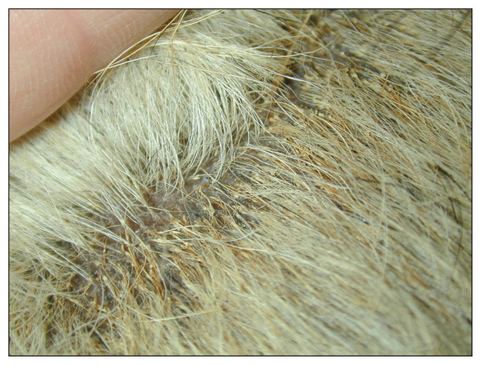
Clinical signs include varying degrees of alopecia, hyperkeratosis, and seborrhea, with follicular casts as a distinctive feature of this inflammatory skin disease (1). When a hair follicle is surrounded by a sheath of keratinaceous debris, this is known as a follicular cast. The cast remains attached to the hair shaft as the hair grows (Figure 1). Pruritus can be variable but is worsened by secondary infection. Early lesions can include both scaling and erythema.
Figure 1.
Follicular casts on a dog.
Clinical signs differ slightly between individuals with long versus short hair coats. Long-coated breeds will present in the early stages of the disease with clinical signs of darkening or lightening of coat color and hair changing from curled or wavy to straight. Hair will then become dull and brittle, and both scale and follicular casts will be noted in the coat. Alopecia will progress over time as the haircoat thins and then hairs are lost. Remaining hairs can be stuck together. Otitis externa can occur, along with the generalized skin lesions. A secondary staphylococcal folliculitis is present in approximately 40% of cases and when present can exacerbate pruritus (1,8).
Dogs with short hair coats will present with annular areas of scale and alopecia. These regions will then coalesce to form larger areas of hair loss. The scales are often white and fine and do not adhere to the skin. These individuals may also present with nodular lesions and will more rarely have secondary pyoderma (1,4).
Lesions most often start on the head or cervical region, along with the pinnae. Follicular casts are common along the lateral margins of the ear pinnae. Lesions will then spread to generalize over the dorsum and begin to involve the tail, trunk, and legs (4). In certain individuals, the legs and paws can be spared. Owners may complain of their dog having an “odor” which is due to the abnormalities/changes in the epidermal lipid layer and the presence of secondary infection.
Certain breeds have been noted to exhibit more severe clinical signs. In 1 study, springer spaniels had more severe clinical signs of alopecia, pyoderma, and seborrhea. In the same study, 57% of springer spaniels had otitis externa compared to 21% of the standard poodles and none of the Akitas. Perhaps there are breed-specific mechanisms of disease development (7).
Diagnosis
A diagnosis of sebaceous adenitis can be suspected based on history, signalment, and physical examination. Differential diagnoses would include staphylococcal folliculitis, demodicosis, dermatophytosis, keratinization defects, follicular dysplasia, endocrinopathies, and nutritional deficiencies (1,4). To rule out some more common dermatologic diseases, skin scrapings should be performed, along with a fungal culture and blood-work. If generalized follicular casts are noted on examination, these are most often associated with demodicosis, vitamin A responsive dermatosis, and sebaceous adenitis. Cytology should also be performed to identify any secondary infection present.
To definitively diagnose sebaceous adenitis, skin biopsies are the diagnostic method of choice. Histopathologic changes vary, depending on the chronicity of the disease. In many cases, a mild to moderate acanthosis, along with moderate to severe hyperkeratosis with follicular plugging, will be noted. Keratin will be present surrounding hair shafts as they emerge (3). Inflammation is variable, depending on the breed and the stage of disease. If inflammation is present, it will be surrounding a sebaceous gland or in the region of a previous gland. In early lesions, there will be nodular granulomatous to pyogranulomatous reactions, sometimes with sebocytes visible within the granulomas. The cellular infiltrate is comprised of histiocytes, neutrophils, lymphocytes and plasma cells. In chronic cases, there will be complete destruction of the sebaceous glands with minimal inflammation. Perifollicular fibrosis can also be present (4). In short-coated individuals, there can be nodular inflammation centered on the isthmus (middle section) of hair follicles (4).
The institute for Genetic Disease Control in Animals (GDC) has an open registry for sebaceous adenitis in the standard poodle. Any individuals who have been bred, who are intended for breeding, or have a diagnosis of sebaceous adenitis, should be registered through an annual skin biopsy. Subclinically affected poodles (those with early signs of the disease on histopathology) can produce affected puppies with clinical signs.
Treatment
Treatment of sebaceous adenitis revolves around removing scale and follicular casts from the skin and coat, improving the quality of the coat and hair regrowth (1,4). Owners should be counselled on the need for lifelong therapy, as the disease cannot be cured. No treatment is effective in 100% of cases, but there are multiple treatment options. Dogs can also be prone to recurrences even when receiving medication and flares in clinical signs during treatment are reported (8). Any secondary infections with bacteria or Malassezia should be treated appropriately.
There are both topical and oral treatments available. Topical treatments include shampoos, humectants, and oil soaks.
Shampoos containing both sulfur and salicylic acid can be used 2 or 3 times per week, allowing a contact time of 10 min before rinsing. A soft brush can be used to help remove some of the scaling. A conditioner can be applied after bathing or a 50 to 75% dilution of propylene glycol, as a spray or rinse after bathing. These sprays have also been used daily and then reduced to 2 or 3 times weekly as maintenance. The propylene glycol acts as a humectant and aids in moisture retention. Baby oil soaks have historically been used to treat sebaceous adenitis. The oil or a 1:1 dilution with water is massaged into the coat and then left for 1 to 6 h. Thereafter, dogs are bathed using shampoo or dish-washing liquid to remove excess oil. These soaks are recommended every 7 to 30 d (4). There are anecdotal reports that phytosphingosine spot-ons or sprays have improved scaling and alopecia.
Oral treatments include omega fatty acids, systemic retinoids, cyclosporine, vitamin A, tetracycline, and niacinamide. The disease is commonly unresponsive to glucocorticoids. Improvement has been observed in dogs on a combination of tetracycline and niacinamide, although there are no studies characterizing the response to this combination. Dogs weighing less than 10 kg, should be given 250 mg of each medication every 8 h, whereas dogs weighing more than 10 kg are given 500 mg of each every 8 h. Side effects with tetracycline and niacinamide can include gastrointestinal upset, anorexia, and lethargy. Liver toxicity has rarely been noted with niacinamide. Eicosapentaenoic acid (EPA) can be used at a dose of 180 mg per 4.5 kg of body weight (BW) and was efficacious in mild to moderately affected dogs (1,4).
Isotretinoin has been effective in the treatment of sebaceous adenitis in multiple breeds of dogs (9). A dose of 1 mg/kg BW is administered once or twice daily, with improvement in clinical signs in 6 wk. The dosing frequency can then be decreased to every other day or the dose itself lowered to 0.5 mg/kg BW once daily. Other retinoids were also effective treatments. Side effects with retinoid therapy include gastrointestinal upset, hepatotoxicity, teratogenicity, and increased serum triglyceride concentrations. Serum biochemistries should be monitored regularly during therapy (4,9).
Vitamin A can be useful as an adjunct treatment of sebaceous adenitis. In 1 study, 15 of 21 dogs had initial improvement in their clinical signs when given doses ranging from 380 to 2667 IU/kg BW per day (mean: 1037 IU/kg BW per day) (10). However, 3 of these dogs later relapsed. Vitamin A caused keratoconjunctivitis sicca in 1 dog out of 24 in the study after 2 y of treatment. Schirmer tear tests are therefore recommended routinely on dogs receiving this therapy (10). Other reports document doses of 10 000 to 30 000 IU vitamin A twice daily producing clinical improvement in 3 mo (4).
In 1 study, cyclosporine at a dose of 5 mg/kg BW daily given for 12 mo decreased clinical signs such as alopecia and severity of follicular casts. Signs recurred when treatment was discontinued, which is to be expected when long-term therapy is needed to control the disease (11). A separate study compared improvement in clinical signs using cyclosporine alone or in combination with topical therapy versus topical therapy as the sole treatment. Topical treatment alone or in combination with cyclosporine reduced scaling more effectively than solely cyclosporine. Alopecia was reduced with all treatments. When using both topical therapy and cyclosporine, there was evidence of a synergistic effect on scaling and alopecia and inflammation of the sebaceous glands is also reduced when a combination of these therapies is used (12).
Sebaceous adenitis generally carries a good prognosis; however, in the study by Tevell et al (7) in Sweden, 14 of 44 dogs were euthanized due to clinical signs. Owners should be made aware that this disease requires lifelong treatment and that flare-ups can occur during therapy. If affected individuals require intense topical treatment, this may be unacceptable to some owners; therefore, oral therapy can be attempted.
Footnotes
The CAVD invites everyone with a professional interest in dermatology to join (www.cavd.ca) and share our passion for dermatology, including continuing education in this dynamic field. Annual membership fees are: $50 for regular members and free for students.
Use of this article is limited to a single copy for personal study. Anyone interested in obtaining reprints should contact the CVMA office (hbroughton@cvma-acmv.org) for additional copies or permission to use this material elsewhere.
The Veterinary Dermatology column is a collaboration of The Canadian Veterinary Journal with the Canadian Academy of Veterinary Dermatology (CAVD). Established in 1986, the CAVD is a not-for-profit organization for veterinarians, and for veterinary technicians technologists, and students with an interest in veterinary dermatology. Being under the umbrella of the World Association of Veterinary Dermatology, the mission of the CAVD is to advance the science and practice of veterinary dermatology in Canada by providing education and resources for veterinary teams, supporting research, and promoting excellence in care for animals affected with skin and ear disease.
References
- 1.Miller WH, Jr, Griffin CE, Campbell KL. Muller & Kirk’s Small Animal Dermatology. 7th ed. St. Louis, Missouri: Elsevier Mosby; 2013. Granulomatous sebaceous adenitis; pp. 695–699. [Google Scholar]
- 2.Scott DW. Granulomatous sebaceous adenitis in dogs. J Am Anim Hosp Assoc. 1986;22:631–634. [Google Scholar]
- 3.Gross TL, Ihrke PJ, Walder E, et al. Skin Diseases of the Dog and Cat. 2nd ed. Ames, Iowa: Blackwell Science; 2005. Sebaceous adenitis; pp. 186–188. [Google Scholar]
- 4.Sousa CA. Sebaceous adenitis. Vet Clin Small Anim. 2006;36:213–249. doi: 10.1016/j.cvsm.2005.09.009. [DOI] [PubMed] [Google Scholar]
- 5.Reichler IM, Hauser B, Schiller I, et al. Sebaceous adenitis in the Akita: Clinical observations, histopathology and hereditary. Vet Dermatol. 2001;12:243–253. doi: 10.1046/j.0959-4493.2001.00251.x. [DOI] [PubMed] [Google Scholar]
- 6.Dunstan RW, Hargis AM. The diagnosis of sebaceous adenitis in standard poodle dogs. In: Bonagura JD, Kirk RW, editors. Kirk’s Current Veterinary Therapy XII. Philadelphia, Pennsylvania: WB Saunders; 1995. pp. 619–622. [Google Scholar]
- 7.Tevell EH, Bergvall K, Egenvall A. Sebaceous adenitis in Swedish dogs, a retrospective study of 104 cases. Acta Vet Scand. 2008;50:11–18. doi: 10.1186/1751-0147-50-11. [DOI] [PMC free article] [PubMed] [Google Scholar]
- 8.Frazer MM, Schick AE, Lewis TP, Jazic E. Sebaceous adenitis in Havanese dogs: A retrospective study of the clinical presentation and incidence. Vet Dermatol. 2011;22:267–274. doi: 10.1111/j.1365-3164.2010.00942.x. [DOI] [PubMed] [Google Scholar]
- 9.White SD, Rosychuk RAW, Scott KV, Hargis AM, Jonas L, Trettien A. Sebaceous adenitis in dogs and results of treatment with isotretinoin and etretinate: 30 cases (1990–1994) J Am Vet Med Assoc. 1995;207:197–200. [PubMed] [Google Scholar]
- 10.Lam ATH, Affolter VK, Outerbridge CA, Gericota B, White SD. Oral vitamin A as an adjunct treatment for canine sebaceous adenitis. Vet Dermatol. 2011;22:305–311. doi: 10.1111/j.1365-3164.2010.00944.x. [DOI] [PubMed] [Google Scholar]
- 11.Linek M, Boss C, Haemmerling R, Hewicker-Trautwein M, Mecklenberg L. Effects of cyclosporine A on clinical and histologic abnormalities in dogs with sebaceous adenitis. J Am Vet Med Assoc. 2005;226:59–64. doi: 10.2460/javma.2005.226.59. [DOI] [PubMed] [Google Scholar]
- 12.Lortz J, Favrot C, Mecklenburg L, et al. A multicentre placebo-controlled clinical trial on the efficacy of oral ciclosporin A in the treatment of canine idiopathic sebaceous adenitis in comparison with conventional topical treatment. Vet Dermatol. 2010;21:593–601. doi: 10.1111/j.1365-3164.2010.00902.x. [DOI] [PubMed] [Google Scholar]


