Abstract
Sex determination, which is based on the existence of dimorphism between specimens of the same species, plays an important role in the process of human identification. In the absence of pelvic elements, the skull appears to be the best sex indicator, and can also be submitted to quantitative or metric assessments. Eleven measurements were taken for this study, four in the sagittal plane and seven in the horizontal, in two groups of 186 skulls each, with 101 from males and 85 from females for those of the sagittal plane, and 100 and 86, respectively, for those concerning the horizontal, of subjects aged between 18 and 94 years at the time of death. The sample belongs to the Osteological and Tomographic Biobank Professor Doctor Eduardo Daruge of the Piracicaba Dental School of the University of Campinas. The aim of this research was to establish a reliable method to determine sex and elaborate mathematical prototypes capable of assisting in investigation or identification activities, in a preliminary study. Of the measures implemented (Lambda-Nasion, Lambda-Rhinion, Lambda-Nasospinale, Rhinion-Nasospinale, Zygomaxillare-Zygomaxillare, Lambda-Incisive Foramen, Lambda-Right Zygomaxillare, Lambda-Left Zygomaxillare, Basion-Incisive Foramen, Basion-Right Zygomaxillare and Basion-Left Zygomaxillare), only the Lambda-Nasospinale and Rhinion-Nasospinale in the sagittal plane, and the Zygomaxillare-Zygomaxillare and Lambda-Incisive Foramen in the horizontal plane, were significantly dimorphic. Two predictive mathematical models of sex were conceived for each pair of them: one of logistic regression and another of conditional inference trees, displaying accuracy rates of 78.5% and 77.42%, and of 68.28% and 72.04%, respectively. The authors concluded that there is the possibility to apply the aforementioned data in forensic anthropology as an auxiliary tool in investigation or identification tasks.
Keywords: Forensic Anthropology; Sex dimorphism; Craniometry,
Keywords: Forensic Dentistry
Introduction
Sexual dimorphism, the main pattern of variation among members of population groups, refers to the volumetric, physiognomic, somatic, anatomical, physiological and structural particularities, which manifest themselves differently in males and females of the same species. (1) It is a phenomenon that has undoubtedly aroused the special interest of forensic medical experts and anthropologists due to its presence in most human bones. (2, 3)
Some skeletal parts (such as those of hands, feet and scapular girdle, sternum, first rib, humerus, ulna, radius, femur, tibia, fibula, patella and mandible) are used to determine sex, although the pelvic girdle and skull are considered to be the most reliable for this purpose. (1, 3, 4) In addition, the skull in particular has enormous forensic potential due to its extraordinary resistance to extreme conjunctures, environmental conditions and natural stages of decomposition, usually being found well preserved and separated from the rest of the skeleton after death. (5)
Several scientific techniques have been used over the course of time to achieve a correct differential sex diagnosis, either through the neuro- and viscero-cranium itself or through their respective images (photographs, radiographs and/or tomographies, preferably digital), ranging from the very classic, common and inexpensive (descriptive and metric of bones and teeth), to the most modern, sophisticated and costly (microscopic observation of pulp tissue or bone architecture, physical and chemical analysis of dental and bone calcified tissues, and DNA examination). In general terms, procedures consisting of qualitative (cranio-scopic), quantitative (cranio-metric) or qualitative-quantitative evaluations are those of first choice, despite the fact that the final decision will depend on the quantity and state of the material questioned, and also the simplicity, practicality, accuracy and cost of those procedures. (1, 6-11) However, it should be mentioned that they appear to be much less effective and accurate in the presence of remnants of sub-adults, due to the lack of or minimal expression of secondary sexual characters. (12)
In view of the foregoing, the present work sought to establish a reliable method for sex determination, using linear measurements performed on dry skulls of adult humans, and to create a mathematical model, from the significantly dimorphic ones, capable of collaborating in investigation or identification activities, in a preliminary study.
MATERIALS AND METHODS
This was a descriptive, cross-sectional and quantitative study of dry skulls of Brazilians belonging to the collection of the Osteological and Tomographic Biobank Professor Doctor Eduardo Daruge of the Piracicaba Dental School of the University of Campinas, which are part of 320 skeletons, 179 from males and 141 from females, of individuals with age, origin and cause of death known. The referred material was donated on March 24, 2015 by Parque Nossa Senhora da Conçeição (Amarais) Cemetery, of the city of Campinas, State of São Paulo, as set out on page 44 of case 06-P-1447/16. In regards to ancestry, the collection includes 188 (58.75%) skeletons of Caucasians (whites), 91 (28.44%) of Mulattoes (mixed races), 40 (12.5%) of Afro-descendants (blacks) and 1 (0.31%) of Amerindian (aborigine).
Of the 320 skulls, only 194 (105 from males and 89 from females) were selected, of subjects who died in the second half of the 20th century, aged between 18 and 94 years at the time of death, with no morphological or pathological abnormalities, neither traces of extensive trauma nor surgical interventions that might interfere with their assessment. The study consisted of eleven measurements (four in the sagittal plane and seven in the horizontal plane) in two groups of 186 skulls each (n=186), with 101 male skulls and 85 female skulls for measurements in the sagittal plane, and 100 and 86, respectively, for those concerning the horizontal plane, because of the impossibility of materialising them on the 194 skulls, as some of the anthropometric points used would be damaged.
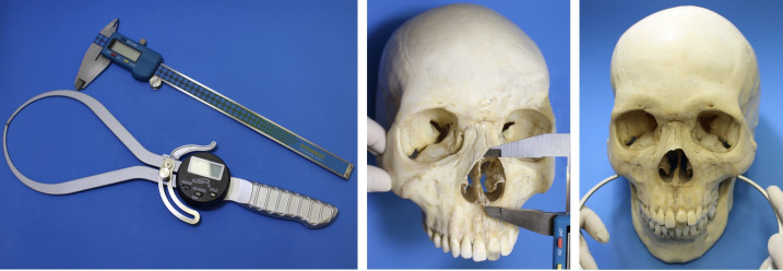
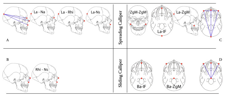
These measurements were carried out by means of a digital sliding calliper with a resolution of 0.01mm (150 mm - Digimess®, São Paulo, Brazil), or a digital spreading calliper with a resolution of 0.01mm (203 mm - Igaging®, California, United States of America), by a single operator as follows: in the first 25 skulls on three different occasions, with an interval of not less than two weeks between them, a requisite for intra-examiner calibration (intra-examination agreement), and in the remaining 161, merely once. Three of the four measurements in the sagittal plane [Lambda-Nasion (La-Na), Lambda-Rhinion (La-Rhi) and Lambda-Nasospinale (La-Ns)] were performed with the aid of the spreading calliper, and one, Rhinion-Nasospinale (Rhi-Ns) of the sliding calliper, while four of the seven in the horizontal plane [Zygomaxillare-Zygomaxillare (ZgM-ZgM), Lambda-Incisive Foramen (La-IF), Lambda-Right Zygomaxillare (La-RZgM) and Lambda-Left Zygomaxillare (La-LZgM)] made with spreading calliper, and three [Basion-Incisive Foramen (Ba-IF), Basion-Right Zygomaxillare (Ba-RZgM) and Basion-Left Zygomaxillare (Ba-LZgM)] with sliding calliper (Figure 1, Table 1 and Figure 2).
Figure 1.
Instruments for measuring and Rhi-Ns and ZgM-ZgM measurements
Table 1. Definition of the cranial landmarks (adapted from Pereira & Mello e Alvim, 1979).
| Lambda (La) | Point at the intersection of the sagittal and lambdoid sutures, in the midline. |
|---|---|
| Nasion (Na) | Point at the intersection of the internasal and nasofrontal sutures, in the midline. |
| Rhinion (Rhi) | Most inferior and anterior point of the internasal suture. |
| Nasospinale (Ns) | Most inferior and anterior point of the inferior margin of nasal aperture, at the anterior nasal spine base. |
| Zygomaxillare (ZgM) | Most inferior point of the zygomaticomaxillary suture. Bilateral (RZgM and LZgM). |
| Incisive Foramen (IF) | Midpoint on the posterior margin of the incisive foramen. |
| Basion (Ba) | Midpoint on the anterior margin of the foramen magnum. |
Figure 2.
Cranial measurements in the sagittal (A and B) and horizontal (C and D) planes
It should be noted that the corresponding project was submitted to the Ethics Committee in Research of the Piracicaba Dental School, and finally approved (protocol nº 138/2014, CAAE nº 38522714.6.0000.5418), complying with Resolution 466/12 about guidelines and regulatory norms of research involving human beings. (13)
STATISTICAL ANALYSIS
The data obtained were entered into a spreadsheet and analysed using R statistical programme. Intra-examiner reliability was evaluated by the intra-class correlation coefficient, with a range from 0.985 to 0.997 for sagittal plane measurements, and from 0.993 to 0.999 for the horizontal values, thus proving the absence of a statistically significant difference amongst the three series of measurements taken.
A scatter plot matrix of a set of independent variables, quantitative (measurements in the sagittal or horizontal planes), and one response or dependent, dichotomous, with only two categories (“male” and “female”), in which each element consisted of a scatter plot of one of the first and the response, allowed to select the most significantly dimorphic variables.
Then, two kinds of statistical models were proposed and customized for this survey: one parametric of logistic regression and one more non-parametric of conditional inference trees (Ctree).
Afterwards, two logistic regression models were elaborated, consisting of a constant (x0=1) and two explanatory variables - La-Ns and Rhi-Ns for the measurements in the sagittal plane, and ZgM-ZgM and La-IF for those in the horizontal - whose mathematical formulae are the result of the substitution of the respective parameters or coefficients (ß0, ß1 and ß2) by the appropriately estimated values. In these equations, results greater than zero are indicative of feminine sex and less than zero of masculine sex.
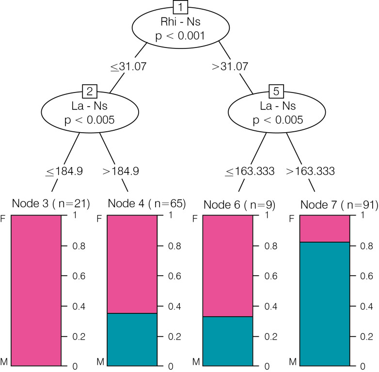
In turn, two models of conditional inference trees were developed, one for each group of measurements, with the same variables as the previous ones, evaluating the corresponding final nodes. Each of these was assigned one of the two categories of the response variable, being designated as category 1 to the one with the highest number of components, and 2 to the one with the lowest number of them.
Hence, the logistic regression model for the group of measures in the sagittal plane had the succeeding expression, as well as the conditional inference trees, the configuration represented in Figure 3.
Figure 3.
Conditional inference trees model for measurements in the sagittal plane
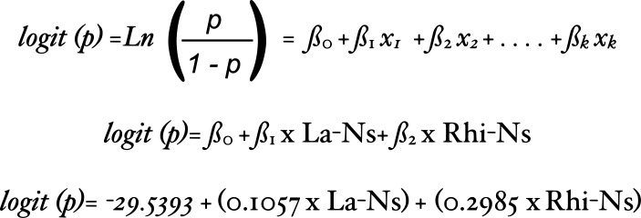 |
| logit (p)= ß0 + ß1 x La-Ns+ ß2 x Rhi-Ns |
| logit (p)= -29.5393 + (0.1057 x La-Ns) + (0.2985 x Rhi-Ns) |
On the other hand, the logistic regression model for the group of measurements in the horizontal plane was formulated as follows:
 |
| logit (p)= ß0 + ß1 x ZgM-ZgM + ß2 x La-IF |
| logit (p)= -24.4233 + (0.1089 x ZgM-ZgM) + (0.0777 x La-IF) |
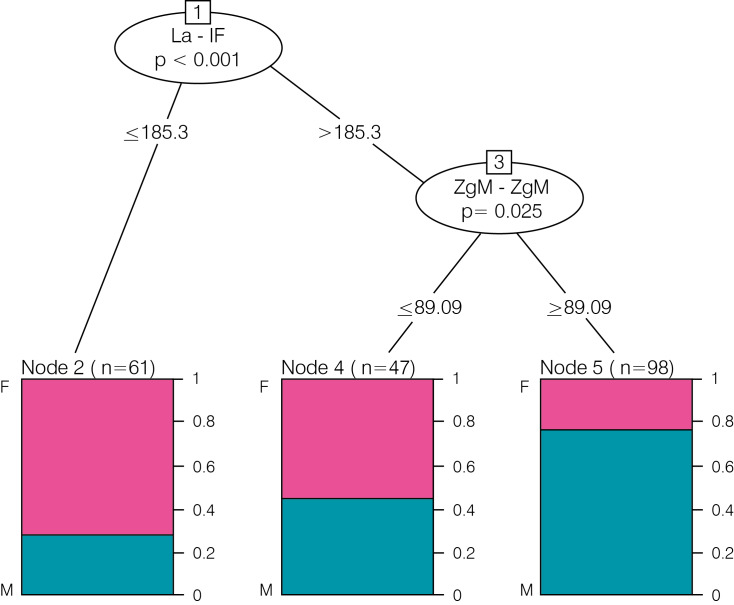
The conditional inference trees model for the aforementioned acquired the configuration reproduced in Figure 4.
Figure 4.
Conditional inference trees model for measurements in the horizontal plane
RESULTS
Once the measurements in the sagittal plane were evaluated through logistic regression model (Table 2), it was possible to verify that:
Table 2. Cataloguing of skulls for measurements in the sagittal plane, by means of the logistic regression model.
| Predicted | ||||
|---|---|---|---|---|
| Observed | F | M | Total | |
| F | 63 | 22 | 85 | |
| M | 18 | 83 | 101 | |
| Total | 81 | 105 | 186 | |
83 male and 63 female skulls were correctly classified (success rate of 78.5%);
the model identified 105 of the 186 skulls examined as male and the remaining 81 as female, with an accuracy of 79.04% (83 of 105) and 77.77% (63 of 81), respectively;
the success rate by sex corresponded to 82.18% of males and 74.12% of females; and
the total classification error was 21.5%, whereas the error by sex reached 17.82% in males and 25.88% in females.
When these measurements were submitted to the conditional inference trees model (Table 3), the subsequent outcomes were obtained:
Table 3. Categorization of skulls for measurements in the sagittal plane, according to conditional inference trees model.
| Predicted | ||||
|---|---|---|---|---|
| Observed | F | M | Total | |
| F | 69 | 16 | 85 | |
| M | 26 | 75 | 101 | |
| Total | 95 | 91 | 186 | |
-
75 male and 69 female skulls were correctly categorized (success rate of 77.42%);
91 skulls were typified as male and 95 as female, with an accuracy of 82.4% (75 of 91) and 72.6% (69 of 95), respectively;
the success rate by sex corresponded to 74.26% of males and 81.18% of females; and
the total classification error was 22.58%, whilst that considered by sex reached 25.74% in males and 18.82% in females.
After evaluating the measurements in the horizontal plane, taking advantage of the logistic regression model (Table 4), it became feasible to assert that:
Table 4. Classification of skulls for measurements in the horizontal plane, using the logistic regression model.
| Predicted | ||||
|---|---|---|---|---|
| Observed | F | M | Total | |
| F | 53 | 33 | 86 | |
| M | 26 | 74 | 100 | |
| Total | 79 | 107 | 186 | |
74 male and 53 female skulls were correctly catalogued (success rate of 68.28%);
the model predicted that 107 of the 186 skulls examined were masculine and the remaining 79 feminine, with an accuracy of 69.1% (74 of 107) and 67.1% (53 of 79), respectively;
the success rate by sex corresponded to 74% of males and 61.63% of females; and
the total classification error was 31.72%, 26% in males and 38.37% in females when considered by sex.
Finally, the measurements were analyzed through conditional inference trees model (Table 5), with the following results:
Table 5. Cataloguing of skulls for measurements in the horizontal plane, by means of the conditional inference trees model.
| Predicted | ||||
|---|---|---|---|---|
| Observed | F | M | Total | |
| F | 69 | 17 | 86 | |
| M | 35 | 65 | 100 | |
| Total | 104 | 82 | 186 | |
65 male and 69 female skulls were correctly classified (success rate of 72.04%);
82 skulls were characterized as male and 104 as female, with an accuracy of 79.2% (65 of 82) and 66.3% (69 of 104), respectively;
the success rate by sex corresponded to 65% of males and 80.23% of females; and
the total classification error was 27.96%, while that considered by sex reached 35% in males and 19.77% in females.
DISCUSSION
It could be stated that the need and eagerness for identification can be traced back to the beginnings of civilisation, since man has always been careful regarding his property and identity rights, in order to differentiate himself from his peers. (14) In view of these precepts, nowadays, the legal scope, social significance and media repercussions of this are no longer discussed. (14, 15)
On this treadmill, sex determination, which is based on the existence of dimorphism between specimens of the same species, arises as a key step to reconstruct the biological profile of human remains, (1, 3, 8, 16) by enabling the reduction of the search universe by 50%. (3, 9, 17) It is also one of the cardinal scopes of anatomists, bioarchaeologists, anthropologists and criminal, forensic medical and odontology experts, when responsible for the anthropological examination of unscathed or fragmented bones. (2, 3)
These contrasting features are evident in most parts of the skeleton, and markedly so in the pelvic girdle and skull, which are considered as reliable sex indicators. (1, 3, 4) It is added that, because of its formidable resistance, the skull is often recovered from crime scene or crash site in a good condition to undergo a detailed appraisal. (3, 5)
Some cranial and mandibular traits must be analyzed in an attempt to arrive at a precise differential sex diagnosis, not forgetting that their degree of expression - usually higher in males - will be influenced by diachronic, mesological, geographical, evolutionary, ontogenic, occupational, socio-economic, behavioural, nutritional, genetic, ethnic, age-related, constitutional, physiological, hormonal, biomechanical, traumatic, pathological and/or surgical factors. (1, 3-5, 9, 10, 15, 16, 18) Therefore, forensic anthropology, a sub-speciality of physical, anatomical or biological anthropology, comes on scene as a supporting science for forensic medicine and justice, especially in cases related to identification. (15)
In general, macroscopic qualitative, non-metric or visual inspection techniques continue to be widely used as a consequence of their universality, practicability, replicability, speed, efficiency and simplicity (not requiring sophisticated equipment), despite demanding capable, experienced and trained professionals, and of their high subjectivity load. (1, 3-5, 8-11, 15, 17, 18) To overcome these disadvantages, minimise the chances of inter-observer error (11) and favour the admissibility of findings in court, (11, 17) an attempt was made to quantify the morphological attributes using ordinal scales with three or five levels, (10, 19) or special devices such as coordinate callipers, (11) which are not always effective in increasing the accuracy rate. In contrast, quantitative or metric techniques rely on standardised linear or angular measurements of distances delimited by landmarks, weighted individually or as indices, as well as in spatial references of superior objectivity, statistical value, population specificity and susceptibility to secular changes, and of lesser challenge in the judicial sphere. (3-5, 7-11, 15, 17, 18) Nevertheless, their use may be restricted in view of the three-dimensionality and natural irregularity of the various body units in question, (20) for which Mandelbrot (21), in 1982, proposed resorting to fractal geometry for morphometric analysis of these structures. In this same goal, Bookstein (22) utilized a geometric morphometric approach with the aim of recording 2D and 3D coordinates on landmarks and semi-landmarks. This is based on the application of new photographic (photogrammetry, digital cameras, microlenses, illuminators, reflectors, tripods, etc.) and computational (images digitization, scanners, computers, and specific software) technologies and accessories, which make it possible to visualise, measure and reproduce the variations in shape and size of biological components. (1, 4, 5, 10, 18) Its main contribution consists of the possibility of practising a more objective, realistic, complete and accurate evaluation, (10) although hampered by the eventual unavailability or lack of economic, operational and/or human resources. (18)
It is worth remembering that Brazilian anthropology directed its first efforts towards the validation (for the national samples) of findings of successive and celebrated European scientific works, capturing the inconveniences that generated to interpolate them and the urgency to coin their own paradigms and tables. For these reasons, in the midst of the uninterrupted and impetuous expansion of statistics in the mid-twentieth century, mathematical archetypes began to be developed that were adapted to the reality of the country. (2)
In a nutshell, adequate statistical analysis requires the application of flexible methodologies capable of leading to easily understandable results. Using this as a guide, it should detail the selection process of variables and indicate the performance prediction of mathematical models. On this occasion, two multivariate tools of uncontroversial utility in health sciences - one of logistic regression and another of conditional inference trees - were expressly designed for the assorting of the skulls studied. The first is a valuable statistical appliance for forecasting a binary dependent variable such as sex, albeit still insufficiently explored in forensic dentistry. (23) In essence, it represents a generalised alternative that has proven to be very adaptable in its hypotheses and able of manipulating both discrete and continuous variables, which request not to be normally distributed, linearly related, or of equal variance within each ensemble. (24) Furthermore, the second is a predictive and exploratory instrument that facilitates the explanation of a numerical response variable (regression) or categorised (classification), through a group of covariables and their possible relations. In fact, it is a versatile scheme with a simple interpretation, with no restrictions as to the type and distribution of the considered variables, either dichotomous (as in this case) or not.
The sample in question consisted of dry skulls of Brazilians belonging to the Osteological and Tomographic Biobank Professor Doctor Eduardo Daruge of the Piracicaba Dental School of the University of Campinas. The collection comprises of 320 skeletons, 179 from males and 141 from females, 188 (58.75%) of Caucasians (whites), 91 (28.44%) of Mulattoes (mixed races), 40 (12.5%) of Afro-descendants (blacks) and 1 (0.31%) of Amerindian (aborigine), of individuals with age, origin and cause of death known. This material was donated by Parque Nossa Senhora da Conceição Cemetery, of the city of Campinas, State of São Paulo, South East Region of Brazil. As to the ancestry, it would be inappropriate to forget that the nation is home to a multi-ethnic society with European, indigenous and, above all, African and Asian influences, (9, 14, 15) since 46% of its population (one of the most miscegenated in the world) are descendants of former slaves from the African continent, (14) and the largest Japanese community in the world is located in and around São Paulo. (14) Given the aforementioned information, there appears to be no physical or genetic stereotype of the Brazilian citizen, which symbolises the synthesis of a long, intricate and very rich ethnocultural history. (25)
The present research focused on cranial measurements performed in the sagittal and horizontal planes in order to verify the presence of sexual dimorphism. Statistically significant linear distances were Lambda-Nasospinale and Rhinion-Nasospinale in the sagittal plane, and Zygomaxillare-Zygomaxillare and Lambda-Incisive Foramen in the horizontal. For each pair of them, a mathematical prototype of logistic regression and one of conditional inference trees were created, with success rates of 78.5% and 77.42%, and of 68.28% and 72.04%, respectively.
The bizygomatic breadth has been referred to as the most dimorphic measurement in conglomerates of white (7) and black (17) South Africans, Greeks, (4) Americans, (8) Indians, (3) Romanians, (26) Scots (27) and Brazilians. (15, 27, 28) Nonetheless, it was excluded from this evaluation because it appears in the measurement battery of Ulbricht et al. (28) for a sample of the same osteological collection.
The Basion-Incisive Foramen distance was statistically significant in two subsets of Brazilians, one in São Paulo (29) and the other in Mato Grosso, (9) but not in this study. In contrast, the Zygomaxillare-Zygomaxillare distance was significant in this, but not in Western Australians (18) and South Eastern Brazilians, (10) whereas the Rhinion-Nasospinale was significant for two of this bone collection, as expected: that of Ulbricht et al. (28) and the present study.
Saini et al. (3) reported the dimorphic character of the Nasion-Nasospinale distance in Indians, which was not measured in this survey, but the Rhinion-Nasospinale (which is known to be included in the former) was, and it was shown to be significant. It should be noted that an analogy has been drawn between the two, since they are associated with the sagittal plane and in the same anatomical region.
For Green & Curnoe (5) and Saini et al. (3), the aforesaid results would be attributed to a succession of biological phenomena and mechanisms, as well as to the applied methodology, sample size and/or ancestry of the human sets assessed. Indeed, the singularities of organic growth and development, which are fundamentally conditioned by genetic and hormonal factors (hereditary patterns, beginning and end of puberty), are responsible for the onset and magnitude of sexual dimorphism in the cranial unit. The traits considered more dimorphic are located in the face and vault, due to the fact that they reach their final volume and shape relatively late in ontogeny. The superior facial region (orbital region) is the first to complete its maturation and is consequently less dimorphic than the nasal, zygomatic, maxillary and mandibular areas. (1, 3, 5) The vault is closely related to cranial capacity (on average 10% higher in men) and should adapt to the progressive increase in brain mass until the age of forty, from which it will decline at a rate of 5% per decade, (16) as the base will grow and mature earlier to support the brain and protect the vital nerves and vessels. (1) Taking all of this information into account, it should be possible to explain the reason for the dissimilar success rates of predictive models of the present study and the lesser reliability of the measurements in the horizontal plane. On the other hand, it is essential to emphasise that modern population groups do not exhibit appreciable or palpable phenotypic or genotypic discrepancies, as is evident from cladistic analyses that seek to trace the genealogy of our species. (14) In essence, they constitute authentic, temporary, dynamic and changeable ethnic crucibles, with certain ancestral peculiarities that are periodically repeated, in total harmony with increasing degrees of human variation and miscegenation for which it is necessary that every procedure of identification is tested and validated for each of them. (14)
In addition to the limitations and controversies inherent in osteometric methods in general and craniometric methods in particular, the one used in this opportunity, although not infallible, (9) proved to be reliable, valid, simple, feasible, accessible, rapid, practical, economical, standardised and reproducible. Likewise, it can achieve results comparable to those of much more abstruse and costly ones, and be useful in cases of fractured skulls. (17)
At last, it should be pointed out that sex determination and human identification are conceptualised as fundamental individual rights. Besides, these activities are the responsibility of Brazilian dental surgeons when invested with the role of odontology experts (items IV and IX of article 6 of the Law 5081/66), who will have the possibility to contribute and expedite these processes, in the pursuit of providing better and more efficient services to the community and to be subject to current legislation. (30)
CONCLUSIONS
Quantitative assessments of the different cranial structures emerge as suitable, useful, valuable, pragmatic, inexpensive and complementary resources for the reconstruction of the individual biological profile. Sex determination as a major step in this arduous and relevant task, is one of the most common and critical problems faced by anatomists, bioarchaeologists, anthropologists and criminal, forensic medical and odontology experts.
A positive identification should never be based on a single technique, according to the anthropological premise of making use as many available means as possible. Additionally, it will be necessary that the above mentioned be tested and validated for each population sample, in light of the notorious level of variation and miscegenation of contemporary human beings.
The Lambda-Nasospinale and Rhinion-Nasospinale distances in the sagittal plane and the Zygomaxillare-Zygomaxillare and Lambda-Incisive Foramen in the horizontal revealed themselves to be significantly dimorphic. For each pair of them, two predictive mathematical models of sex were developed, one of logistic regression and one of conditional inference trees, with success rates of 78.5% and 77.42%, and of 68.28% and 72.04%, respectively.
Footnotes
The authors declare that they have no conflict of interest.
References
- 1.Menéndez L, Lotto F. Comparación de técnicas para determinar sexo en poblaciones humanas: estimaciones diferenciales a partir de la pelvis y el cráneo en una muestra de San Juan, Argentina. Rev. Cienc. Morfolog. 2013;15(1):12–21. [Google Scholar]
- 2.Costa AA, Pereira MA, Ramos DIA, Meléndez BVC, Silva RF, Velos GSM, et al. Determinação do gênero por meio de medidas craniométricas e sua importância pericial. Rev. Med. LegalDir. 2005;1(3):18–23. [Google Scholar]
- 3.Saini V, Srivastava R, Rai R, Shamal SN, Singh TB, Tripathi SK. An Osteometric Study of Northern Indian Populations for Sexual Dimorphism in Craniofacial Region. J Forensic Sci. 2011;56(3):700–5. 10.1111/j.1556-4029.2011.01707.x [DOI] [PubMed] [Google Scholar]
- 4.Kranioti EF, Garcia-Vargas S, Michalodimitrakis M. Dimorfismo sexual del cráneo en la población actual de Creta. Boletin Galego de Medicina Legal. 2009;16:37–43. [Google Scholar]
- 5.Green H, Curnoe D. Sexual dimorphism in Southeast Asian crania: A geometric morphometric approach. Homo. 2009;60:517–34. 10.1016/j.jchb.2009.09.001 [DOI] [PubMed] [Google Scholar]
- 6.Pereira CB, Mello e Alvim M. Manual para estudos craniométricos e cranioscópicos. Santa Maria: Universidade Federal de Santa Maria – RS; 1979. [Google Scholar]
- 7.Steyn M, Işcan MY. Sexual dimorphism in the crania and mandibles of South African whites. Forensic Sci Int. 1998;98:9–16. 10.1016/S0379-0738(98)00120-0 [DOI] [PubMed] [Google Scholar]
- 8.Spradley MK, Jantz RL. Sex Estimation in Forensic Anthropology: Skull Versus Postcranial Elements. J Forensic Sci. 2011;56(2):289–96. 10.1111/j.1556-4029.2010.01635.x [DOI] [PubMed] [Google Scholar]
- 9.Nascimento Correia Lima N, Oliveira OF, Sassi C, Picapedra A, Francesquini Júnior L, Eduardo Daruge Júnior E. Sex determination by linear measurements of palatal bones and skull base. J Forensic Odontostomatol. 2012;30(1):37–43. [PMC free article] [PubMed] [Google Scholar]
- 10.Jurda M, Urbanová P. Sex and ancestry assessment of Brazilian crania using semi-automatic mesh processing tools. Leg Med (Tokyo). 2016;23:34–43. 10.1016/j.legalmed.2016.09.004 [DOI] [PubMed] [Google Scholar]
- 11.Casado AM. Quantifying Sexual Dimorphism in the Human Cranium: A Preliminary Analysis of a Novel Method. J Forensic Sci. 2017;62(5):1259–65. 10.1111/1556-4029.13441 [DOI] [PubMed] [Google Scholar]
- 12.Scheuer L. Application of Osteology to Forensic Medicine. Clin Anat. 2002;15:297–312. 10.1002/ca.10028 [DOI] [PubMed] [Google Scholar]
- 13.Brasil. Resolução 466, de 12 de dezembro de 2012. Ministério da Saúde. [updated 2016 jun 6]. Available from: bvsms.saude.gov.br /bvs/saudelegis/.../res0466 _12_12_2012.html
- 14.Sassi C, Picapedra A, Lima L, Massa F, Gargano V, Francesquini Jr. L, Daruge Jr. E Contribución de la antropología dental en la determinación de la identidad uruguaya. Actas odontológicas 2013; 10(1):29-34.
- 15.Fortes de Oliveira O, Tinoco RLR, Daruge Júnior E, Sayuri A, Silveira TD, Silva RHA, et al. Sexual Dimorphism in Brazilian Human Skulls: Discriminant Function Analysis. J Forensic Odontostomatol. 2012;30(2):26–33. [PMC free article] [PubMed] [Google Scholar]
- 16.Eboh DE, Okoro EC, Iteire KA. A cross-sectional anthropometric study of cranial capacity among Ukwuani people of South Nigeria. Malays J Med Sci. 2016;23(5):72–82. 10.21315/mjms2016.23.5.10 [DOI] [PMC free article] [PubMed] [Google Scholar]
- 17.Dayal MR, Spocter MA, Bidmos MA. An assessment of sex using the skull of South Africans by discriminat function analysis. Homo. 2008;59(3):209–21. 10.1016/j.jchb.2007.01.001 [DOI] [PubMed] [Google Scholar]
- 18.Franklin D, Cardini A, Flavel A, Kuliukas A. The application of traditional and geometric morphometric analyses for forensic quantification of sexual dimorphism: preliminary investigations in a Western Australian population. Int J Legal Med. 2012;126(4):549–58. 10.1007/s00414-012-0684-8 [DOI] [PubMed] [Google Scholar]
- 19.Defrise-Gussenhoven E. A masculinity-femininity scale based on a discriminant function. Acta Genet. 1966;16:198–08. 10.1159/000151964 [DOI] [PubMed] [Google Scholar]
- 20.Schiwy-Bochat KH. The roughness of the supranasal region-a morphological sex trait. Forensic Sci Int. 2001;117(1-2):7–13. 10.1016/S0379-0738(00)00434-5 [DOI] [PubMed] [Google Scholar]
- 21.Mandelbrot BB. The Fractal Geometry of Nature. San Francisco. Freeman; 1982 [Google Scholar]
- 22.Bookstein FL. Morphometric tools for landmark data: geometry and biology. Cambridge. Cambridge University Press; 1991. [Google Scholar]
- 23.Christensen R. Log-linear models and logistic regression. New York. Stringer; 1997 [Google Scholar]
- 24.Spicer J. Making Sense of Multivariate Data Analysis. An Intuitive Approach. Thousand Oaks. SAGE Publications; 2004. [Google Scholar]
- 25.Sassi C, Picapedra A, Caria PHF, Groppo F, Francesquini L, Jr, Daruge E, Jr, et al. Comparación antropométrica entre mandíbulas de las poblaciones uruguaya y brasileña. Int J Morphol. 2012;30(2):379–87. 10.4067/S0717-95022012000200003 [DOI] [Google Scholar]
- 26.Marinescu M, Panaitescu V, Rosu M, Maru N. Punga A. Sexual dimorphism of crania in a Romanian population: Discriminant function analysis approach for sex estimation. Rev Med Leg. 2014;22:21–6. 10.4323/rjlm.2014.21 [DOI] [Google Scholar]
- 27.Lopez Capp TT. Análise da variabilidade métrica dos parâmetros de Antropologia Forense para estimativa do sexo de duas populações: escocesa e brasileira. [Tese]. São Paulo: Faculdade de Odontologia, Universidade de São Paulo; 2017. [Google Scholar]
- 28.Ulbricht V, Martins Schmidt C, Groppo FC. Daruge Jr.E, Queluz DP, Francesquini Jr. L. Sex Estimation in Brazilian Sample: Qualitative or Quantitative Methodology? BJOS. 2017;15(3):1–9. [Google Scholar]
- 29.Francesquini Júnior L, Francesquini MA, De La Cruz BM, Pereira SDR, Ambrosano GMB, Barbosa CMR, et al. Identification of sex using cranial base measurements. J Forensic Odontostomatol. 2007;25(1):7–11. [PubMed] [Google Scholar]
- 30.Francesquini L, Jr, Francesquini MA, Daruge E, Ambrosano GMB, Bosqueiro MR. Verificação do grau do conhecimento do cirurgião-dentista sobre perícia de identificação humana pelos dentes. Revista do CROMG. 2001;7(2):113–9. [Google Scholar]


