Abstract
Aim
The aim of this study was to evaluate and compare the accuracy of two age estimation methods in Indian children by using the open apex method proposed by Cameriere et al and the London Atlas of Tooth Development.
Materials and method
Three hundred and thirty five archived digitised panoramic radiographs of healthy children (165 males and 170 females) in the age group of 5 to 15.99 years were retrieved and analysed. The observations were entered in the SPSS software (Version 19.0). The paired t-test and independent samples t-test were applied to assess the differences between chronological and estimated age in both genders.
Results and conclusion
Inter-observer reliability was found to be excellent with Cronbach Alpha to be 1.000 and 0.997 for Cameriere’s and London Atlas age estimation methods respectively. The difference of 0.59 years (SD ±1.32 years) was highly significant and indicated a consistent underestimation of age using Cameriere’s method. While, applying the London Atlas, the difference of -0.03 years (SD± 0.69 years) was not significant and indicated a little overestimation of age. No significant difference was observed for both genders with the methods. Our results revealed that the methods are reliable for age estimation in Indian children, however, the London Atlas is simpler to use and is more accurate than Cameriere’s method.
Keywords: Age estimation,
Keywords: Indian children,
Keywords: Cameriere’s method,
Keywords: London Atlas,
Keywords: Forensic odontology
Introduction
Children are the future of any nation, however, in recent times, increasing trend of juvenile crimes, escalating cases of immigrants and child abuse are being reported globally, making the estimation of age in children more significant medico-legally. Furthermore, exact age assessment in the paediatric population becomes mandatory in various fields like forensic medicine, endocrinology and orthodontic treatment planning. (1, 2) Though there are different methods of age estimation like the secondary sexual characteristics, the biomarkers, bone and dental development, most of the techniques are expensive and are not very accurate. (3)
Dental age, however, is, considered to be a reliable, easy and quick method of age assessment in children as there is minimal variability observed due to the calcification rate that is not much affected by environmental factors. (4-6) Furthermore, teeth are the most indestructible mineralised structures which survive for several years, hence, examination of teeth is considered the most reliable method of age estimation. (3)
Amongst all the imaging methods, the method which is cost effective, uncomplicated and gives an excellent overview of the dental maturity is orthopantomography. (7) Some recently introduced methods of age estimation which are more precise, reliable and use panoramic radiographs for age assessment, include Cameriere’s open apices method in children (8) and the London Atlas of Human Tooth Development and Eruption. (9)
Cameriere et al, 2006 (8) developed a linear regression formula to assess the chronological age in a European population. Popularly recognised as Cameriere’s equation, it estimates age by measuring the open apices of seven permanent mandibular teeth on the left side of the jaw. While, an innovative and simple approach to age estimation was introduced by AlQahtani et al in 2010, (9) who developed a comprehensive atlas of human tooth development to determine age between 28 weeks intrauterine and 23 years. This method utilised the Moorrees et al’s (4) and Bengston’s (10) tooth developmental stages. There is no study done in an Indian paediatric population which compares the accuracy of Cameriere’s with the London Atlas method of age estimation. Thus, the present study was carried out to estimate the accuracy of Cameriere’s formula and the London Atlas of Tooth Development in assessing the actual age of Indian children. The second objective was to analyse if there was any difference in the accuracy of these two methods. Additionally, the study aimed to determine if there was a difference between the dental age of male and female subjects using Cameriere’s equation and the London Atlas of Tooth Development.
MATERIALS AND METHODS
This was a cross-sectional retrospective study which examined the digitised orthopantomographs (OPGs) retrieved from the archives of Department of Oral Medicine and Radiology. Only best quality radiographs were selected of 335 healthy Indian children aged between 5.00 and 15.99 years. All the subjects were categorised into 11 age groups with equal distribution of 15 males and 15 females in each age group, except Group 5 which had 20 females (Table 1).
Table 1. Age and gender distribution in the sample studied.
| Groups (of Sample studied) | Age (years) | Boys(n) | Girls(n) | Total (n) |
|---|---|---|---|---|
| 1 | 5.5.99 | 15 | 15 | 30 |
| 2 | 6.6.99 | 15 | 15 | 30 |
| 3 | 7.7.99 | 15 | 15 | 30 |
| 4 | 8.8.99 | 15 | 15 | 30 |
| 5 | 9.9.99 | 15 | 20 | 35 |
| 6 | 10.10.99 | 15 | 15 | 30 |
| 7 | 11.11.99 | 15 | 15 | 30 |
| 8 | 12.12.99 | 15 | 15 | 30 |
| 9 | 13.13.99 | 15 | 15 | 30 |
| 10 | 14.14.99 | 15 | 15 | 30 |
| 11 | 15.15.99 | 15 | 15 | 30 |
| Total | 165 | 170 | 335 |
The uniformity in age and gender distribution was maintained purposely to achieve accuracy across the age range and gender. All the radiographs were taken for routine diagnostic and treatment purposes; no radiographs were taken particularly for this study. Poor quality unclear radiographs, as well as those showing pathology, dental anomalies, previous orthodontic treatment, severe dental caries, periapical cysts, grossly destructed teeth and crowns, were excluded from the study. The chronological age of participants was calculated by subtracting the date of the birth from the date on which radiographs were taken. Coding was done for all participants, and the observer was blinded to their actual age. Only the subject’s gender was revealed to the examiner.
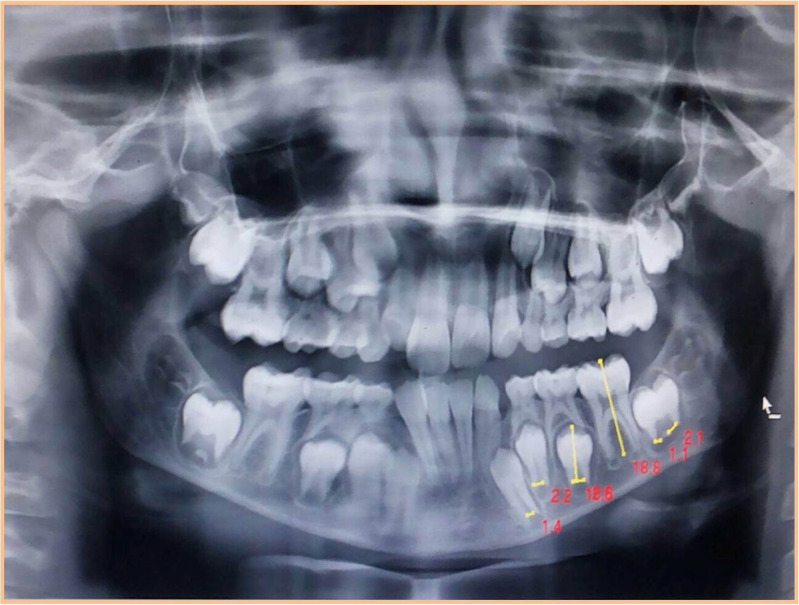
While using Cameriere’s method, all seven left permanent mandibular teeth were examined. In single rooted teeth, the distance between the inner side of the pulp canal at the apex was measured (Ai, i= 1,..5). In multirooted teeth, the sum of the distances between the inner sides of the pulp of the two apices was calculated (Ai, i=6,7). Normalised measurements were obtained by dividing the measurement of the apices by the tooth length from the highest cusp tip to the lowest root tip (Li, i=1,….,7) (Figure 1).
Figure 1.
Cameriere’s method of measuring open apices and tooth length (on Adobe Photoshop)
Finally, dental maturity was calculated using the normalised measurements of the open apices of the seven left permanent mandibular teeth, xi, i=1,…..,7, their sum, s, and the number of teeth (N0), with root development complete. Dental age in Indian children was calculated by putting all the values in Cameriere’s equation formulated according to linear regression model:
| Dental age = 8.971+0.375g +1.631X5 +0.674 No-1.034s-0.176s. No, |
where g stands for gender, X5 = A5/L5, s = sum of normalised open apices, and N0 = number of teeth showing complete root development.
When the London Atlas of Tooth Development was used for age estimation, OPGs were examined to assess the development stages for all primary and permanent teeth on the right side of both upper and lower jaws. Subsequently, the dental age of the individual was calculated by using available software on the website: http://www.atlas.dentistry.qmul.ac.uk. The tables were filled by observing specific figures of the development stage and level of alveolar eruption of the tooth and matching these with the panoramic radiographs of each participant; the dental age calculator feature automatically displayed the dental age.
To test inter-observer reproducibility, a random sample of 35 panoramic radiographs were examined by two observers at fifteen days interval. Reliability analysis showed inter-observer Cronbach Alpha to be 1.000 and 0.997 for Cameriere’s and London Atlas age estimations respectively, suggesting excellent agreement. All the calculated values obtained from both age estimation methods, were entered in an excel file and subjected to SPSS (Statistical Package for the Social Sciences) software, (version 19.0) analysis.
Dental age (Estimated age) was subtracted from the chronological age (Actual age): a positive result indicated underestimation and a negative one indicated overestimation of age. A paired t-test was applied for each of the two methods with a significance level of P< 0.001, to calculate the bias which is the mean difference between the predicted and chronological ages.
RESULTS
Cameriere’s Method
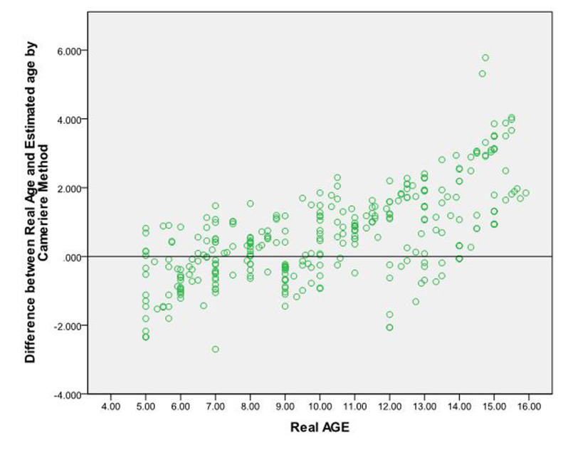
The mean chronological age of the entire sample was 10.238 ± 3.160 years. The mean estimated age of the whole sample using Cameriere’s method was 9.639 ± 2.486 years. The difference of 0.59 years (SD ±1.32 years was highly significant and indicated a consistent underestimation of age. The bias was highly significant in all age groups (Table 2).
Table 2. Paired t-test for Cameriere’s method showing the bias for the sample population.
| Measure of accuracy | N | Mean | ± SD | SE mean | Significance | 95% CI Lower | 95% CI Upper |
|---|---|---|---|---|---|---|---|
| Bias | 335 | 0.59773 | 1.32365 | 0.07232 | 0.000** | 0.4555 | 0.7399 |
| Absolute Difference | 335 | 1.11048 | 0.93464 | 0.05106 | 0.000** | 1.1010 | 1.2109 |
**p<0.001; Highly significant SD = Standard deviation, SE = Standard error, CI = Confidence interval
Independent samples t-test was applied to measure the differences between male and female participants. No statistically significant difference (p = 0.154) was observed between males and females. The mean difference between the actual and predicted ages was less in male subjects as compared to female subjects (Table 3).
Table 3. Independent samples t-test for Cameriere’s method to express bias for gender.
| Measure of accuracy | Sex | N | Mean | ± SD | SE mean | Significance |
|---|---|---|---|---|---|---|
| Bias | M | 165 | 0.4931 | 1.2961 | 0.1009 | 0.154 |
| F | 170 | 0.6994 | 1.3459 | 0.1032 | ||
| Absolute Difference | M | 165 | 1.0755 | 0.8723 | 0.0679 | 0.500 |
| F | 170 | 1.1445 | 0.9929 | 0.0761 |
F = Female, M =male, SD = Standard Deviation, SE = Standard error
Figure 2 depicts the paired t-test results for the entire sample as well as for each year interval. Age was significantly overestimated in the children between the age of 5 to 9 years, on the other hand, in children of age range from 11 to 15 years, it was significantly underestimated.
Figure 2.
Paired t-test for Cameriere’s method showing the bias for the sample population
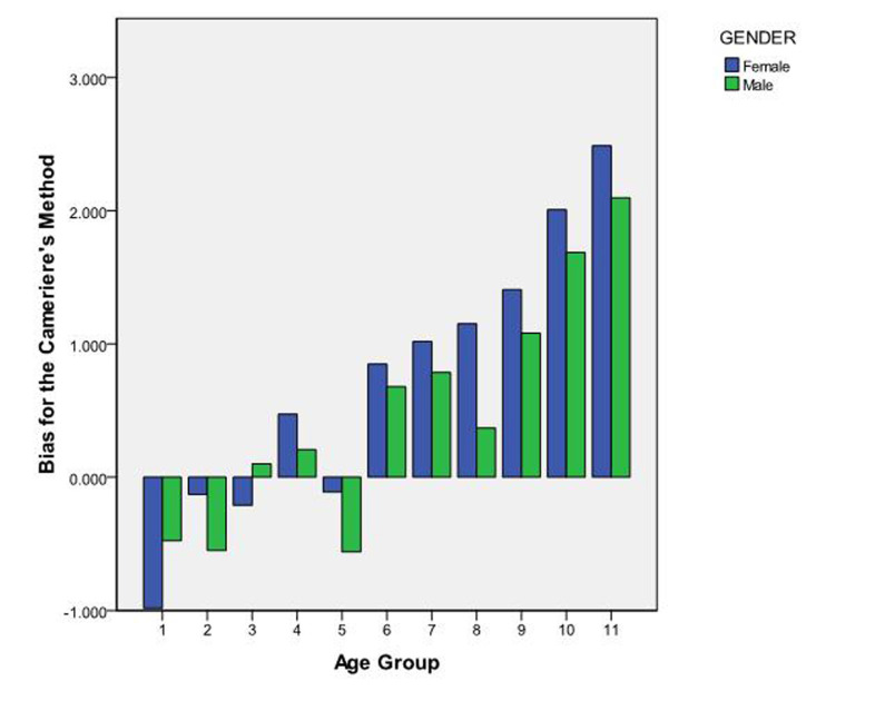
The paired t-test results for male and female participants for each year interval are observed in Figure 3. Non-significant overestimation of age was seen in age groups 1,2,3 and 5 in both genders. While underestimation of age was seen in age groups (4,6,7,8,9,10,11) for both males and females. However, in the age group 3 (7-7.99 years), males showed underestimation of age.
Figure 3.
Independent samples t-test for Cameriere’s method to express bias for gender
London Atlas of Tooth Development
The mean chronological age of the entire sample was 10.238 ± 3.160 years, while the mean estimated age of the whole sample was 10.267 ± 3.033 years when the London Atlas was applied to determine the age of the subjects. The difference of -0.03 years (SD± 0.69 years) was not significant and indicated a little overestimation. The bias was statistically significant in all the age groups (Table 4). To analyse and compare the differences between both genders, independent samples t-test was used. No statistically significant difference (p = 0.321) was observed between male and female participants (Table 5).The mean difference between the chronological and estimated ages was less in female subjects than in male subjects.
Table 4. Paired t-test for the London Atlas method showing the bias for the sample population.
| Measure of accuracy | N | Mean | ± SD | SE mean | Significance | 95% CI Lower | 95% CI Upper |
|---|---|---|---|---|---|---|---|
| Bias | 335 | -0.02955 | 0.69944 | 0.03821 | 0.440 | -0.10472 | 0.04562 |
| Absolute Difference | 335 | 0.54048 | 0.44397 | 0.02426 | 0.000** | 0.49276 | 0.58819 |
**p<0.001; Highly significant SD = Standard deviation, SE= Standard error, CI= Confidence Interval
Table 5. Independent samples t-test for the London Atlas method to express bias for gender.
| Measure of accuracy | Sex | N | Mean | ± SD | SE mean | Significance |
|---|---|---|---|---|---|---|
| Bias | M | 165 | -0.06812 | 0.616293 | 0.047978 | 0.321 |
| F | 170 | 0.00788 | 0.771636 | 0.059182 | ||
| Absolute Difference | M | 165 | 0.49794 | 0.367464 | 0.028607 | 0.084 |
| F | 170 | 0.58176 | 0.505007 | 0.038732 |
F = Female, M= Male, SD= Standard deviation, SE = Standard error
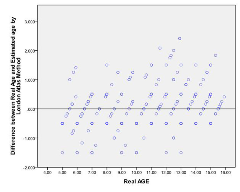
The paired t-test was applied to estimate the accuracy of age intervals of the entire sample. Differences between the actual and estimated age for the entire sample at each year interval are illustrated in Figure 4. Applying the London Atlas method of age assessment to the study sample, there was non-significant underestimation and overestimation in all age groups, hence it was found to be more accurate.
Figure 4.
The London Atlas method showing bias for age estimation in different age groups
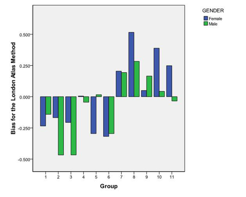
Paired t-test applied was used to test the accuracy of different age intervals in both male and female subjects as depicted in Figure 5. While non-significant, overestimation of age was noticed for both genders in age groups 1 to 6 and underestimation was observed in age groups 7 to 11. However, overestimation of age was seen in males in the age group 11 (15-15.99 years), while, underestimation of age was observed in males in the age group 5 (9-9.99 years).
Figure 5.
The London Atlas method showing bias in different age groups in male and female subjects
DISCUSSION
In view of the UN convention on the Rights of the Child, (11) age estimation in children and adolescents becomes the prime concern for the forensic fraternity and is one of the most relevant issues today in forensic medicine. According to its preamble, a child has the right to be registered and granted a nationality and according to article 12 of its constitution, the child has the right to express their opinions in accordance with their age and maturity. (11) Juvenile delinquency is a serious concern for India as recent years have witnessed a rapid rise in criminal cases involving minors. Furthermore, violation of the child’s basic rights like child labour, other forms of exploitation, non-registration of births are prevalent in India. Thus, it becomes more important to estimate accurate age in juveniles in the Court of Law. Thus, we chose children between 5 and 15.99 years of age as our target study population. Different age estimation methods have been tested in Indian children, for instance, a study conducted on Indian children concluded that Willem’s method was the most accurate followed by Demirjian and Chaillet’s methods. (12)
Various studies on different populations have assessed the accuracy of Cameriere’s method using the measurement of open apices in teeth. This method proved to be reasonably accurate in all the populations including Italian, European, Indian and Saudi Arabian children. (7, 13-15) While the London Atlas has also been found to be fairly precise in estimating age in different populations, Cameriere’s linear regression equation has been tested for accuracy in an Indian population earlier, where the results revealed that an open apex method in children was highly accurate with the morphological variables explaining 88.5% of the variations in predicted age. (13) However, to the best of our knowledge, no research has been undertaken so far in Indian subjects assessing the difference in the accuracy of age estimation applying these two non-invasive radiographic techniques. Thus, we compared the accuracy of both these methods in an Indian population (age 5-15.99 years) visiting the OPD of Oral Medicine and Radiology Department.
Cameriere et al, 2006 (8) tested the stepwise multiple regression formula in Italian white children and observed that age can be predicted more accurately and efficiently by using this method. Moreover, when tested and compared with Willems and Demirjian method in White Italian, Spanish and Croatian children, it was again found to be a more reliable and precise method for age assessment in young children. (13) The study emphasised the suitability of the sum of normalised open apices (s) and number (N0) of teeth with complete root development as accurate morphological parameters for determining age in juveniles. On the other hand, in a sample of Italian children between 11 and 16 years of age, the authors compared four age estimation methods.i.e. Demirjian, Willems, Cameriere and Haavikko and observed that Cameriere’s method underestimated the age by one year for both genders, while other methods were found to be more accurate. (16) The results of our study are more congruent with the latter study as, in the current study, a statistically significant underestimation of age was observed for all age intervals. Our results are also more similar to other age estimation studies carried out in different populations like Saudi, Iranian, Turkish and American children, where Cameriere’s formula invariably underestimated the age. (15, 17-19)
However, no significant difference was found between male and female subjects and the underestimation of age was uniform between the genders in several studies using Cameriere’s formula. (15, 17, 18) The present study was in agreement with the results of these studies and showed no statistically significant difference between the genders. Contrast results were observed in Mexican and Bosnian Herzegovinian populations, where overestimation of age was reported in females. (20, 21)
The London Atlas of Human Tooth Development determines age based on tooth development stage and the level of alveolar eruption. The use of software (http://www.atlas.dentistry.qmul.ac.uk) makes the technique convenient and practicable. (9) When the London Atlas was compared with the Schour and Massler estimation chart and Ubelaker estimation chart, it was found to be reasonably accurate and no significant difference was found for most age groups, except some. The study sample included white and Bangladeshi populations and the authors observed that tooth formation showed minimum variation during infancy but revealed most variability after the age of 16 years. (9)
Alshihri et al, 2015 (22, 23) assessed the suitability of the London Atlas of Human Tooth Development for age estimation in Saudi Arabian children and adolescents and found a significant difference between mean estimated and actual age. Further, there was a significant difference in the accuracy of age estimation between males and females. In females, the frequency of overestimation of age was higher as compared to males, emphasising the requirement of development of designate charts for each gender. This signified that hormonal changes during growth or puberty affect the tooth formation stages. (24) On the other hand, the results of a cross-sectional study carried out in Iranian children indicated high accuracy with no significant differences between the mean chronological age and mean dental ages using Smith’s method and the London Atlas but suggested the latter to be simpler to use. (25) Our findings were in congruence with the results of the latter study.
When the London Atlas of Human Tooth Development was used in Indian children, the results of the present study observed non-significant underestimation and overestimation in all age groups. The difference of -0.03 years was not significant and indicated a little overestimation, implying that it was more accurate than Cameriere’s stepwise linear regression equation. The London Atlas was also applied to estimate age in a Portuguese population and no significant difference was observed between male and female subjects either. (26) The observations of our study are in agreement and indicated no statistically significant difference between male and female participants. The mean difference between the chronological and estimated ages was less in female subjects than in male subjects.
In Saudi children, there was a significant difference between the dental and actual age of the subjects when the London Atlas was used for age estimation. (22) While, underestimation of age was a common finding in Saudi (22) as well as American populations, (19) overestimation of age was noticed in two different studies conducted in Portugal. (26, 27) In contrast to above studies, using the London Atlas, the results of the present study observed non-significant underestimation and overestimation in all age groups.
With the London Atlas, age is shown as an average.e.g.10.5 represents the mean of an age range from 10.00 to 10.99 years. While, when using Cameriere’s formula, 10.5 implies 10 years and 6 months. Thus, there is more likelihood of error of six months with the London Atlas and the bias between predicted and actual age may be overstated. (15)
In the present study, results from both the methods did not show any significant difference between male and female participants. Though overestimation and underestimation of age was observed in all age groups with both the methods, a non-significant difference was found when the London Atlas method was applied, implying it to be a more accurate technique. It has certain advantages, using both the upper and lower jaws, inclusion of both deciduous and permanent dentition developmental stages and observing the level of alveolar bone eruption of the teeth, implying that it is more accurate.
CONCLUSIONS
Considering the results of the present study, both Cameriere’s method of open apices and the London Atlas of Human Tooth Development are reliable for accurate dental age estimation in Indian children. While Cameriere’s method requires precise calculations and relies more on the expertise of the observer, the London Atlas method is relatively convenient and simple to use. The latter method uses software programme (available in different languages) to make accurate calculations.
Footnotes
The authors declare that they have no conflict of interest.
References
- 1.Agarwal D. Juvenile delinquency in India- Latest trends and entailing amendments in Juvenile Justice Act. People: Int J of Social Sciences. 2018;3(3):1365–83. 10.20319/pijss.2018.33.13651383 [DOI] [Google Scholar]
- 2.Berndt DC, Despotovic T, Mund MT, Filippi A. The role of the dentist in modern forensic age determination. Schweiz Monatsschr Zahnmed. 2008;118:1073–88. [in French, German] [PubMed] [Google Scholar]
- 3.AlQahtani SJ, Hector MP, Liversidge HM. Accuracy of dental age estimation charts: Schour and Massler, ubelaker and the London Atlas. Am J Phys Anthropol. 2014;154:70–8. 10.1002/ajpa.22473 [DOI] [PubMed] [Google Scholar]
- 4.Moorrees CF, Fanning EA, Hunt EE., Jr Age variation of formation stages for ten permanent teeth. J Dent Res. 1963;42:1490–502. 10.1177/00220345630420062701 [DOI] [PubMed] [Google Scholar]
- 5.Gleiser I, Hunt EE. The permanent mandibular first molar, its calcification, eruption and decay. Am J Phys Anthropol. 1955;13:253–83. 10.1002/ajpa.1330130206 [DOI] [PubMed] [Google Scholar]
- 6.Nolla C. The development of permanent teeth. ASDC J Dent Child. 1960;27:254–66. [Google Scholar]
- 7.Butti AC, Clivio A, Ferraroni M, Spada E, Testa A, Salvato A. Haavikko’s method to assess dental age in Italian children. Eur J Orthod. 2009;31:150–5. 10.1093/ejo/cjn081 [DOI] [PubMed] [Google Scholar]
- 8.Cameriere R, Ferrante L, Cingolani M. Age estimation in children by measurement of open apices in teeth. Int J Legal Med. 2006;120:49–52. 10.1007/s00414-005-0047-9 [DOI] [PubMed] [Google Scholar]
- 9.AlQahtani SJ, Hector MP, Liversidge HM. Brief communication: the London Atlas of human tooth development and eruption. Am J Phys Anthropol. 2010;142:481–90. 10.1002/ajpa.21258 [DOI] [PubMed] [Google Scholar]
- 10.Bengston RG. A study of the time of eruption and root development of the permanent teeth between six and thirteen years. Dent Res Grad Study. 1935;35:3–9. [Google Scholar]
- 11.Children’s Rights Alliance. Summary of the UN Convention on the Rights of the Child,(2013)https://www.childrensrights.ie/sites/default/files/information_sheets/files/SummaryUNCRC.pdf.
- 12.Hegde S, Patodia A, Shah K, Dixit U. The applicability of the Demirjian, Willems and Chaillet standards to age estimation of 5-15 year old Indian children. J Forensic Odontostomatol. 2019;37(1):40–50. [PMC free article] [PubMed] [Google Scholar]
- 13.Cameriere R, Ferrante L, Liversidge HM, Prieto JL, Brkic H. Accuracy of age estimation in children using radiograph of developing teeth. Forensic Sci Int. 2008;176:173–7. 10.1016/j.forsciint.2007.09.001 [DOI] [PubMed] [Google Scholar]
- 14.Sharma P, Wadhwan V, Prakash R, Goel S, Aggarwal P. Age estimation in children by measurement of open apices in teeth: a study in North Indian population. Aust J Forensic Sci. 2016;48:592–600. 10.1080/00450618.2015.1112426 [DOI] [Google Scholar]
- 15.Alsudairi DM, AlQahtani SJ. Testing and comparing the accuracy of two dental age estimation methods on saudi children: Measurement of open apices in teeth and the London Atlas of tooth development. Forensic Sci Int. 2019;295:226.e1–9. 10.1016/j.forsciint.2018.11.011 [DOI] [PubMed] [Google Scholar]
- 16.Pinchi V, Norelli GA, Pradella F, Vitale G, Rugo D, Nieri M. Comparison of the applicability of four odontological methods for age estimation of the 14 years legal threshold in a sample of Italian adolescents. J Forensic Odontostomatol. 2012;30(2):17–25. [PMC free article] [PubMed] [Google Scholar]
- 17.Javadinejad S, Sekhavati H, Ghafari R. A comparison of the accuracy of four age estimation methods based on panoramic radiography of developing teeth. J Dent Res Dent Clin Dent Prospects. 2015;9:72–8. 10.15171/joddd.2015.015 [DOI] [PMC free article] [PubMed] [Google Scholar]
- 18.Gulsahi A, Tirali RE, Cehreli SB, De Luca S, Ferrante L, Cameriere R. The reliability of Cameriere’s method in Turkish children: a preliminary report. Forensic Sci Int. 2015;249:319.e1. 10.1016/j.forsciint.2015.01.031 [DOI] [PubMed] [Google Scholar]
- 19.Santana SA, Bethard JD, Moore TL. Accuracy of dental age in non-adults: a comparison of two methods for age estimation using radiographs of developing teeth. J Forensic Sci. 2017;62:1320–5. 10.1111/1556-4029.13434 [DOI] [PubMed] [Google Scholar]
- 20.De Luca S, Giorgio SD, Butti AC, Biagi R, Cingolani M, Cameriere R. Age estimation in children by measurement of open apices in tooth roots: study of a Mexican sample. Forensic Sci Int. 2012; 221;155 e1-7. [DOI] [PubMed]
- 21.Galić I, Vodanovic M, Cameriere R, Nakas E, Galic E, Selimovic E, et al. Accuracy of Cameriere, Haavikko, and Willems radiographic methods on age estimation on Bosnian-Herzegovian children age groups 6-13. Int J Legal Med. 2011;125:315–21. 10.1007/s00414-010-0515-8 [DOI] [PubMed] [Google Scholar]
- 22.Alshihri AM, Kruger E, Tennant M. Dental age assessment of Western Saudi children and adolescents. Saudi Dent J. 2015;27:131–6. 10.1016/j.sdentj.2015.01.002 [DOI] [PMC free article] [PubMed] [Google Scholar]
- 23.Alshihri A, Kurger E, Tennant M. Integrating standard methods of age estimation in Western Saudi children and adolescents. Eur J Forensic Sci. 2016;3:157–62. 10.5455/ejfs.201936 [DOI] [Google Scholar]
- 24.Blenkin M, Taylor J. Age estimation charts for a modern Australian population. Forensic Sci Int. 2012;221(1-3):106–12. 10.1016/j.forsciint.2012.04.013 [DOI] [PubMed] [Google Scholar]
- 25.Ghafari R, Ghodousi A, Poordavar E. Comparison of the accuracy of the London atlas and Smith method in dental age estimation in 5-15.99-year-old Iranians using the panoramic view. Int J Legal Med. 2019;133:189–95. [DOI] [PubMed] [Google Scholar]
- 26.Pavlović S, Pereira PC, Vargas de Sousa Santos RF. Age estimation in Portugese population: the application of the London Atlas of tooth development and eruption. Forensic Sci Int. 2017;272:97–103. 10.1016/j.forsciint.2017.01.011 [DOI] [PubMed] [Google Scholar]
- 27.Cesario C, Santos R, Pestana D, Pereira PC. Medico-legal age estimation in a sub-adult Portugese population: validation of Atlas Schour and Massler and London. J Civil Legal Sci. 2016;5:196. [Google Scholar]


