. 2021 Feb 12;10:e61279. doi: 10.7554/eLife.61279

Figure 6. Cerebral blood flow (CBF) time profiles acquired with laser Doppler flowmetry (LDF) and arterial spin labeling (ASL)-magnetic resonance imaging (MRI).

On the left, 21-min CBF time profiles acquired with LDF in the somatosensory cortex are displayed for wild-type (WT) and transgenic Swedish Dutch Iowa (Tg-SwDI) mice. In the middle, 21-min CBF time profiles are displayed that are acquired with ASL-MRI in the left somatosensory cortex, after baseline correction, so the profiles can be compared to the LDF time profiles. On the right are the same profiles as in the middle, without baseline correction.

Figure 6.

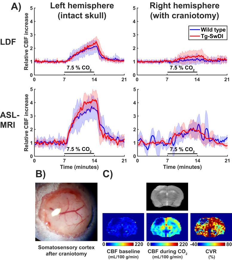
Figure 6—figure supplement 1. Cerebrovascular reactivity (CVR) measurements with laser Doppler flowmetry (LDF) and arterial spin labeling-magnetic resonance imaging (ASL-MRI) with and without skull removal.

Figure 6—figure supplement 1.

(A) CVR time profiles acquired either with LDF (top row) or ASL-MRI (bottom row). The left column shows the CVR time profiles from the intact, left hemisphere, the right column the profiles from the right hemisphere after craniotomy. (B) A photograph of the somatosensory cortex of a representative mouse after craniotomy. (C) On the top row, a T2 weighted image that was acquired with MRI after craniotomy. On the bottom row, cerebral blood flow (CBF) and CVR images that were acquired in the same scan session. Note that the left hemisphere is displayed on the right, according to radiological convention.