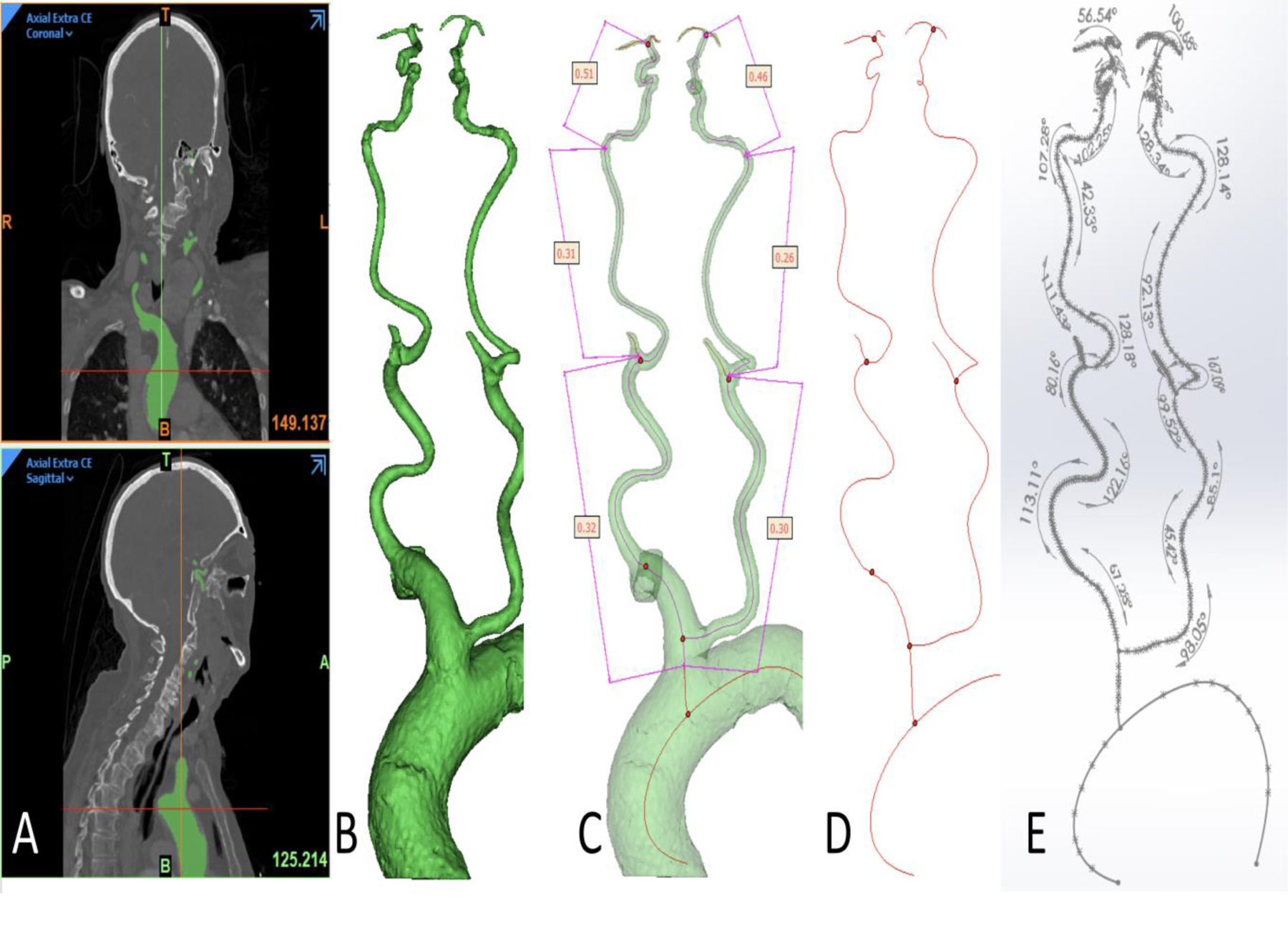
Figure 1.

Image processing and measurement of tortuosity.
(A) Segmentation was performed using cranio-cervical CT angiogram.
(B) A 3D reconstruction was obtained including aortic arch, bilateral common and internal carotid arteries (ICA) till ICA bifurcation with length of different segments.
(C) Tortuosity index being taken bilaterally for the following 3 segments: common carotid artery, cervical ICA and intracranial ICA.
(D) A center line was obtained and exported to SolidWorks software(Dassault Systemes, Vélizy-Villacoublay, France)..
(E) Angle of each curve was calculated on SolidWorks (Dassault Systemes, Vélizy-Villacoublay, France).
