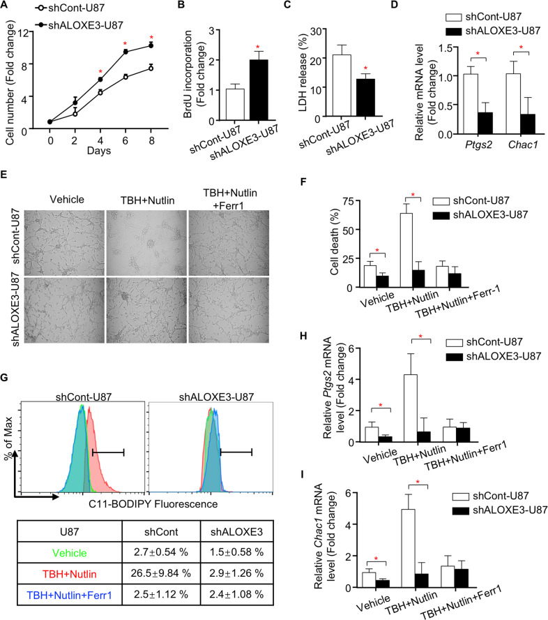
Fig. 3. GBM cells with ALOXE3 silencing are resistant to p53-induced ferroptosis.
A–D shALOXE3-U87 and shCont-U87 cells were used (n = 6). A Cell number was determined by trypan blue assay at indicated time points. B BrdU incorporation of cells. C LDH release of cells. D Relative mRNA levels of Ptgs2 and Chac1 normalized with GAPDH in cells. E–I shALOXE3-U87 and shCont-U87 cells were pre-treated with Nutlin-3a for 12 h and followed with TBH and Nutlin-3a ± Ferr1 treatment for additional 8 h (n = 6). E Representative images of cells. F Trypan blue staining for cell death. G C11-BODIPY staining for lipid peroxidation. H, I Relative mRNA levels of (H) Ptgs2 and (I) Chac1 normalized with GAPDH in cells. All data are represented as the mean ± s.e.m. *p < 0.05 (Student’s t test).

