Abstract
Background:
Hunterian ligation has been adapted for complex intracranial aneurysm repair when other, more modern techniques are insufficient. Before drastic alteration of cerebral blood flow dynamics, intraoperative challenges and consideration of blood flow dynamics must be completed to ensure adequate perfusion postligation. On satisfaction, ligation may proceed; however, subtle changes related to hypoperfusion may not be immediately observed during intraoperative challenge under general anesthesia and/or before onset of the vasospasm window.
Case Description:
In this report, we describe a patient who presented with a Hunt-Hess Grade III subarachnoid hemorrhage (SAH), with a right internal carotid artery (ICA) occlusion and a ruptured giant left ICA aneurysm. Endovascular treatment of the aneurysm was aborted because the nominal, 9 mm diameter of the ICA was too large for any intracranial balloon or stent. Three days later, she underwent a left-sided “insurance” extracranial-tointracranial arterial bypass (EIAB) using the superficial temporal artery simultaneously with hunterian ligation of the left ICA following reassuring results on intraoperative occlusion challenge. Over several days, her neurologic condition declined concurrent with the vasospasm window, and a right-sided EIAB was required to augment vascular supply. Following a protracted hospital course, the patient became progressively more independent and is currently residing in an assisted living facility.
Conclusion:
We illustrate an ultimately successful microsurgical treatment option in the setting of acute SAH that highlights the importance of cerebrovascular reserve and blood flow replacement in the setting of a compromised circle of Willis, especially during the vasospasm window.
Keywords: Extracranial-to-intracranial arterial bypass, Hunterian ligation, Hypoperfusion, Intracranial aneurysm, Microvascular neurosurgery, Subarachnoid hemorrhage

INTRODUCTION
Hunterian ligations were popularized in the 1800s by John Hunter who demonstrated a “safe and reproducible means of ligating peripheral arteries.”[4] Since then, neurosurgeons have adapted the technique for the treatment of intracranial aneurysms without sufficient open or endovascular options. Hunterian ligations are performed after reassuring intraoperative occlusion challenges while monitoring electroencephalography (EEG), somatosensory evoked potentials (SSEPs), and motor evoked potentials (MEPs).[2] Despite reassuring challenges, hypoperfusion is possible and swift recognition may prevent significant postoperative deficits. When occlusion tests are equivocal, an “insurance’ extracranial-to-intracranial arterial bypass is performed contemporaneously. Here, we illustrate a successful microsurgical treatment of a giant cavernous carotid aneurysm in the setting of an acute subarachnoid hemorrhage (SAH). We highlight the importance of cerebrovascular reserve in the setting of a compromised circle of Willis, especially during the vasospasm window, despite reassuring intraoperative challenges.
CASE DESCRIPTION
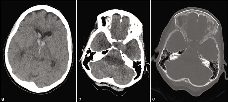
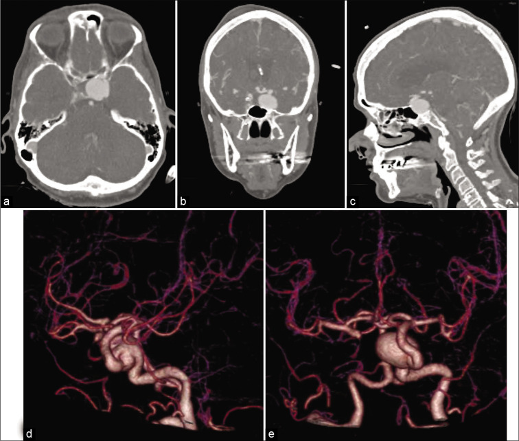
Our patient is a 71-year-old woman on aspirin (81 mg, daily) with a history of the right internal carotid artery (ICA) occlusion and a giant left cavernous carotid aneurysm that had been followed conservatively by an outside physician. She presented to an outside hospital with a thunderclap headache, nausea, vomiting, meningismus, and confusion consistent with Hunt-Hess Grade III SAH. She was transferred to our facility where noncontrast computed tomography (CT) revealed a Fisher Grade 4 SAH as well as bony erosion and intradural extension of the left cavernous carotid aneurysm [Figure 1a-c]. A ventriculostomy catheter was placed, and subsequent CT angiography (CTA) confirmed the left cavernous carotid aneurysm and right ICA occlusion [Figure 2a-e]. The patient was admitted to the neurosurgical intensive care unit and placed on SAH protocol, which consisted of levetiracetam (weight-based dosing), nimodipine (60 mg every 4 h), strict blood pressure management, daily transcranial Doppler imaging, and volume maintenance.
Figure 1:
Noncontrast computed tomography (CT) scan of the head showing intraventricular hemorrhage in the lateral and third ventricles (a); noncontrast CT scan showing left cavernous carotid aneurysm (b); bone window showing bony erosion in the left cavernous sinus secondary to cavernous carotid aneurysm (c).
Figure 2:
Axial (a), coronal (b), and sagittal (c) contrast-enhanced CT images showing left cavernous carotid aneurysm; 3D CT angiography reconstructions of the left cavernous carotid aneurysm (d and e).
On hospital day (HD) 1, she was taken for a diagnostic cerebral angiogram (DSA) to further characterize her vascular lesions. A giant left cavernous carotid aneurysm measuring approximately 24×17 mm was observed. No other vascular lesions were revealed, and SAH protocol was continued. On HD 4, she was taken for Pipeline (Medtronic, Dublin, Ireland) embolization of the left cavernous ICA aneurysm. Flow diversion could not be performed because the nominal diameter of the petrocavernous carotid artery measured close to 9 mm, making it too large for any available adjunctive balloons or intracranial stents [Figure 3a and b]. Therefore, microsurgical options were considered; and on HD 7, she was taken to the operating room. Following occlusion of the left ICA and hypotension (mean arterial pressure, 50–60 mmHg) induced for 40–45 min without electrophysiologic changes observed on EEG, SSEP, or MEP testing, a left-sided superficial temporal artery-to-middle cerebral artery (STA-MCA) “insurance” bypass followed by hunterian ligation of the left ICA was performed [Video 1]. Of note, the occlusion was maintained longer than the typical 20–30 min in an attempt to add additional stress, similar to what may occur as a result of vasospasm in the following days; and given the reassuring results, it was felt that a low-flow bypass was sufficient.
Figure 3:
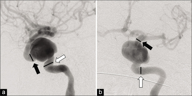
Lateral digital subtraction angiographic image showing nominal diameters of the parent vessel of 8.67 mm (white arrow) and 6.67 mm (black arrow) (a); anteroposterior digital subtraction angiographic image showing nominal parent artery diameters of 6 mm (white arrow) and 5.1 mm (black arrow) (b).
Postoperatively, the patient was at her neurological baseline level, apart from trace weakness in her lower extremities. Through HD 7, the patient’s mentation slowly declined. Perfusion CT imaging demonstrated a lack of vascular reserve in the left hemisphere on acetazolamide challenge.[7] A DSA [Figure 4a and b] confirmed patency of the left-sided bypass, and EEG showed no seizure activity. Induced hypertension failed to improve mentation, and given concerns about overlap of clinical decline within the SAH vasospasm window, flow augmentation appeared to be an appropriate option.
Figure 4:
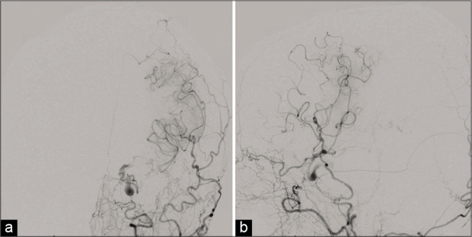
Anteroposterior (a) and lateral (b) digital subtraction angiographic images showing patency of the left-sided superficial temporal artery-to-middle cerebral artery bypass.
On HD 8, a right-sided external carotid artery-to-MCA high-flow bypass using an interpositional radial artery graft was performed [Video 1]. The high-flow option was ultimately chosen over an additional low-flow procedure due to the tenuous nature of the patient’s condition and need for aggressive revascularization.
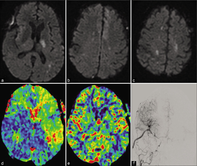
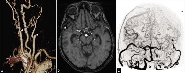
Postoperatively, the patient experienced a protracted hospital course with a poor neurological examination, despite only a few scattered areas of restricted diffusion in the left thalamus and bilateral watershed regions on magnetic resonance (MR) imaging. In addition, changes in flow dynamics consistent with patent bilateral bypasses were observed, including filling of the left-sided circulation from the right-sided high-flow bypass [Figure 5a-f]. Intermittent periods of increased responsiveness and appropriate verbal reactions to stimuli were observed. She was discharged to long-term care on HD 29 and continued to make steady progress in functional status. Postoperative CTA showed patency of both bypasses; MR imaging at 6 months showed no chronic ischemic changes; MR and CTA performed at 7 months showed patency of the bypasses as well as compensatory changes in the right vertebral artery [Figure 6a-c]. At 9 months, the patient had continued to make significant progress. She was experiencing mild cognitive difficulties and was ambulating independently, despite a recent fall and broken hip with an mRS score of 2.
Figure 5:
Diffusion restriction imaging showing ischemic regions of the left thalamus, bilateral anterior cerebral artery territory, and bilateral watershed regions (a-c). Computed tomography perfusion imaging showing time to peak in the right cerebral circulation (d) with preserved cerebral blood volume (e). Diagnostic angiogram showing partial left anterior circulation filling from the right side (f).
Figure 6:
3D reconstruction of CT of the neck showing bilateral carotid occlusions and patent proximal bypasses bilaterally at 7 months. White hollow arrow points to the proximal anastomosis of the radial artery interposition graft. Solid arrows indicate bilateral internal carotid artery occlusions. Large right dominant vertebral artery. (a) Magnetic resonance angiography at 7 months showing patency of the bypasses bilaterally (b); Immediate postoperative 4D CT angiography reconstruction showing bilateral patency of the bypasses (c).
DISCUSSION
Advancements in intracranial aneurysm treatments have expanded tremendously since the first reports of hunterian ligations; however, hunterian ligation remains the best option for a small subset of patients. Our patient had a ruptured giant left cavernous carotid aneurysm that was not amenable to endovascular intervention as well as a preexisting contralateral ICA occlusion that highlighted the importance of flow replacement during ligation of a major cerebral artery. This is especially true when factoring in the percentage of blood flow typically supplied by each major intracranial artery. In the typical intracranial circulation, average percentages of unilateral blood flow are 36% in the ICA and 14% in the vertebral artery;[8] however, our patient’s chronic ICA occlusion likely contributed to changes in flow dynamics where the posterior circulation carried an excess load sufficient to temporarily supply the additional requirement. Furthermore, expectant management was not an option as the risk of rebleed in untreated aneurysms has been shown to be 4–12% over the first 24 hours and 1–2% daily for the first 14 days, with rebleed mortality as high as 80%.[1,6] Therefore, the patient underwent hunterian ligation with an “insurance” low-flow STA-MCA bypass, which can be expected to initially provide approximately 30 ml/min of cerebral blood flow,[5] following reassuring intraoperative challenges. These challenges were performed under general anesthesia, which may have resulted in reduced cerebral metabolic rate of oxygen (CMRO2), leading to a false-negative result. In addition, although the circle of Willis and collateral circulation may have initially provided sufficient vascular supply, this was not adequate as the patient entered the maximal vasospasm window. Therefore, the patient required additional flow augmentation through a contralateral high-flow bypass, which can be expected to provide approximately 130 ml/min of cerebral blood flow.[3] In retrospect, the choice of a low-flow (STA-MCA) bypass for replacement of the left ICA in this case was suboptimal. Although it is important to note that the concurrent timing with the vasospasm window likely contributed, at least in part to the patient’s slow decline, more weight should have been placed on the preoperative DSA in which the left ICA appears to be the primary blood supply to the anterior circulation in both cerebral hemispheres. A more practical and appropriate alternative would have been to plan initially for the use of a high-flow left ICA-MCA bypass. In an awake patient, balloon test occlusion may have been helpful in evaluating the potential hemodynamic effects of sacrificing the ICA; the test may be difficult to carry out in critically ill patients or those who are not fully cooperative. Fortunately, our patient slowly made a good recovery following delayed high-flow bypass to the right hemisphere. This case illustrates that hypoperfusion, despite reassuring challenges, may still occur and must be addressed before irreversible ischemia and permanent deficits. In acute SAH, the vasospasm window must also be considered as it is a known cause of delayed cerebral ischemia. The authors suggest that an initial approach with a high-flow bypass would have potentially negated the effects of vasospasm and prevented the patient’s decline and need for additional procedures.
Video Annotations
0:50 – Initial digital subtraction angiography findings
2:07 – Left bypass
4:35 – Hunterian ligation of the left internal carotid artery
5:25 – Perfusion deficits/worsening
5:35 – Right bypass
8:51 – Left side filling from the right bypass.
CONCLUSION
This case illustrates the importance of considering blood flow replacement when selecting whether to utilize low-flow versus high-flow bypass in procedures that require acute occlusion (hunterian ligation) of a major cerebral artery. In addition, it highlights the limitations of using intraoperative electrophysiologic monitoring to access collateral blood flow, especially when concurrent with the vasospasm window following acute SAH. Although hunterian ligation is reserved for cases with limited options due to an elevated risk profile, outcomes can be positive if managed appropriately. In patients with reduced cerebrovascular reserve, it is imperative that clinicians be able to recognize signs of hypoperfusion and be prepared to intervene appropriately to avoid irreversible ischemia and suboptimal outcomes.
Footnotes
How to cite this article: Housley SB, Vakharia K, Waqas M, Siddiqui AH. Cerebral hypoperfusion necessitating additional bypass following hunterian ligation of the internal carotid artery despite reassuring intraoperative challenges: Video case report. Surg Neurol Int 2021;12:22.
Contributor Information
Steven B. Housley, Email: shousley@ubns.com.
Kunal Vakharia, Email: kunalvakharia5@gmail.com.
Muhammad Waqas, Email: mwaqas@ubns.com.
Adnan H. Siddiqui, Email: asiddiqui@ubns.com.
Declaration of patient consent
The authors certify that they have obtained all appropriate patient consent.
Financial support and sponsorship
Nil.
Conflicts of interest
There are no conflicts of interest.
Videos available on:
REFERENCES
- 1.Ameen AA, Illingworth R. Anti-fibrinolytic treatment in the pre-operative management of subarachnoid haemorrhage caused by ruptured intracranial aneurysm. J Neurol Neurosurg Psychiatry. 1981;44:220–6. doi: 10.1136/jnnp.44.3.220. [DOI] [PMC free article] [PubMed] [Google Scholar]
- 2.Barnett DW, Barrow DL, Joseph GJ. Combined extracranialintracranial bypass and intraoperative balloon occlusion for the treatment of intracavernous and proximal carotid artery aneurysms. Neurosurgery. 1994;35:92–8. doi: 10.1227/00006123-199407000-00014. [DOI] [PubMed] [Google Scholar]
- 3.Morton RP, Moore AE, Barber J, Tariq F, Hare K, Ghodke B, et al. Monitoring flow in extracranial-intracranial bypass grafts using duplex ultrasonography: A single-center experience in 80 grafts over 8 years. Neurosurgery. 2014;74:62–70. doi: 10.1227/NEU.0000000000000198. [DOI] [PubMed] [Google Scholar]
- 4.Polevaya NV, Kalani MY, Steinberg GK, Tse VC. The transition from hunterian ligation to intracranial aneurysm clips: A historical perspective. Neurosurg Focus. 2006;20:E3. doi: 10.3171/foc.2006.20.6.3. [DOI] [PubMed] [Google Scholar]
- 5.Sia SF, Morgan MK. High flow extracranial-to-intracranial brain bypass surgery. J Clin Neurosci. 2013;20:1–5. doi: 10.1016/j.jocn.2012.05.007. [DOI] [PubMed] [Google Scholar]
- 6.Starke RM, Connolly ES., Jr Participants in the International Multi-Disciplinary Consensus Conference on the Critical Care Management of Subarachnoid Hemorrhage. Rebleeding after aneurysmal subarachnoid hemorrhage. Neurocrit Care. 2011;15:241–6. doi: 10.1007/s12028-011-9581-0. [DOI] [PubMed] [Google Scholar]
- 7.Vagal AS, Leach JL, Fernandez-Ulloa M, Zuccarello M. The acetazolamide challenge: Techniques and applications in the evaluation of chronic cerebral ischemia. AJNR Am J Neuroradiol. 2009;30:876–84. doi: 10.3174/ajnr.A1538. [DOI] [PMC free article] [PubMed] [Google Scholar]
- 8.Zarrinkoob L, Ambarki K, Wåhlin A, Birgander R, Eklund A, Malm J. Blood flow distribution in cerebral arteries. J Cereb Blood Flow Metab. 2015;35:648–54. doi: 10.1038/jcbfm.2014.241. [DOI] [PMC free article] [PubMed] [Google Scholar]
Associated Data
This section collects any data citations, data availability statements, or supplementary materials included in this article.


