



Abstract
Introduction:
Sarcoidosis is a multi-system disease reported to occur with a higher incidence in Alberta than many other health jurisdictions within and outside of Canada. The reasons for this higher incidence are currently not known. Exposure to beryllium can result in a clinically and radiologically identical disease to sarcoidosis. The purpose of our study was to identify patterns with potential occupational or environmental exposures to beryllium amongst individuals with sarcoidosis in Alberta through a tertiary referral center.
Methods:
A prospective observational study was carried out at the University of Alberta Hospital. Patients with confirmed sarcoidosis (stages 0-4) were recruited from subspecialty clinics (Respirology, Cardiology, Neurology and Occupational Health). A predetermined list of industries thought to involve potentially relevant exposures for the development of sarcoidosis was used to capture current and previous exposure history. Results were entered into a database and where possible verified by comparing with existing electronic medical records (including histories, physical examination, diagnostic imaging and physiology).
Results:
A total of 45 patients were recruited, 25 men and 20 women. Of these, 84% of participants reported working in or being exposed to an industry/environment suspected of contributing to development of sarcoidosis over their lifetime. The most frequently reported exposures were within farming and agriculture (27%), oil and gas (20%), metalworking and handling animals (18%).
Conclusions:
Amongst this cohort, a high proportion reported working with a potentially relevant exposure. Individuals being assessed for sarcoidosis should have their most responsible physician elicit a detailed work and environmental history. (Sarcoidosis Vasc Diffuse Lung Dis 2020; 37 (4): e2020014)
Keywords: sarcoidosis, occupational exposure, environmental exposure, beryllium, epidemiology
Introduction
Sarcoidosis is a multisystem disease that incorporates the pathologic hallmark of noncaseating granulomata. Sarcoidosis affects the lungs in about 90% of patients, but can also affect the eye, heart, nervous system, lymph nodes, skin and other organ systems (1-5). Outcomes can vary widely between patients and symptoms can resolve spontaneously, remain in remission for years, or lead to multisystem organ failure or sudden cardiac death (1-4). Alberta has a population of over 4 million individuals. According to information from the Government of Alberta, rates of this disorder appear to be rising in recent years within Alberta (6). In 2015, the incidence rate for females was 7.8 and 11.2 for males (each per 100,000; not age-standardized). For comparison, the American Thoracic Society Statement on sarcoidosis from 1999 reported incidence rates of 5.9 (men) and 6.3 (women) per 100,000 person years in the United States. Additionally, although some studies of sarcoidosis show a higher prevalence amongst women (2,7), in Alberta rates for women have increased only 33% over a 10-year period whereas rates for men have increased 157% over the same period (6). While no one cause of sarcoidosis has been identified, occupational and/or environmental exposures have been suggested to contribute to the development of this disease. Furthermore, the spectrum of clinical phenotypes has led researchers to believe that there may be more than one exposure that may lead to immunologic sensitization (1).
There has been little success in identifying exactly which exposures may lead to sarcoidosis in which individuals, although beryllium exposure has been shown to cause a disease clinically and radiologically similar to sarcoidosis called chronic beryllium disease (CBD) (8,9). Beryllium is a metal with many useful properties when used as an alloy in a variety of industries, including aerospace, computers, and the oil and gas industry (10). Up to 16% of individuals exposed to beryllium become sensitized through the cutaneous and / or inhalational route (8,9). Inhalation of beryllium particulates can result in the development of a “sarcoid-like illness” which, when biopsied, also reveals noncaseating granulomata. Other exposures which have been suggested as potential links to sarcoidosis, or mortality due to sarcoidosis, include: agricultural dust, wood-burning (11), metalworking, health care and teaching (12,13). Given the recent rise in rates of sarcoidosis in Alberta, the purpose of this study was to identify patterns in occupational or environmental exposures among patients diagnosed with sarcoidosis in this province.
Methods
A prospective observational study was undertaken. Recruitment of patients attending relevant ambulatory clinics in two main tertiary care hospitals in the Edmonton region (University of Alberta Hospital and Royal Alexandra Hospital). The subspecialty clinics all had a pre-existing interest in sarcoidosis, and collaborated in clinical research with the provision of clinical care to patients with this disorder. As opposed to other large studies, such as the ACCESS study (A Case Control Etiologic Study of Sarcoidosis) which only recruited subjects from one subspecialty group (Respirologists) (12), our study broadly evaluated potential subjects to recruit from subspecialty clinics including Respirologists, Neurologists, Cardiologists and Occupational Medicine Specialists and their affiliated clinics. This was reflective of patterns of subspecialty referral for patients in Northern Alberta.
Recruitment was carried out over 12 months (May 2016-May 2017) and included all adult subjects (18 years of age and older) screened through our common electronic medical record. They were approached and willing to participate in the study with biopsy-confirmed sarcoidosis (all radiographic stages), except for those with Löfgren’s Syndrome where only a clinical review with classic radiographic findings were sufficient (no biopsy required). Pathologic samples confirmed sarcoidosis in the appropriate clinical context and the presence of typical noncaseating granulomata. Patterns of industries and / or environmental exposures thought to be relevant to the development of sarcoidosis, were identified utilizing prior studies which reviewed similar environmental and occupational exposures (12).
Exclusion criteria comprised active malignancy and/or currently receiving chemotherapy, as well as those expected to die within the next 6 months due to any cause. Additionally, individuals with a history of lung or other solid organ transplant or hematologic transplant were excluded. Active tuberculosis was excluded in all research participants.
All patients with confirmed sarcoidosis attending the relevant clinics were identified prior to their visit through a review of patient files by their primary clinician. Individuals were informed about the study by clinic staff at the time of their attendance through the study research assistant (RA) and asked if they were willing to meet after their clinic visit. If agreed to, the RA met with the subject following their clinic visit and the study procedures discussed with them. After providing informed consent, clinical information was reviewed by the RA with the subject. Participants were then given a list of occupations and industries with potential for exposure to agents associated with an increased risk of sarcoidosis both from expert input and available literature (14). This included lifetime exposures both at the workplace and otherwise (not limited to just at the time of recruitment). Blood was drawn and stored for biobanking at the time of recruitment for future work. Clinical information including thoracic staging along with other diagnostic imaging studies performed as part of clinical care, along with progress to date was obtained through review of the clinical record. The EQ-5D quality of life index was performed, along with screening for neurologic involvement with the 9-hole peg test (15,16) and Montreal Cognitive Assessment (MoCA) (17).
Study information was collected and entered into the REDCap (20) electronic data capture tools hosted and supported by the Faculty of Medicine and Dentistry at the University of Alberta. Verification of data entered was performed by two individuals (JP, DV) for accuracy. Data were exported from REDCap to Excel (MS Excel, 2016) for further analysis.
Institutional ethics approval was provided by the University of Alberta Research Ethics Board (Study ID: Pro00061653) and operational approval through the University of Alberta Hospital (Alberta Health Services [AHS]). The latter included access for research staff to the AHS supported electronic medical record system, e-Clinician (Epic-based system, Wisconsin).
Results
Overall, 49 patients from the various clinics were approached, one excluded due to coexisting (active) malignancy and three chose not to participate (no particular reason provided). A total of 45 subjects (25 males / 20 females) were recruited for the study. The majority were white Caucasian by self-identified ethnicity (91%). Based on thoracic diagnostic imaging, the majority of recruited subjects were females with stage I disease and males with stage II disease. There was also a significant proportion (27%) who resided in rural and remote communities - outside of urban / suburban areas mainly captured with other large sarcoidosis cohorts previously published.
Available information was gathered from the shared electronic medical record hosted by Alberta Health Services (Epic-based system, Wisconsin). All subjects also had electrocardiograms (ECGs) and echocardiograms reviewed, along with cardiac MR and 24 hour holter monitor tests when available. All subjects had neurologic imaging including computed tomography (CT) Heads and magnetic resonance imaging (MRI) Brains reviewed when available.
Subjects were reassessed at 6 and 12 months. There were no deaths or loss to follow up over the study duration. No adverse change was observed in this cohort during the 12-month time period.
The majority of subjects had sarcoidosis involvement of the respiratory tract (98% of participants). In addition, approximately 1/3 (31%) had cardiac involvement and 1/5 (18%) had neurologic involvement (Table 1). Some patients had active disease; some had dormant disease (latter primarily from the respiratory and occupational medicine clinics). The number of months between a diagnosis of sarcoidosis and study recruitment ranged from 1 month (6 subjects) to 444 months (the latter being a single outlier followed since their diagnosis in the 1970s with inactive disease but significant prior morbidity). The median number of months from diagnosis was 20 months, with 12 subjects (1/4) recruited within 4 months of diagnosis. Whereas all patients with a neurologic presentation had neurological deficits, none of the recruited subjects with other organ system presentation had evidence of neurologic compromise through clinical review and formal bedside neurologic screening tests.
Table 1.
Baseline Demographics of Sarcoidosis Recruits
| Gender (F/M) 20/25 | |||
| Age at recruitment (years) 50.6 +/- 13.6 | |||
| Body Mass Index (kg/m2) 31.5 +/- 6.7 | |||
| % of total | |||
| Region of residence | Urban | 21 | 47 |
| Suburban | 12 | 27 | |
| Rural/remote | 12 | 27 | |
| Ethnicity | White | 41 | 91 |
| South Asian | 2 | 4 | |
| Black | 1 | 2 | |
| Mixed | 1 | 2 | |
| Organ systems involved | respiratory | 44 | 98 |
| cardiac | 14 | 31 | |
| neurological | 8 | 18 | |
| ocular | 6 | 13 | |
| sarcoidosis arthritis | 5 | 11 | |
| skin | 4 | 9 | |
The majority of subjects recruited into this cohort were radiographic stage I and II thoracic disease, as determined by computed tomography (CT) of the chest (Figure 1).
Fig. 1.
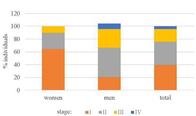
Thoracic Radiographic Staging
Lifetime industry/occupation and environmental exposure (Table 2) incorporated a variety of areas with some overlap – including farming and agriculture (27%), oil and gas (20%), metalworking (18%) and animal handling (18%) constituting the most frequently reported exposures (Table 2). Although farming and agriculture had equal representation of males and females in our cohort, all individuals in the oil and gas industry were males.
Table 2.
Lifetime Exposure History by Job Title/Category
| Job Title / Exposure Category* | Females (%) | Males (%) | Total (%) |
| Farming/agricultural work | 6 (30) | 6 (24) | 12 (26.7) |
| Oil and gas | 0 | 9 (36) | 9 (20) |
| Metalworking | 1 (5) | 7 (28) | 8 (17.8) |
| Animal handler/veterinarian | 4 (20) | 4 (16) | 8 (17.8) |
| Construction | 1 (5) | 6 (24) | 7 (15.6) |
| None of the above | 4 (20) | 3 (12) | 7 (15.6) |
| Health care sector | 5 (25) | 1 (4) | 6 (13.3) |
| Lumber/wood products | 0 | 5 (20) | 5 (11.1) |
| Mining | 1 (5) | 2 (8) | 3 (6.7) |
| Chemical industry | 0 | 3 (12) | 3 (6.7) |
| Stone, clay, glass or concrete | 0 | 3 (12) | 3 (6.7) |
| Metal industry | 1 (5) | 2 (8) | 3 (6.7) |
| Manufacturing of heating equipment | 1 (5) | 2 (8) | 3 (6.7) |
| Repairing electrical equipment | 0 | 3 (12) | 3 (6.7) |
| Pulp and paper industry | 0 | 2 (8) | 2 (4.4) |
| Manufacturing of industrial equipment | 0 | 2 (8) | 2 (4.4) |
| Manufacturing of automotive electrical equipment | 0 | 2 (8) | 2 (4.4) |
| Repairing/rebuilding automotive electrical equipment | 0 | 2 (8) | 2 (4.4) |
| Manufacturing/rebuilding of non-electrical vehicle parts | 0 | 2 (8) | 2 (4.4) |
| Armed forces | 1 (5) | 0 | 1 (2.2) |
| Firefighting | 0 | 1 (4) | 1 (2.2) |
*Adapted from Henneberger, P., et al. (14)
Quality of Life as determined by the EQ-5D did not show any difference between baseline and at the 6 and 12 month intervals of follow up utilized in this study (Figure 2).
Fig. 2.

Patient Quality of Life Status (EQ-5DTM) at Baseline and at 6** and 12-Month* Interviews. (*In one subject, 6-month value was used for 12-month data point as no information could be obtained for final interview. **Two additional data points missing from 6-month interviews).
Discussion
The Sarcoidosis Study evaluated a group of higher risk individuals with sarcoidosis in relation to occupational and environmental exposures. We found a striking proportion of individuals with potentially relevant occupational and / or environmental exposures that were missed during their initial clinical assessments. As well, new environmental exposures have more recently been elucidated (26, 27).
In comparison to other recent cohorts from Northern Europe (28, 29), our demographics in Northern Alberta constitute a unique population due to the predominant industries found in this region. The oil and gas industry captured within the list of industries queried was particularly unique in our study, as all positive respondents were male. This may explain some of the unusual provincial demographics that were previously noted through Alberta Health (6). Interestingly, recent work on sarcoidosis that was published from Ontario, Canada has also shown a relative increase in male preponderance spanning two decades (31). This Central Canadian province-wide study utilized health administrative database. It is well known that over the latter half of their study period which was during the economic boom in Alberta, many Ontarians traveled back and forth to work in Northern Alberta, often living in large camps during extended work periods spanning several weeks. Many Ontarian residents have been seen by specialist clinicians at the site of our tertiary referral center for different acute and chronic conditions including sarcoidosis (clinical observation - with only limited specialist follow up feasible). Individuals frequently traveled back to their home of residence (home province of Ontario) during their subsequent extended periods of time away from work. However it would have been impossible to capture this work-related information with the methodology used (this health administrative database is not linked to individuals’ place of work).
Another recent study was published on the largest cohort of sarcoidosis patients from Belgium to date (31). Of the 234 subjects in this study, a similar male preponderance was also noted (60%), however the industries captured were slightly different than found in our cohort; close to half of the Belgian subjects were white-collar workers (31). The construction and chemical industries appeared to be over-represented in their cohort.
It is not at all surprising that no change in quality of life was noted in our study over the short time frame of monitoring (12 months), as sarcoidosis can present and evolve over many years.
Study limitations included reliance on subject recall for prior occupational and environmental exposures (however, this would result in more negatives as has been noted in asbestos-related studies) specific to environmental exposures both within and external to the workplace (21-25). However, this is often the case with occupational histories pertinent to respiratory health in many similar studies (22). Inevitably using job title to identify relevant workplace exposures will have led to us missing some accidental exposures for occupations where exposure was not anticipated. However, we believe the approach taken using a standardised list with occupations and industries known to be at risk was most practical in the circumstances.
TB has been noted with noncaseating granulomata and can be difficult to distinguish from sarcoidosis. Therefore a high degree of clinical suspicion is required to exclude this infectious etiology and biopsies require culture to correlate with cytomorphology (32, 33).
Beryllium sensitization has been revealed as a common issue in certain industries, however, the regulatory standards vary significantly between different countries. Within Canada’s Provinces and Territories the American Conference of Governmental Industrial Hygienists Threshold Limit Value (ACGIH TLV) is used. The threshold limit value of a chemical substance is believed to be a level to which a worker can be exposed day after day for a working lifetime without adverse effects. The primary tool to objectively assess exposure with subsequent sensitization, based on a detailed history, is utilization of the beryllium lymphocyte proliferation test (8,9,18,19) (BeLPT) either through blood and / or bronchoscopy with bronchoalveolar lavage (BAL). The technical aspects of this assay require centralization of this study to primarily one site (QA/QC) through the National Jewish Hospital (Denver). Further research on this would be supported by better availability of testing for beryllium sensitization. However, this is complex and expensive. Indeed, the collected fresh blood or BAL sample requires processing within twenty-four hours to ensure limitation of falsely negative results, which are otherwise common without strict adherence to transport protocol. An alternative, utilising testing for a genetic polymorphism known to be associated with CBD, and which has already been used in Alberta, can be a valuable diagnostic tool, but is not as specific as a BeLPT (8,9,18,19).
We plan to carry out a prospective trial to more formally evaluate beryllium sensitization when resources are available both for funding and for accurate testing. Genetic associations should be evaluated further, and individuals considered for pre-screening prior to work in high risk occupations.
Imaging techniques utilized in the past for thoracic staging have relied on plain chest radiographs, however the literature supporting this is older. Given new techniques with low radiation exposure, we chose to use low dose high resolution computed tomography of the chest (HRCT Chest) (34, 35) to stage individuals accordingly, as is done within our clinical environments.
Sarcoidosis remains a heterogeneous disease with occupational and environmental factors that need to be taken into account. How assessments are done within clinical settings, particularly within a single payer system as exists in Canada, could include more formal access to Industrial Hygienists or a dedicated nurse. This done within a more structured inter-disciplinary clinic to ensure a more comprehensive assessment of exposures can be captured as part of the patient assessments and better structured patient-oriented care, which will mitigate the many “misses” for important occupational exposures in particular as noted in other occupational lung diseases (36). We need to better understand environmental and occupational exposures related to its occurrence through further prospective clinical trials in larger cohorts. Additionally, more formal inter-disciplinary clinical case conferences to rule out unusual presentations of TB (which can occasionally present with noncaseating granulomata), and drug-induced sarcoid-like reactions which may have inadvertently have been missed, would be important to consider. Without a complete occupational and environmental history being taken for all patients with sarcoidosis, relevant exposures will continue to be missed, and with them opportunities for prevention.
Acknowledgements
Ms. Iris de Guzman for administrative support. Drs. Lisa Maier and Sebastian Straube for manuscript review
Funding Sources:
Alberta Health Services, InterMune (investigator-initiated grant), In-kind support from physicians and secretarial services
References
- 1.Judson MA. The clinical features of sarcoidosis: a comprehensive review. Clin Rev Allergy Immunol. 2015;49:63–78. doi: 10.1007/s12016-014-8450-y. [DOI] [PubMed] [Google Scholar]
- 2.The Joint Statement of the American Thoracic Society (ATS), the European Respiratory Society (ER) and the World Association of Sarcoidosis and Other Granulomatous Disorders (WASOG) Statement on sarcoidosis. Am J Respir Crit Care Med. 1999;160:736–755. doi: 10.1164/ajrccm.160.2.ats4-99. [DOI] [PubMed] [Google Scholar]
- 3.Neville E, Walker AN, James DG. Prognostic factors predicting the outcome of sarcoidosis: an analysis of 818 patients. Q J Med. 1983;52:525–533. [PubMed] [Google Scholar]
- 4.Hillerdal G, Nöu E, Osterman K, Schmekel B. Sarcoidosis: epidemiology and prognosis. Am Rev Respir Dis. 1984;130:29–32. doi: 10.1164/arrd.1984.130.1.29. [DOI] [PubMed] [Google Scholar]
- 5.Sones M, Israel HL. Course and prognosis of sarcoidosis. Am J Med. 1960;29:84–93. doi: 10.1016/0002-9343(60)90009-7. [DOI] [PubMed] [Google Scholar]
- 6.Alberta Health. Sarcoidosis in Alberta. Government of Alberta; 2016. Health Trends Alberta. [Google Scholar]
- 7.Cozier YC, Berman JS, Palmer JR, Boggs DA, Serlin DM, Rosenberg L. Sarcoidosis in black women in the United States. Data from the black women’s health study. Chest. 2011;139:144–150. doi: 10.1378/chest.10-0413. [DOI] [PMC free article] [PubMed] [Google Scholar]
- 8.Cherry N, Beach J, Burstyn I, Parboosingh J, Schouchen J, Senthilselvan A, Svenson L, Tamminga J, Yiannakoulias N. Genetic susceptibility to beryllium: a case-referent study of men and women of working age with sarcoidosis or other chronic lung disease. Occup Environ Med. 2015;72:21–27. doi: 10.1136/oemed-2014-102359. [DOI] [PubMed] [Google Scholar]
- 9.Műller-Quernheim J, Gaede KI, Fireman E, Zissel G. Diagnoses of chronic beryllium disease within cohorts of sarcoidosis patients. Eur Respir J. 2006;27:1190–1195. doi: 10.1183/09031936.06.00112205. [DOI] [PubMed] [Google Scholar]
- 10.Balmes JR, Abraham JL, Dweik RA, Fireman E, Fontenot AP, Maier LA, Műller-Quernheim J, Ostiguy G, Pepper LD, Saltini C, Schuler CR, Takaro TK, Wambach PF. An official American Thoracic Society statement: diagnosis and management of beryllium sensitivity and chronic beryllium disease. Am J Respir Crit Care Med. 2014;190:e34–e59. doi: 10.1164/rccm.201409-1722ST. [DOI] [PubMed] [Google Scholar]
- 11.Kreider ME, Christie JD, Thompson B, Newman L, Rose C, Barnard J, Bresnitz E, Judson MA, Lackland DT, Rossman MD. Relationship of environmental exposures to the clinical phenotype of sarcoidosis. Chest. 2005;128:207–215. doi: 10.1378/chest.128.1.207. [DOI] [PubMed] [Google Scholar]
- 12.Newman LS, Rose CS, Bresnitz EA, Rossman MD, Barnard J, Frederick M, Terrin ML, Weinberger SE, Moller DR, McLennan G, Hunninghake G, DePalo L, Baughman RP, Iannuzzi MC, Judson MA, Knatterud GL, Thompson BW, Teirstein AS, Yeager H, Jr, Johns CJ, Rabin DL, Rybicki BA, Cherniack R. A case control etiologic study of sarcoidosis: environmental and occupational risk factors. Am J Respir Crit Care Med. 2004;170:1324–1330. doi: 10.1164/rccm.200402-249OC. [DOI] [PubMed] [Google Scholar]
- 13.Liu H, Patel D, Welch AM, Wilson C, Mroz MM, Li L, Rose CS, Van Dyke M, Swigris JJ, Hamzeh N, Maier LA. Association between occupational exposures and sarcoidosis: an analysis from death certificates in the United States, 1988-1999. Chest. 2016;150:289–298. doi: 10.1016/j.chest.2016.01.020. [DOI] [PMC free article] [PubMed] [Google Scholar]
- 14.Henneberger PK, Goe SK, Miller WE, Doney B, Groce DW. Industries in the United States with airborne beryllium exposure and estimates of the number of current workers potentially exposed. J Occup Environ Hyg. 2004;1:648–659. doi: 10.1080/15459620490502233. [DOI] [PubMed] [Google Scholar]
- 15.Feys P, Lamers I, Francis G, Benedict R, Phillips G, LaRocca N, Hudson LD, Rudick R. The nine-hole peg test as a manual dexterity performance measure for multiple sclerosis. Mult Scler. 2017;23:711–720. doi: 10.1177/1352458517690824. [DOI] [PMC free article] [PubMed] [Google Scholar]
- 16.Oxford Grice K, Vogel KA, Le V, Mitchell A, Muniz S, Vollmer MA. Adult norms for a commercially available nine-hole peg test for finger dexterity. Am J Occup Ther. 2003;57:570–573. doi: 10.5014/ajot.57.5.570. [DOI] [PubMed] [Google Scholar]
- 17.Nasreddine ZS, Phillips NA, Bédirian V, Charbonneau S, Whitehead V, Collin I, Cummings JL, Chertkow H. The Montreal Cognitive Assessment, MoCA: a brief screening tool for mild cognitive impairment. J Am Geriatr Soc. 2005;53:695–699. doi: 10.1111/j.1532-5415.2005.53221.x. [DOI] [PubMed] [Google Scholar]
- 18.Newman LS, Kreiss K, King TE, Jr, Seay S, Campbell PA. Pathologic and immunologic alterations in early stages of beryllium disease: re-examination of disease definition and natural history. Amer Rev Resp Dis. 1989;139:1479–1486. doi: 10.1164/ajrccm/139.6.1479. [DOI] [PubMed] [Google Scholar]
- 19.Lang L. Beryllium: a chronic problem. Environ Health Perspect. 1994;102:526–531. doi: 10.1289/ehp.94102526. [DOI] [PMC free article] [PubMed] [Google Scholar]
- 20.Harris PA, Taylor R, Thielke R, Payne J, Gonzalez N, Conde JG. Research electronic data capture (REDCap) - a metadata-driven methodology and workflow process for providing translational research informatics support. J Biomed Inform. 2009;42:377–381. doi: 10.1016/j.jbi.2008.08.010. [DOI] [PMC free article] [PubMed] [Google Scholar]
- 21.Ahrens W, Jöckel KH, Brochard P, Bolm-Audorff U, Grossgarten K, Iwatsubo Y, Orlowski E, Pohlabeln H, Berrino F. Retrospective assessment of asbestos exposure-I. case-control analysis in a study of lung cancer: efficiency of job-specific questionnaires and job exposure matrices. Int J Epidemiol. 1993;22:S83–S95. doi: 10.1093/ije/22.supplement_2.s83. [DOI] [PubMed] [Google Scholar]
- 22.Teschke K, Olshan AF, Daniels JL, DeRoos AJ, Parks CG, Schulz M, Vaughan TL. Occupational exposure assessment in case-control studies: opportunities for improvement. Occup Environ Med. 2002;59:575–594. doi: 10.1136/oem.59.9.575. [DOI] [PMC free article] [PubMed] [Google Scholar]
- 23.Peto J, Doll R, Hermon C, Binns W, Clayton R, Goffe T. Relationship of mortality to measures of environmental asbestos pollution in an asbestos textile factory. Ann Occup Hyg. 1985;29:305–355. doi: 10.1093/annhyg/29.3.305. [DOI] [PubMed] [Google Scholar]
- 24.Doll R. Mortality from lung cancer in asbestos workers. Br J Ind Med. 1955;12:81–86. doi: 10.1136/oem.12.2.81. [DOI] [PMC free article] [PubMed] [Google Scholar]
- 25.Peto J, Doll R, Howard SV, Kinlen LJ, Lewinsohn HC. A mortality study among workers in an English asbestos factory. Br J Ind Med. 1977;34:169–173. doi: 10.1136/oem.34.3.169. [DOI] [PMC free article] [PubMed] [Google Scholar]
- 26.Judson MA. Environmental Risk Factors for Sarcoidosis. Front Immunol. 2020;11:1340. doi: 10.3389/fimmu.2020.01340. [DOI] [PMC free article] [PubMed] [Google Scholar]
- 27.Caruana LB, Redwine GD, Rohde RE, Russian CJ. A prospective study of patients diagnosed with sarcoidosis: factors – environmental exposure, health assessment, and genetic outlooks. Sarcoidosis Vasculitis Diffuse Lung Disease. 2019;36:228–242. doi: 10.36141/svdld.v36i3.7112. [DOI] [PMC free article] [PubMed] [Google Scholar]
- 28.Beijer E, Meek B, Bossuyt X, Peters S, Vermeulen RCH, Kromhout H, Veltkamp M. Immunoreactivity to metal and silica associates with sarcoidosis in Dutch patients. Respir Res. 2020;21:141. doi: 10.1186/s12931-020-01409-w. [DOI] [PMC free article] [PubMed] [Google Scholar]
- 29.Larsson J, Graff P, Bryngelsson I-L, Vihlborg P. Sarcoidosis and increased risk of comorbidities and mortality in Sweden : Sarcoidosis - Comorbidities and Mortality. Sarcoidosis Vasculitis Diffuse Lung Disease. 2020 doi: 10.36141/svdld.v37i2.9142. [DOI] [PMC free article] [PubMed] [Google Scholar]
- 30.Fidler LM, Balter M, Fisher JH, To T, Stanbrook MB, Gershon A. Epidemiology and health outcomes of sarcoidosis in a universal healthcare population: a cohort study. Eur Respir J. 2019;54(4) doi: 10.1183/13993003.00444-2019. [DOI] [PubMed] [Google Scholar]
- 31.De Ridder J, Ronsmans S, Vanderschueren S, Wuyts W, Yserbyt J. Clinical characteristics of sarcoidosis patients in Belgium. Acta Clinica Belgica. 2020:1–8. doi: 10.1080/17843286.2020.1821493. [DOI] [PubMed] [Google Scholar]
- 32.Muthu V, Gupta N, Dhooria S, et al. Role of cytomorphology in the diagnosis of sarcoidosis in subjects undergoing endobronchial ultrasound-guided transbronchial needle aspiration. Sarcoidosis Vasculitis Diffuse Lung Disease. 2019;36(3):209–16. doi: 10.36141/svdld.v36i3.8175. [DOI] [PMC free article] [PubMed] [Google Scholar]
- 33.Rajagopala S, Shankari S, Kancherla R, Ramanathan RP, Balalakshmoji D. Miliary Sarcoidosis: does it exist? A case series and systematic review of literature. Sarcoidosis Vasculitis Diffuse Lung Disease. 2020;37:53–5. doi: 10.36141/svdld.v37i1.7837. [DOI] [PMC free article] [PubMed] [Google Scholar]
- 34.Bergin CJ, Bell DY, Coblentz CL, et al. Sarcoidosis: correlation of pulmonary parenchymal pattern at CT with results of pulmonary function tests. Radiology. 1989 Jun;171(3):619–24. doi: 10.1148/radiology.171.3.2717731. [DOI] [PubMed] [Google Scholar]
- 35.Russo JJ, Nery PB, Ha AC, et al. Sensitivity and specificity of chest imaging for sarcoidosis screening in patients with cardiac presentations. Sarcoidosis Vasculitis Diffuse Lung Disease. 2019;36:18–4. doi: 10.36141/svdld.v36i1.6865. [DOI] [PMC free article] [PubMed] [Google Scholar]
- 36.Tarlo SM. Occupational lung diseases. Canadian Journal of Respiratory, Critical Care, and Sleep Medicine. 2020 Apr 1;4(sup1):S6–8. [Google Scholar]