Mouse model studies are frequently used in oral microbiome research, particularly to investigate diseases such as periodontitis and caries, as well as other related systemic diseases. We have reported here the details of the development of a curated reference database to characterize the oral microbial community in laboratory and some wild mice.
KEYWORDS: 16S rRNA, Streptococcus danieliae, Apodemus sylvaticus, database, mouse models, Mus musculus, oral microbiology, oral microbiome
ABSTRACT
A curated murine oral microbiome database to be used as a reference for mouse-based studies has been constructed using a combination of bacterial culture, 16S rRNA gene amplicon, and whole-genome sequencing. The database comprises a collection of nearly full-length 16S rRNA gene sequences from cultured isolates and draft genomes from representative taxa collected from a range of sources, including specific-pathogen-free laboratory mice, wild Mus musculus domesticus mice, and formerly wild wood mouse Apodemus sylvaticus. At present, it comprises 103 mouse oral taxa (MOT) spanning four phyla—Firmicutes, Proteobacteria, Actinobacteria, and Bacteroidetes—including 12 novel undescribed species-level taxa. The key observations from this study are (i) the low diversity and predominantly culturable nature of the laboratory mouse oral microbiome and (ii) the identification of three major murine-specific oral bacterial lineages, namely, Streptococcus danieliae (MOT10), Lactobacillus murinus (MOT93), and Gemella species 2 (MOT43), which is one of the novel, still-unnamed taxa. Of these, S. danieliae is of particular interest, since it is a major component of the oral microbiome from all strains of healthy and periodontally diseased laboratory mice, as well as being present in wild mice. It is expected that this well-characterized database should be a useful resource for in vitro experimentation and mouse model studies in the field of oral microbiology.
IMPORTANCE Mouse model studies are frequently used in oral microbiome research, particularly to investigate diseases such as periodontitis and caries, as well as other related systemic diseases. We have reported here the details of the development of a curated reference database to characterize the oral microbial community in laboratory and some wild mice. The genomic information and findings reported here can help improve the outcomes and accuracy of host-microbe experimental studies that use murine models to understand health and disease. Work is also under way to make the reference data sets publicly available on a web server to enable easy access and downloading for researchers across the world.
INTRODUCTION
Mouse models play a crucial role in microbiome research, particularly in the investigation of the interactions between the host and the resident microbiota in health and disease (1, 2). Oral microbial population characterization in wild-type and mutant laboratory mouse strains have ranged from historical studies that involved identification of cultured isolates based on phenotypic characterization (3–6) to more recent molecular based studies focused on 16S rRNA gene sequencing (2, 7, 8). These studies have all demonstrated the mouse bacterial community to have a simple and relatively stable composition, with a major proportion of cultivable components, particularly in the specific-pathogen-free (SPF) laboratory mouse strains.
Despite this inherent simplicity of the mouse oral microbiome, it has significant relevance as a model to investigate and understand the mechanisms of human oral diseases such as periodontitis (7, 9, 10). A primary reason for this has been the parallels observed in the nature of the initiation and development of disease in experimental studies, specifically the development of a dysbiotic microbiome (characterized by increased total microbial loads) and soft tissue destruction with gingival inflammation (2, 11, 12), which are often comparable to that seen among humans (13, 14). Also, since the microbial genera observed are often similar to the predominant ones seen in humans (15), these animal models are also useful for understanding host-microbiota interactions and homeostasis mechanisms in health and disease.
However, the lack of adequate information in the public domain about mouse oral bacterial isolates from various sources, as well as poorly curated 16S rRNA gene sequences in the public databases, may lead to non- or misidentification of the organisms, which could thereby affect the outcome of such studies. A host-specific curated reference database for murine oral microbial populations in the public domain would enable researchers to accurately, reliably, and consistently identify the bacterial communities in experimental samples. Databases provide an improved and more accurate characterization of bacterial communities and allow easy comparison of work from different laboratories.
Similar databases have already been developed to characterize the oral microbiome in humans (15, 16) and other mammalian host species such as cats and dogs (17, 18). More recently, researchers have characterized and generated a database for the mouse intestinal bacterial community (19, 20), as well as chronicled the collection of genes in the mouse gut metagenome (21).
Here, we report a curated and well-characterized database of the oral bacterial population in mice, with representative genome sequences, which should greatly benefit researchers as a reference for oral microbiome studies in health and disease using laboratory mouse models.
RESULTS
Assignment of mouse oral taxa.
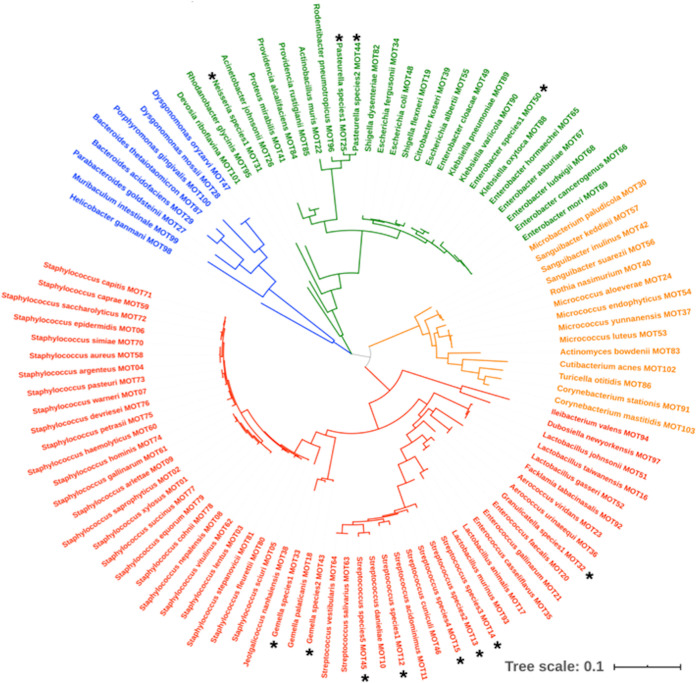
To date, 325 16S rRNA gene sequences from murine oral bacterial isolates have been analyzed and found to constitute 103 mouse oral taxa (MOT) (Table 1 and Fig. 1). Twelve of the assigned MOTs (12.36%) represent novel, previously unidentified species that need further characterization in order to be assigned a formal species name. Representative 16S rRNA gene sequences for these novel taxa have been submitted to the National Center for Biotechnology Information (NCBI) database and are available under accession numbers MN095260 to MN095271. The 16S rRNA gene sequences for all the isolates analyzed in this study are available to download under accession numbers MW175535 to MW175859.
TABLE 1.
Details of bacterial species and MOTs included in the murine oral microbiome databasea
| Taxonomy ID | Species ID | Type strain | 16S rRNA gene accession no. | Source host |
|---|---|---|---|---|
| MOT01 | Staphylococcus xylosus | ATCC 29971 | D83374 | Mus musculus /Apodemus sylvaticus |
| MOT02 | Staphylococcus saprophyticus | ATCC 15305 | NR_115607 | Mus musculus /Apodemus sylvaticus |
| MOT03 | Staphylococcus lentus | ATCC 29070 | D83370 | Mus musculus /Apodemus sylvaticus |
| MOT04 | Staphylococcus argenteus | MSHR1132 | FR821777 | Mus musculus |
| MOT05 | Staphylococcus sciuri | DSM 20345T | AJ421446 | Mus musculus /Apodemus sylvaticus |
| MOT06 | Staphylococcus epidermidis | ATCC 14990 | D83363 | Mus musculus |
| MOT07 | Staphylococcus warneri | ATCC 27836 | L37603 | Mus musculus |
| MOT08 | Staphylococcus nepalensis | DSM 15150 | AJ517414 | Mus musculus /Apodemus sylvaticus |
| MOT09 | Staphylococcus arlettae | ATCC 43957 | AB009933 | Mus musculus /Apodemus sylvaticus |
| MOT10 | Streptococcus danieliae | DSM 22233 | GQ456229 | Mus musculus |
| MOT11 | Streptococcus acidominimus | LMG 17755 | JX986969 | Mus musculus |
| MOT12 | Streptococcus species 1 | MN095260 | Mus musculus | |
| MOT13 | Streptococcus species 2 | MN095261 | Mus musculus | |
| MOT14 | Streptococcus species 3 | MN095262 | Apodemus sylvaticus | |
| MOT15 | Streptococcus species 4 | MN095263 | Mus musculus | |
| MOT16 | Lactobacillus taiwanensis | BCRC 17755 | EU487512 | Mus musculus |
| MOT17 | Lactobacillus animalis | NBRC 15882 | AB326350 | Mus musculus /Apodemus sylvaticus |
| MOT18 | Gemella palaticanis | CCUG 39489 | Y17280 | Mus musculus |
| MOT19 | Shigella flexneri | ATCC 29903 | X96963 | Mus musculus |
| MOT20 | Enterococcus faecalis | JCM 5803 | AB012212 | Mus musculus |
| MOT21 | Enterococcus gallinarum | ATCC 49573 | AF039900 | Mus musculus |
| MOT22 | Muribacter muris | NCTC 12432 | AY362894 | Mus musculus |
| MOT23 | Aerococcus viridans | NCTC 8251 | AB680262 | Mus musculus |
| MOT24 | Micrococcus aloeverae | DSM 27472 | KF524364 | Mus musculus |
| MOT25 | Pasteurella species 1 | MN095264 | Apodemus sylvaticus | |
| MOT26 | Acinetobacter johnsonii | ATCC 17909 | Z93440 | Mus musculus |
| MOT27 | Parabacteroides goldsteinii | WAL 12034 | AY974070 | Mus musculus |
| MOT28 | Dysgonomonas mossii | CCUG 43457 | AJ319867 | Mus musculus |
| MOT29 | Bacteroides acidifaciens | JCM 10556 | AB021164 | Mus musculus |
| MOT30 | Microbacterium paludicola | DSM 16915 | AJ853909 | Mus musculus |
| MOT31 | Neisseria species 1 | MN095265 | Apodemus sylvaticus | |
| MOT32 | Granulicatella species 1 | MN095266 | Apodemus sylvaticus | |
| MOT33 | Gemella species 1 | MN095267 | Apodemus sylvaticus | |
| MOT34 | Escherichia fergusonii | ATCC 35469 | AF530475 | Mus musculus |
| MOT35 | Enterococcus casseliflavus | ATCC 700327 | AF039903 | Mus musculus |
| MOT36 | Aerococcus urinaeequi | ATCC 29723 | D87677 | Mus musculus |
| MOT37 | Micrococcus yunnanensis | YIM 65004 | FJ214355 | Mus musculus |
| MOT38 | Jeotgalicoccus nanhaiensis | JSM 077023 | FJ237390 | Mus musculus |
| MOT39 | Citrobacter koseri | ATCC 27028 | HQ992945 | Mus musculus |
| MOT40 | Rothia nasimurium | CCUG 35957 | AJ131121 | Mus musculus |
| MOT41 | Proteus mirabilis | ATCC 29906 | DQ885256 | Mus musculus |
| MOT42 | Sanguibacter inulinus | NCFB 3024 | X79451 | Mus musculus |
| MOT43 | Gemella species 2 | MN095268 | Mus musculus | |
| MOT44 | Pasteurella species 2 | MN095269 | Apodemus sylvaticus | |
| MOT45 | Streptococcus species 5 | MN095270 | Apodemus sylvaticus | |
| MOT46 | Streptococcus cuniculi | CCUG 65085 | HG793791 | Apodemus sylvaticus |
| MOT47 | Dysgonomonas oryzarvi | JCM 16859 | AB547446 | Mus musculus |
| MOT48 | Escherichia coli | ATCC 11775 | X80725 | Mus musculus |
| MOT49 | Enterobacter cloacae | ATCC 13047 | AJ251469 | Mus musculus |
| MOT50 | Enterobacter species 1 | MN095271 | Mus musculus | |
| MOT51 | Lactobacillus johnsonii | ATCC 33200 | AJ002515 | Mus musculus |
| MOT52 | Lactobacillus gasseri | ATCC 33323 | AF519171 | Mus musculus |
| MOT53 | Micrococcus luteus | DSM 20030 | AJ536198 | Mus musculus |
| MOT54 | Micrococcus endophyticus | DSM 17945 | EU005372 | Mus musculus |
| MOT55 | Escherichia albertii | CCUG 46494 | AJ508775 | Mus musculus |
| MOT56 | Sanguibacter suarezii | ATCC 51766 | X79452 | Mus musculus |
| MOT57 | Sanguibacter keddieii | ATCC 51767 | X79450 | Mus musculus |
| MOT58 | Staphylococcus aureus | ATCC 12600 | L36472 | Mus musculus |
| MOT59 | Staphylococcus caprae | ATCC 35538 | AB009935 | Mus musculus |
| MOT60 | Staphylococcus haemolyticus | ATCC 29970 | X66100 | Mus musculus |
| MOT61 | Staphylococcus gallinarum | ATCC 35539 | D83366 | Mus musculus /Apodemus sylvaticus |
| MOT62 | Staphylococcus vitulinus | ATCC 51145 | AB009946 | Mus musculus /Apodemus sylvaticus |
| MOT63 | Streptococcus salivarius | ATCC 7073 | AY188352 | Mus musculus |
| MOT64 | Streptococcus vestibularis | NCTC 12167 | AY188353 | Mus musculus |
| MOT65 | Enterobacter hormaechei | ATCC 49162 | AJ508302 | Mus musculus |
| MOT66 | Enterobacter cancerogenus | ATCC 33241 | Z96078 | Mus musculus |
| MOT67 | Enterobacter asburiae | ATCC 35953 | AB004744 | Mus musculus |
| MOT68 | Enterobacter ludwigii | CCUG 51323 | AJ853891 | Mus musculus |
| MOT69 | Enterobacter mori | LMG 25706 | EU721605 | Mus musculus |
| MOT70 | Staphylococcus simiae | LMG 22723 | AY727530 | Mus musculus |
| MOT71 | Staphylococcus capitis | ATCC 27840 | L37599 | Mus musculus |
| MOT72 | Staphylococcus saccharolyticus | ATCC 14953 | L37602 | Mus musculus |
| MOT73 | Staphylococcus pasteuri | ATCC 51129 | AB009944 | Mus musculus |
| MOT74 | Staphylococcus hominis | ATCC 27844 | X66101 | Mus musculus |
| MOT75 | Staphylococcus petrasii | CCUG 62727 | JX139845 | Mus musculus |
| MOT76 | Staphylococcus devriesei | CCUG 58238 | FJ389206 | Mus musculus |
| MOT77 | Staphylococcus succinus | ATCC 700337 | AF004220 | Mus musculus /Apodemus sylvaticus |
| MOT78 | Staphylococcus cohnii | ATCC 29974 | D83361 | Mus musculus /Apodemus sylvaticus |
| MOT79 | Staphylococcus equorum | ATCC 43958 | AB009939 | Mus musculus /Apodemus sylvaticus |
| MOT80 | Staphylococcus fleuretti | ATCC BAA-274 | AB233330 | Mus musculus /Apodemus sylvaticus |
| MOT81 | Staphylococcus stepanovicii | CCM 7717 | GQ222244 | Mus musculus /Apodemus sylvaticus |
| MOT82 | Shigella dysenteriae | ATCC 13313 | X96966 | Mus musculus |
| MOT83 | Actinomyces bowdenii | DSM 15435 | AJ234039 | Wild Mus musculus |
| MOT84 | Providencia alcalifaciens | ATCC 9886T | AJ301684 | Wild Mus musculus |
| MOT85 | Providencia rustigianii | DSM 4541 | AM040489 | Wild Mus musculus |
| MOT86 | Turicella otitidis | DSM 8821 | X73976 | Mus musculus |
| MOT87 | Bacteroides thetaiotaomicron | NCTC 10582 | NR_074277 | Mus musculus |
| MOT88 | Klebsiella oxytoca | ATCC 13182 | AF129440 | Mus musculus |
| MOT89 | Klebsiella pneumoniae | ATCC 13883 | X87276 | Mus musculus |
| MOT90 | Klebsiella variicola | DSM 15968 | AJ783916 | Mus musculus |
| MOT91 | Corynebacterium stationis | DSM 20302 | FJ172667 | Mus musculus |
| MOT92 | Facklamia tabacinasalis | ATCC 700838 | Y17820 | Mus musculus |
| MOT93 | Lactobacillus murinus | ATCC 35020 | AJ621554 | Mus musculus |
| MOT94 | Ileibacterium valens | DSM 103668T | NR_156909 | Mus musculus |
| MOT95 | Rhodanobacter glycinis | NBRC 105007 | EU912469 | Mus musculus |
| MOT96 | Rodentibacter pneumotropicus | ATCC 35149 | M75083 | Mus musculus |
| MOT97 | Dubosiella newyorkensis | DSM 103457T | NR_156910 | Mus musculus |
| MOT98 | Helicobacter ganmani | CCUG 43526 | AF000221 | Mus musculus |
| MOT99 | Muribaculum intestinale | DSM 28989 | KR364784 | Mus musculus |
| MOT100 | Porphyromonas gingivalis | ATCC 33277 | AF414809 | Mus musculus |
| MOT101 | Devosia riboflavina | ATCC 9526 | AJ549086 | Mus musculus |
| MOT102 | Cutibacterium acnes | ATCC 6919 | X53218 | Mus musculus |
| MOT103 | Corynebacterium mastitidis | DSM 44356 | Y09806 | Wild Mus musculus |
Accession numbers are provided for 16S rRNA gene sequences of the type strains of named species.
FIG 1.
Taxonomic diversity in the murine oral microbiome database. The maximum-likelihood tree describes the phylogenetic relationships between the 103 mouse oral taxa (MOTs) identified in the murine oral microbiome database. Black asterisks indicate the MOTs of novel, previously unidentified species. Phylum clusters are color coded: Firmicutes (red), Proteobacteria (green), Bacteroidetes (blue), and Actinobacteria (orange).
Diversity of the murine oral bacterial community.
The mouse oral taxa are distributed across four bacterial phyla (Fig. 1): Firmicutes (54 taxa), Proteobacteria (27 taxa), Actinobacteria (14 taxa), and Bacteroidetes (8 taxa). The Firmicutes phylum, having the greatest number of taxa, is represented by species of the Streptococcus, Staphylococcus, Lactobacillus, Gemella, Enterococcus, Aerococcus, Jeotgalicoccus, Granulicatella, Facklamia, Dubosiella, and Ileibacterium genera. Proteobacteria are comprised of members of the Enterobacter, Klebsiella, Shigella, Escherichia, Pasteurella, Providencia, Proteus, Actinobacillus, Acinetobacter, Neisseria, Rodentibacter, Rhodanobacter, and Devosia genera. The Bacteroidetes phylum comprises the Bacteroides, Parabacteroides, Porphyromonas, Helicobacter, Muribaculum, and Dysgonomonas genera, while Actinobacteria are represented by the Micrococcus, Sanguibacter, Microbacterium, Rothia, Corynebacterium, Actinomyces, Cutibacterium, and Turicella genera. At the species level, the species Streptococcus danieliae MOT10 was most frequently observed among the samples tested. The periodontal pathogen Porphyromonas gingivalis, albeit never isolated in the laboratory from the murine swab samples, was identified among the amplicon sequencing data of the 16S rRNA gene. The species has been observed in the wild mouse species and occasionally in experimental SPF laboratory mice, but never in healthy, untreated SPF mice.
Laboratory versus wild mouse oral bacterial community.
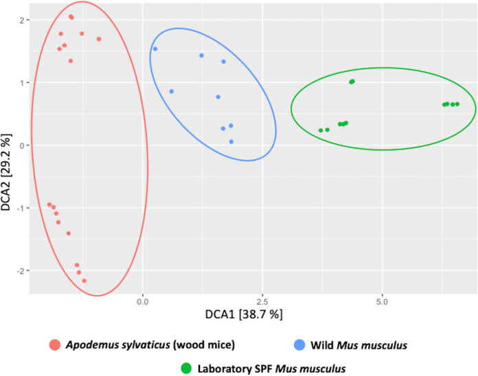
The SPF laboratory mice included in this study represented diverse background strains (C57BL/6J, C3H/Orl, CD-1, BALB/c) and were also obtained from a range of diverse sources (commercially purchased, in-house colonies, conventionalized from germfree mice, and genetic knockouts). In addition, samples were also collected from the formerly wild wood mouse Apodemus sylvaticus and the wild house mouse Mus musculus domesticus. Beta-diversity analyses of the oral microbial populations of the sampled mice (Fig. 2) showed distinct separation in the clustering of all three groups of mice. However, a number of shared species have also been observed between them, including Lactobacillus murinus (MOT93) and Streptococcus danieliae (MOT10) (Table 1).
FIG 2.
Beta-diversity analyses of the oral microbiome of various mouse species. Beta-diversity analyses of oral microbial populations of SPF laboratory mice (green), wild Mus musculus domesticus mice (blue), and wood mice Apodemus sylvaticus (red) using the 16S rRNA gene amplicon community data targeting the V1-V2 region were performed.
Culture versus 16S rRNA gene community profiling.
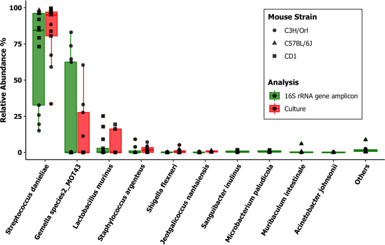
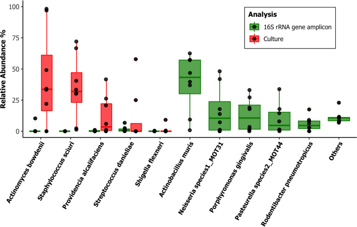
Custom reference data sets for community profiling analysis were constructed using the current version of the database and used for data analysis. Analysis of the Illumina MiSeq amplicon sequencing data (the V1-V2 region of the 16S rRNA gene) in SPF mouse samples using the custom reference data set revealed that 98% of the total number of reads could be assigned to species level. Further comparative analysis of culturing data with this amplicon sequencing data set revealed a significant consensus in the SPF mouse samples, with all of the cultured species being represented at comparable levels with the next-generation sequencing (NGS) data (Fig. 3). Among the wild Mus musculus domesticus samples, 95% of the reads were identified to the species level using the custom reference data set, even though very little consensus has been observed with the culturing data in terms of abundance of individual species (Fig. 4). This could be explained by the fact that the species that show significantly higher abundance in the NGS data set (Muribacter muris, Neisseria species 1, and Porphyromonas gingivalis) are predominantly slow growers and therefore could have been missed being detected in the 48-h culturing protocol followed in this study.
FIG 3.
Relative abundances in the SPF laboratory mice (n = 13). We compared oral microbial population analyses in three background strains of SPF mice by laboratory culture (red) and Illumina MiSeq 16S rRNA gene amplicon sequencing (green) amplifying the V1-V2 region of the 16S rRNA gene. The amplicon sequencing data were analyzed using DADA2 v1.8 against a custom-made taxonomy reference data set from this database. The top 10 most relatively abundant taxa identified after normalization of counts are listed.
FIG 4.
Relative abundances in the wild house mouse Mus musculus domesticus (n = 8). We compared oral microbial population analyses in the wild house mouse Mus musculus domesticus by laboratory culture (red) and Illumina MiSeq 16S rRNA gene amplicon sequencing (green) amplifying the V1-V2 region of the 16S rRNA gene. The amplicon sequencing data were analyzed using DADA2 v1.8 against a custom-made taxonomy reference data set from this database. The top 10 most relatively abundant taxa identified after normalization of counts are listed.
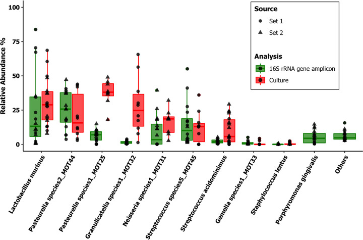
In the samples from the formerly wild wood mouse Apodemus sylvaticus, the proportion of reads positively identified using the custom data set fell to 83%, even though a higher consensus between culturing and NGS was observed compared to the wild Mus musculus domesticus, despite the increased diversity (Fig. 5).
FIG 5.
Relative abundances in the wood mouse Apodemus sylvaticus (n = 16). We compared oral microbial population analyses in the formerly wild wood mouse Apodemus sylvaticus, sampled from two sources (sets 1 and 2), by laboratory culture (red) and Illumina MiSeq 16S rRNA gene amplicon sequencing (green) amplifying the V1-V2 region of the 16S rRNA gene. The amplicon sequencing data were analyzed using DADA2 v1.8 against a custom-made taxonomy reference data set from this database. The top 10 most relatively abundant taxa identified after normalization of counts are listed.
Current versions of the reference data sets have been constructed for both the mothur and DADA2 pipelines and are available to download at https://figshare.com/s/2470f05ab77cdf40b2f8.
Mouse oral bacterial genomes.
Draft genomes of 55 murine oral bacterial isolates have been generated, sourced from all the mouse groups included in this study (Table 2). Representative genome sequences have been uploaded to the NCBI bacterial genome database and are publicly available at NCBI BioProject PRJNA671681. After assembly, the genomes had a mean contig number of 135 (±134) and GC% ratios ranging from 28 to 73%.
TABLE 2.
Details of the draft microbial genomes sequenced as part of the development of the murine oral microbiome databasea
| Sample ID | Species ID | Taxonomy ID | Total length (bp) | GC% | Source | Accession no. |
|---|---|---|---|---|---|---|
| 19428wA1_WT6 | Staphylococcus xylosus | MOT01 | 2806698 | 32.68 | SPF C3H/Orl | JADGLO000000000 |
| 19428wB1_WM06 | Staphylococcus saprophyticus | MOT02 | 2920674 | 32.48 | Apodemus sylvaticus | JADGLJ000000000 |
| 3_3BalbC2O2_S3 | Staphylococcus lentus | MOT03 | 2925394 | 32.36 | SPF BALB/C | JADGLT000000000 |
| 19428wC1_HT5 | Staphylococcus lentus | MOT03 | 2910603 | 31.73 | SPF C3H/Orl | JADGLE000000000 |
| 19428wG1_AE2 | Staphylococcus lentus | MOT03 | 2856564 | 31.81 | SPF C57BL/6J | JADGMD000000000 |
| ACT.1 | Staphylococcus sciuri | MOT05 | 2737620 | 32.5 | SPF C3H/Orl | JADGMB000000000 |
| 19428wE1_BL01 | Staphylococcus sciuri | MOT05 | 2833136 | 32.42 | SPF C57BL/6J | JADGMG000000000 |
| 19428wF1_P912 | Staphylococcus warneri | MOT07 | 2628448 | 32.57 | SPF C3H/Orl | JADGMF000000000 |
| 19428wH1_IOV5 | Staphylococcus arlettae | MOT09 | 2600982 | 33.35 | SPF C3H/Orl | JADGMC000000000 |
| 1_1BalbC1O2_S1 | Streptococcus danieliae | MOT10 | 1356823 | 44.6 | SPF BALB/c | JADGLV000000000 |
| STR.1 | Streptococcus danieliae | MOT10 | 1969143 | 44.5 | SPF C3H/Orl | JADGKG000000000 |
| CCW311.1 | Streptococcus danieliae | MOT10 | 1616067 | 45.03 | SPF C57BL/6J | JADGKO000000000 |
| 5_7CXCR22O2_S5 | Streptococcus danieliae | MOT10 | 2927540 | 43.29 | SPF BALB/c | JADGLY000000000 |
| 19428wB2_HT4 | Streptococcus acidominimus | MOT11 | 2221121 | 42.77 | SPF C3H/Orl | JADGLI000000000 |
| 6_8CXCR23O2_S6 | Streptococcus acidominimus | MOT11 | 2509022 | 43.7 | SPF BALB/c | JADGLR000000000 |
| 19428wC2_LYSM12 | Streptococcus species 1 | MOT12 | 2417981 | 40.73 | SPF C3H/Orl | JADGLD000000000 |
| 19428wD3_AN2 | Streptococcus species 2 | MOT13 | 2324918 | 42.8 | SPF C3H/Orl | JADGKY000000000 |
| CIP106318T.1 | Gemella palaticanis | MOT18 | 1720464 | 27.55 | Culture collection, type strain | JADGKN000000000 |
| 19428wH2_1568 | Shigella flexneri | MOT19 | 5100000 | 50.71 | SPF C3H/Orl | JADGKR000000000 |
| 19428wA3_CC11 | Enterococcus faecalis | MOT20 | 6434324 | 39.02 | SPF C3H/Orl | JADGLM000000000 |
| 19428wB3_C2 | Enterococcus gallinarum | MOT21 | 3661725 | 40.12 | SPF C3H/Orl | JADGLH000000000 |
| ENT.1 | Enterococcus gallinarum | MOT21 | 3793521 | 40 | SPF C3H/Orl | JADGKL000000000 |
| 19428wC3_WT12 | Muribacter muris | MOT22 | 2520282 | 44.93 | SPF C3H/Orl | JADGLC000000000 |
| 7_18CXCR221ANO_S7 | Muribacter muris | MOT22 | 2535311 | 45.96 | SPF BALB/c | JADGLQ000000000 |
| 19428wE3_DK2 | Micrococcus aloeverae | MOT24 | 2559064 | 72.93 | SPF C57BL/6J | JADGLX000000000 |
| 19428wF3_WM03 | Pasteurella species 1 | MOT25 | 2264212 | 39.91 | Apodemus sylvaticus | JADGKV000000000 |
| 19428wG3_P14 | Parabacteroides goldsteinii | MOT27 | 6779962 | 43.44 | SPF C3H/Orl | JADGKT000000000 |
| 19428wE4_P11 | Dysgonomonas mossii | MOT28 | 4110280 | 37.31 | SPF C3H/Orl | JADGKW000000000 |
| 8_33DSS_C5_S8 | Bacteroides acidifaciens | MOT29 | 5228201 | 44.3 | SPF C3H/Orl | JADGLP000000000 |
| 19428wH3_P2318 | Bacteroides acidifaciens | MOT29 | 5116387 | 43.26 | SPF C3H/Orl | JADGKQ000000000 |
| 19428wA4_P1012 | Microbacterium paludicola | MOT30 | 2809652 | 70.53 | SPF C3H/Orl | JADGLL000000000 |
| 19428wB4_WF04 | Neisseria species 1 | MOT31 | 2568531 | 53.59 | Apodemus sylvaticus | JADGLG000000000 |
| 19428wC4_WM01 | Granulicatella species 1 | MOT32 | 2003757 | 33.14 | Apodemus sylvaticus | JADGLB000000000 |
| 19428wG4_IOV6 | Micrococcus yunnanensis | MOT37 | 2475752 | 73.07 | SPF C3H/Orl | JADGKS000000000 |
| 19428wE5_W307 | Jeotgalicoccus nanhaiensis | MOT38 | 2244448 | 41.43 | SPF C57BL/6J | JADGLW000000000 |
| 19428wH4_BL23 | Citrobacter koseri | MOT39 | 4821534 | 53.81 | SPF C57BL/6J | JADGKP000000000 |
| 19428wA5_irhom_31 | Rothia nasimurium | MOT40 | 2677034 | 59.02 | SPF C57BL/6J | JADGLK000000000 |
| 19428wB5_irhom_Sw | Proteus mirabilis | MOT41 | 3939005 | 38.79 | SPF C57BL/6J | JADGLF000000000 |
| DSM100099.1 | Sanguibacter inulinus | MOT42 | 4257809 | 70.83 | Culture Collection; Type Strain | JADGKM000000000 |
| 19428wG2_WT2a | Gemella species 2 | MOT43 | 1404407 | 30.33 | SPF C3H/Orl | JADGKU000000000 |
| GH3.1 | Gemella species 2 | MOT43 | 1563132 | 28.2 | SPF C3H/Orl | JADGKK000000000 |
| GL1.1 | Gemella species 2 | MOT43 | 1299399 | 30.25 | SPF C3H/Orl | JADGKJ000000000 |
| 19428wA2_WM07 | Streptococcus species 5 | MOT45 | 1714730 | 41.26 | Apodemus sylvaticus | JADGLN000000000 |
| 19428wD2_WM131 | Streptococcus cuniculi | MOT46 | 2407100 | 42.7 | Apodemus sylvaticus | JADGKZ000000000 |
| 19428wD4_HM13 | Escherichia coli | MOT48 | 5021976 | 50.62 | SPF C3H/Orl | JADGMH000000000 |
| 19428wE2_CC3 | Lactobacillus johnsonii | MOT51 | 1903680 | 34.48 | SPF C3H/Orl | JADGKX000000000 |
| 4_5BalbC4O2_S4 | Staphylococcus aureus | MOT58 | 2825768 | 33.69 | SPF BALB/c | JADGLS000000000 |
| 19428wD1_P36 | Staphylococcus aureus | MOT58 | 2692071 | 32.75 | SPF C3H/Orl | JADGLA000000000 |
| STF.1 | Staphylococcus aureus | MOT58 | 2699934 | 32.8 | SPF C3H/Orl | JADGKH000000000 |
| GW2.1 | Staphylococcus capitis | MOT71 | 2450948 | 32.8 | SPF C3H/Orl | JADGMA000000000 |
| WMus004.1 | Actinomyces bowdenii | MOT83 | 3174558 | 71.17 | Wild Mus musculus domesticus | JADGKF000000000 |
| WMus003.1 | Providencia alcalifaciens | MOT84 | 3909646 | 41.88 | Wild Mus musculus domesticus | JADGLZ000000000 |
| 2_2BalbC1BO2_S2 | Lactobacillus murinus | MOT93 | 2341418 | 40.54 | SPF BALB/c | JADGLU000000000 |
| 19428wF2_HT12 | Lactobacillus murinus | MOT93 | 2254694 | 39.68 | SPF C3H/Orl | JADGME000000000 |
| LAC.1 | Lactobacillus murinus | MOT93 | 2089346 | 40.1 | SPF C3H/Orl | JADGKI000000000 |
The 55 draft genomes are available to download from BioProject (PRJNA671681), while the individual accession numbers have been provided in the table itself.
Predominant bacterial genera in laboratory mice.
The following three genera were found to be of particular interest due to their presence in a wide range of samples tested.
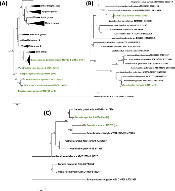
(i) Streptococcus. S. danieliae MOT10 was the most commonly detected species in all SPF laboratory mouse groups from multiple sources and mouse strains (Fig. 6A). This species was also isolated from the wild Mus musculus domesticus. Streptococcus species 5 MOT45 was isolated from the wood mouse A. sylvaticus (Fig. 5) and was found to be very closely related to, but distinct from, S. danieliae (16S rRNA gene sequence similarity, 98.4%) forming a murine S. danieliae cluster.
FIG 6.
Phylogenetic diversity of the predominant bacterial genera in laboratory mice. Maximum-likelihood phylogenetic trees represent the diversity in murine oral bacterial isolates for the three most abundant genera: Streptococcus (A), Lactobacillus (B), and Gemella (C). Each tree has been rooted with an outlier and constructed using 100 bootstrap replicates. The branches in green refer to the taxonomic IDs in each genus of the murine isolates from this study.
Some isolates of S. acidominimus MOT11 were observed in a group of SPF mice belonging to the BALB/c strain, whereas a few novel streptococcal species were also isolated in single occurrences (Streptococcus species 1, 2, and 3; MOTs 12 to 14).
(ii) Lactobacillus. With only two exceptions, all of the lactobacilli in mice were identified as L. murinus MOT93 (Fig. 6B). The two exceptions belonged to the L. taiwanensis-gasseri-johnsonii cluster at >99% identity and could not be distinguished based on the 16S rRNA gene sequence analysis. In silico DNA-DNA hybridization gene sequence analysis identified them as L. johnsonii MOT51 at 72.1% genome identity.
(iii) Gemella. All Gemella isolates from laboratory mice were found to belong to a novel, as yet unnamed, species, Gemella species 2 MOT43 (Fig. 6C). The nearest phylogenetic neighbor is the canine oral species, Gemella palaticanis at 97.4% 16S rRNA gene sequence identity. Another single, novel Gemella isolate was observed in A. sylvaticus wood mice and designated Gemella species 1 MOT33.
DISCUSSION
We report here the details of a curated murine database to represent the diversity of the oral microbiome in laboratory and wild mouse populations.
The low diversity of the mouse oral bacterial community, especially in SPF laboratory mice, has particularly stood out in this characterization. The presence of such a small and specifically targeted population makes accurate species level identification especially significant for improving the outcomes of studies. In addition, the collection of draft genomes of the representative MOTs from the database is also of great benefit for microbial population studies in mice. This repository of genomes should enable the generation of quality reference data sets for metagenomic and metatranscriptomic analyses of mouse-based oral studies.
A comparative analysis of the culture and 16S rRNA gene community profiling data has further confirmed the low diversity of the lab mouse oral microbiome (see Fig. S1 in the supplemental material), which is dominated by four species-level taxa. We have reported here only a representative subset of the samples we have sequenced in order to demonstrate the development of the database. However, over the course of multiple experimental studies, we have shown the consistency in the results of these population analyses (2, 7). The diversity of oral species-level taxa, in comparison, was found to be higher in the wild mouse species. Such variations in the microbial diversity and load within host species has been reported in the gut microbiome of other organisms and has largely been attributed to the influence of diet and environment (22, 23).
Alpha-diversity analyses of the oral microbiome of various mouse species. Alpha-diversity analyses were carried out using Shannon and Simpson diversity indices comparing the oral microbial populations of SPF laboratory mice (green), wild Mus musculus domesticus (blue) mice, and formerly wild wood mice, Apodemus sylvaticus (red). Download FIG S1, TIF file, 2.5 MB (2.5MB, tif) .
Copyright © 2021 Joseph et al.
This content is distributed under the terms of the Creative Commons Attribution 4.0 International license.
Another observation of this study has been the variations observed between different strains of the laboratory mice; we have previously also observed this between batches of the same strain of mice obtained from commercial sources (unpublished data), which has also been reported in some older studies (24). Recently, Abusleme et al. also reported the variations observed in the oral microbial communities in C57BL/6 mice purchased from two major commercial animal suppliers and the increased stability of a particular type of population when the mice were cohoused (8). This raises a very important issue of having a well-curated reference database, as well as internal controls, to ensure accuracy in results in microbiome studies.
Among the three predominant host-specific MOTs identified, S. danieliae MOT10 is of particular interest due to its dominance of the oral bacterial community in the SPF laboratory mice. A recently proposed species of the Streptococcus genus, S. danieliae MOT 10 was originally described based on a murine cecal isolate for which the authors suggested an oral/upper respiratory tract origin (25). There have been few other reports of the organism so far (26–28), all of them of murine origin. The organism has also been reported to be one of the key drivers in the establishment of the oral microbial community in laboratory SPF mice after the eruption of teeth (8). Gemella species 2 has been isolated from oral samples of multiple strains of laboratory mice and needs further characterization to be taxonomically identified as an official bacterial species. Of these three species, L. murinus is the only species that was part of the Altered Schaedler Formula, the community of microbes that were used to originally colonize gnotobiotic mice to develop SPF laboratory mice as we know them today (29, 30). Similar examples of host specificity have also been reported in other mouse microbiomes, including the colonization of segmented filamentous bacteria (31) and Muribaculaceae (19) in the mouse gut microbiome. This specificity also strongly indicates to the phenomenon of host-microbe coevolution and fitness characteristics (32).
In addition, there has been an increasing interest in recent times in the relevance of wild rodent models in research (33, 34), particularly for understanding natural progressions of certain diseases in relation to the microbiome. A study involving microbial transfer of wild mice into laboratory mice has demonstrated the role of the natural or wild microbiota in disease resistance and protective immune mechanisms (35). Further, a more recent study by the same group also showed that laboratory mice bred from such wild mice (referred to as wildlings) exhibited the elevated microbial diversity of the parent wild mice accompanied by a stability to perturbations such as antibiotics and diet, implying that a natural or wild immunity could be more comparable to that seen in the diversity of human illnesses (36). Hence, we considered it pertinent to include the bacterial taxa from some of these wild mouse samples (formerly wild wood mice [Apodemus sylvaticus] and wild Mus musculus domesticus mice) in the database, in addition to cultured oral isolates which could be used for in vivo and in vitro experimentations in the future. We fully appreciate that at present we report this from a limited number of sources and therefore representing only a fraction of the actual diversity observed in the wild, but we have plans to further expand this resource with the inclusion of more diverse sources based on availability.
It is also pertinent here to point out the presence of certain taxa in this murine oral microbial population potentially of intestinal origin, which could be attributed to the coprophagic behavior in murine populations. Compared to the mouse intestinal bacterial collection database, we notice the presence of some shared taxa with our database, particularly species belonging to the order Lactobacillales (19). Equally, oral bacteria might also be found in the gut, particularly lactobacilli because of their aciduric nature. However, despite this overlap, the oral microbiome in mice remains distinct, far simpler, and less diverse than in the murine gut microbiome (see Fig. S2 and S3), as we have demonstrated by amplicon sequencing and characterization of a limited subset of murine fecal samples.
Beta-diversity analyses of the oral and gut microbiome of various mouse species. Beta diversity analyses of the oral and fecal microbial populations of SPF laboratory mice (n = 8) and the wood mouse Apodemus sylvaticus (n = 12) were carried out using the 16S rRNA gene amplicon community data targeting the V1-V2 region of the gene. The sequences were analyzed using the DADA2 v1.8 pipeline, and the assembled ASVs were assigned taxonomy using the SILVA rRNA reference database v138.1. Download FIG S2, TIF file, 1.5 MB (1.5MB, tif) .
Copyright © 2021 Joseph et al.
This content is distributed under the terms of the Creative Commons Attribution 4.0 International license.
It is necessary to stress that this study primarily describes the development of a framework for this murine oral microbial database, which by its nature remains a work in progress. Work is under way with collaborators at the Forsyth Institute, Cambridge, MA, to make the final version of this database publicly available as the Mouse Oral Microbiome Database (MOMD), on the lines of the Human Oral Microbiome Database (HOMD) (16). This will enable the addition of sequences from other researchers in the field; these will be curated and made publicly available. Meanwhile, the cultured isolates analyzed for this database can be made available to interested researchers on request by contacting the authors. Further work is also being undertaken using sequence analysis and cloning to characterize the uncultured as well as the identified but unnamed novel species, including their taxonomic nomenclature, for the further expansion of the database.
We hope that this should enable researchers across the world to access and develop suitable reference data sets for both culture and culture independent studies of the murine oral microbiome.
MATERIALS AND METHODS
Mouse sampling and ethics.
All animal experiments were conducted in accredited facilities in accordance with the UK Animals (Scientific Procedures) Act 1986 (Home Office license number 7006844). Fieldwork for the wild Mus musculus domesticus work was approved by the Animal Welfare Ethical Review Body (AWERB) at the Department of Zoology, University of Oxford.
Conventional SPF C3H/Orl and BALB/c mice were maintained in individually ventilated cages (IVCs) at the animal care facilities of Queen Mary University of London. Conventional SPF C57BL/6J mice were maintained in IVCs at the animal care facilities of King’s College London. SPF C57BL/6J and CD-1 mice were also purchased commercially from Charles River Laboratories UK. In all, 191 SPF mice have been sampled over the course of 6 years for various experimental studies, and isolates obtained from the swabbing of these mice have been used for the analysis and development of this database.
One set (set 1) of formerly wild, now laboratory-bred, A. sylvaticus wood mice sampled in 2014 (n = 20) was originally established from wild wood mice that were live trapped from the UK woodlands and are now housed at the University of Edinburgh. Wild wood mice sampled in January 2017 (n = 39) were housed at the facilities at Fera Science (Sand Hutton, York), and the colony began with wild-caught wood mice from the grounds of the Institute; these animals had been laboratory-bred for 20 years and are referred to as set 2.
Wild house mice (Mus musculus domesticus [n = 21]) were sampled on the island of Skokholm in Wales, UK. These mice were captured temporarily in traps, sampled, and then released back into the wild.
Bacterial culturing.
The murine oral cavity was swabbed for 30 s, using sterile fine-tip rayon swabs (VWR International), while the animal was held in a scruff. The swab was then placed in a tube containing 100 μl of reduced John’s transport medium (see Text S1 in the supplemental material). Swabs from wild mice were stored at −20°C before being transported on ice to the laboratory. Serial dilutions of the suspension were spread onto blood agar plates containing 5% defibrinated horse blood (TCS Biosciences, UK) and incubated for aerobic and anaerobic (80% N2, 10% H2, and 10% CO2) growth at 37°C for 48 h in a Don Whitley anaerobic chamber. The CFUs of predominant cultivable bacteria on each plate were counted. On an average, four to six different colony types could be identified on each blood agar plate. Every different colony morphology type observed was isolated and purified by subculture by restreaking the samples twice on fresh blood agar plates. Once the purity was established, the cultured isolate was cryopreserved for storage in Microbank bead tubes (Prolab Diagnostics) in duplicate. Briefly, a loopful of pure culture was aseptically transferred into the manufacturer’s cryopreservative liquid containing beads, the tube was inverted four to five times for emulsification and allowed to stand for 2 min. Any excess liquid was aseptically removed, and the bead tube was then frozen at −70°C.
Composition of John’s Transport medium. Download Text S1, DOCX file, 0.01 MB (13.5KB, docx) .
Copyright © 2021 Joseph et al.
This content is distributed under the terms of the Creative Commons Attribution 4.0 International license.
16S rRNA gene amplification and sequencing.
Genomic DNA for each isolated bacterial strain was extracted by using a GenElute bacterial DNA kit (Sigma-Aldrich), following the Gram-positive protocol according to manufacturer’s instructions, and used as a template for PCR. The 16S rRNA gene in the bacterial strains was amplified using modified versions of the universal 27FYM and 1492R 16S rRNA gene primers with built-in redundancies (see Text S2), using Phusion Green Hot Start II High Fidelity PCR Master Mix (Thermo Fisher Scientific). PCR conditions were as follows: initial denaturation at 98°C for 30 s; followed by 25 cycles of 98°C for 10 s, 47°C for 45 s, and 72°C for 30 s; followed in turn by a final extension at 72°C for 10 min. The amplified products were purified using Macherey-Nagel NucleoSpin gel and PCR Clean-Up (Fisher Scientific), followed by Sanger sequencing using the universal M13 primers M13 uni(-21) and M13 rev(-29) (Eurofins Genomics). For certain samples, internal primers for the 16S rRNA gene (342R, 357F, 519R, 907R, 926F, 1100R, 1114F, and 1392R) were also used for sequencing to improve the sequence coverage accuracy. All primer sequences have been provided in Text S2.
Modified version of the universal primers of the 16S rRNA gene. Download Text S2, DOCX file, 0.01 MB (13.1KB, docx) .
Copyright © 2021 Joseph et al.
This content is distributed under the terms of the Creative Commons Attribution 4.0 International license.
Allocation of mouse oral taxa.
The forward and reverse sequences for each sample were assembled using the CAP3 assembly tool (37). Sequences were identified by BLAST interrogation of the NCBI nucleotide database. A sequence identity threshold of 98.5% was used for assignation to species, which is consistent with current recommendations (38) and the value used previously for the related human and canine oral databases (17, 18). Sequences were aligned by means of the CLUSTALW algorithm in Bioedit (39), and maximum-likelihood phylogenetic trees for each genus were constructed using MEGA7 (40) with 100 bootstrap replicates. Each species-level taxon was assigned an MOT number. For taxa that were not identified as validly proposed species at 98.5% identity, a novel species-level designation was assigned as “Genusname_species1,” and a new MOT number was allocated. For isolates that could not be definitively distinguished by 16S rRNA gene sequence analysis, each of the possible matching species was assigned a unique MOT ID in order to ensure maximum diversity capture. Assignment of taxa was also carried out using 16S rRNA gene amplicons selected from the MiSeq sequencing data that were not assigned an ID based on the existing reference database and then following the same BLAST protocol as described above. Visualization and annotation of the final phylogenetic tree of the identified 103 MOTs was performed using the web interface of iTOL v4 (41).
MiSeq 16S rRNA gene sequencing library preparation and DNA sequence analysis.
Whole genomic DNA was extracted from the above swabs using the DNeasy PowerSoil kit (Qiagen) according to the manufacturer’s instructions. Cell lysis was performed in the PowerBead tubes provided with the kit by bead beating on a vortex at maximum speed for 20 min. PCRs were performed with Phusion Green Hot Start II High Fidelity PCR Master Mix (Thermo Scientific) targeting the V1-V2 variable regions of the 16S rRNA gene using fusion primers 27F-YM (AGAGTTTGATYMTGGCTCA) and 338R-R (TGCTGCCTCCCGTAGRAG) combined with MiSeq adaptors and barcodes to achieve a double indexing system. The PCR conditions were as follows: initial denaturation for 5 min at 95°C; followed by 25 cycles of 95°C for 45 s, 53°C for 45 s, and 72°C for 45 s; followed in turn by a final extension of 72°C for 5 min. The amplified PCR products were cleaned and normalized in equimolar amounts by using a Sequal Prep normalization plate kit (Thermo Fisher Scientific). Extraction kit controls and PCR negative controls were included in the amplification plates, as well as sequencing pools. Pooled amplicons were sequenced at the Barts and the London Genome Centre using an Illumina MiSeq 2 × 250 flow cell for paired-end sequencing. The generated reads were quality checked, filtered, trimmed, denoised, dereplicated, and assembled into amplicon sequence variants (ASVs) using the DADA2 v1.8 pipeline (42). The assembled ASVs were then assigned taxonomy at the genus and species level using a custom-formatted reference database constructed using the taxa included in this database. Compared to the mean of total reads in the murine swab samples, the negative controls generated a very low percentage of reads (<0.1% in the PCR control and <1% in the kit control). Once this was confirmed, the negative samples were eliminated from further analyses. The generated ASV counts were normalized for sequencing depth by using the median of ratios method in the DeSeq2 (43) package in R, followed by beta-diversity and relative abundance analyses of the microbial population. Graphical analysis and plots were created using the R packages phyloseq (44) and ggplot2 (45). The raw sequencing reads have been uploaded to the NCBI SRA database (accession no. PRJNA642845).
Bacterial genome sequencing.
A selection of bacterial isolates from the database were cultured, purity checked, and genomic DNA extracted using the GenElute Bacterial Genomic DNA kit (Sigma-Aldrich). These isolates are representatives of the cultured microbial population observed in various murine studies and include multiple candidates of the more commonly observed Streptococcus, Lactobacillus, and Staphylococcus species-level taxa. Genomic DNA libraries were prepared by MicrobesNG UK using Nextera XT Library Prep kit (Illumina, San Diego, CA) and sequenced on the Illumina HiSeq using a 250-bp paired-end protocol. Reads were adapter trimmed using Trimmomatic 0.30 with a sliding window quality cutoff of Q15 (46). De novo assembly was performed on samples using SPAdes version 3.7 (47), and contigs were annotated using Prokka 1.11 (48). Further genome analysis and annotation was also performed using the RAST server (https://rast.nmpdr.org/) (49). Phylogenetic distance and relatedness of certain isolates were determined using the genome-to-genome distance calculator, in the form of an in silico DNA-DNA hybridization (50).
Data availability.
The 16S rRNA gene V1-V2 region amplicon sequencing data from this study are available from the NCBI SRA database under accession no. PRJNA642845. Draft genome sequences of representative murine oral bacterial isolates are available to download from NCBI BioProject no. PRJNA671681. 16S rRNA gene sequences of the novel, unnamed bacterial isolates from this study are available under NCBI accession numbers MN095260 to MN095271. The 16S rRNA gene sequences for all the isolates analyzed in this study are available under NCBI accession numbers MW175535 to MW175859. Custom taxonomy reference data sets of the database for NGS analysis using mothur and DADA2 pipelines are available to download from https://figshare.com/s/2470f05ab77cdf40b2f8.
Internal primers of the 16S rRNA gene used for primer walking. Download Text S3, DOCX file, 0.01 MB (13.4KB, docx) .
Copyright © 2021 Joseph et al.
This content is distributed under the terms of the Creative Commons Attribution 4.0 International license.
MiSeq 16S rRNA gene sequencing library preparation and DNA sequence analysis of murine fecal samples. Download Text S4, DOCX file, 0.01 MB (13.9KB, docx) .
Copyright © 2021 Joseph et al.
This content is distributed under the terms of the Creative Commons Attribution 4.0 International license.
Relative abundances in the gut microbiome of various mouse species. The relative abundances of the fecal microbial population in SPF laboratory mice (C3H/Orl and C57BL/6J) (A) and the formerly wild wood mouse, Apodemus sylvaticus (B), sampled from two sources (set 1 and set 2), were analyzed using Illumina MiSeq sequencing technology by amplifying the V1-V2 region of the 16S rRNA gene sequences. The amplicon sequencing data were analyzed using the DADA2 v1.8 pipeline, and the assembled ASVs were assigned taxonomy using the SILVA rRNA reference database v138.1. The top 10 most relatively abundant taxa are listed. Download FIG S3, TIF file, 2.7 MB (2.7MB, tif) .
Copyright © 2021 Joseph et al.
This content is distributed under the terms of the Creative Commons Attribution 4.0 International license.
ACKNOWLEDGMENTS
This study was funded by the UKRI Medical Research Council (award MR/P012175/2). W.G.W. is supported by NIH-NIDCR (grant R37 DE016937). The wild Mus musculus work was funded by a NERC fellowship NEL011867/1 to S.C.L.K. The funders had no role in study design, data collection and interpretation, or the decision to submit the work for publication.
We thank D. Randall and F. Filomena for providing access to their murine bacterial isolates for inclusion of data in this study. We thank Melanie Clerc at the University of Edinburgh and the staff at Fera for assistance in collecting the wood mouse samples.
S.J., W.G.W., and M.A.C. contributed to conception, design, data acquisition, and analysis and interpretation and drafted and critically revised the manuscript. J.A.-O. contributed to data acquisition, analysis, and interpretation and drafted and critically revised the manuscript. A.H., E.H., R.S., S.C.L.K., and A.B.P. contributed to data acquisition and critically reviewed the manuscript. All authors gave final approval and agree to be accountable for all aspects of the work.
We declare there are no competing interests.
REFERENCES
- 1.Greer A, Irie K, Hashim A, Leroux BG, Chang AM, Curtis MA, Darveau RP. 2016. Site-specific neutrophil migration and CXCL2 expression in periodontal tissue. J Dent Res 95:946–952. doi: 10.1177/0022034516641036. [DOI] [PMC free article] [PubMed] [Google Scholar]
- 2.Payne MA, Hashim A, Alsam A, Joseph S, Aduse-Opoku J, Wade WG, Curtis MA. 2019. Horizontal and vertical transfer of oral microbial dysbiosis and periodontal disease. J Dent Res 98:1503–1510. doi: 10.1177/0022034519877150. [DOI] [PubMed] [Google Scholar]
- 3.Marcotte H, Lavoie MC. 1997. Comparison of the indigenous oral microbiota and immunoglobulin responses of athymic (nu/nu) and euthymic (nu/+) mice. Oral Microbiol Immunol 12:141–147. doi: 10.1111/j.1399-302x.1997.tb00370.x. [DOI] [PubMed] [Google Scholar]
- 4.Gadbois T, Marcotte H, Rodrigue L, Coulombe C, Goyette N, Lavoie MC. 1993. Distribution of the resident oral bacterial populations in different strains of mice. Microb Ecol Health Dis 6:245–251. doi: 10.3109/08910609309141333. [DOI] [Google Scholar]
- 5.Wolff LF, Krupp MJ, Liljemark WF. 1985. Microbial changes associated with advancing periodontitis in STR/N mice. J Periodontal Res 20:378–385. doi: 10.1111/j.1600-0765.1985.tb00449.x. [DOI] [PubMed] [Google Scholar]
- 6.Trudel L, St-Amand L, Bareil M, Cardinal P, Lavoie MC. 1986. Bacteriology of the oral cavity of BALB/c mice. Can J Microbiol 32:673–678. doi: 10.1139/m86-124. [DOI] [PubMed] [Google Scholar]
- 7.Hajishengallis G, Liang S, Payne MA, Hashim A, Jotwani R, Eskan MA, McIntosh ML, Alsam A, Kirkwood KL, Lambris JD, Darveau RP, Curtis MA. 2011. Low-abundance biofilm species orchestrates inflammatory periodontal disease through the commensal microbiota and complement. Cell Host Microbe 10:497–506. doi: 10.1016/j.chom.2011.10.006. [DOI] [PMC free article] [PubMed] [Google Scholar]
- 8.Abusleme L, O’Gorman H, Dutzan N, Greenwell-Wild T, Moutsopoulos NM. 2020. Establishment and stability of the murine oral microbiome. J Dent Res 99:721–729. doi: 10.1177/0022034520915485. [DOI] [PMC free article] [PubMed] [Google Scholar]
- 9.Curtis MA, Zenobia C, Darveau RP. 2011. The relationship of the oral microbiotia to periodontal health and disease. Cell Host Microbe 10:302–306. doi: 10.1016/j.chom.2011.09.008. [DOI] [PMC free article] [PubMed] [Google Scholar]
- 10.Dutzan N, Kajikawa T, Abusleme L, Greenwell-Wild T, Zuazo CE, Ikeuchi T, Brenchley L, Abe T, Hurabielle C, Martin D, Morell RJ, Freeman AF, Lazarevic V, Trinchieri G, Diaz PI, Holland SM, Belkaid Y, Hajishengallis G, Moutsopoulos NM. 2018. A dysbiotic microbiome triggers TH17 cells to mediate oral mucosal immunopathology in mice and humans. Sci Transl Med 10:eaat0797. doi: 10.1126/scitranslmed.aat0797. [DOI] [PMC free article] [PubMed] [Google Scholar]
- 11.Polak D, Wilensky A, Shapira L, Halabi A, Goldstein D, Weiss EI, Houri-Haddad Y. 2009. Mouse model of experimental periodontitis induced by Porphyromonas gingivalis/Fusobacterium nucleatum infection: bone loss and host response. J Clin Periodontol 36:406–410. doi: 10.1111/j.1600-051X.2009.01393.x. [DOI] [PubMed] [Google Scholar]
- 12.Graves DT, Fine D, Teng Y-TA, Van Dyke TE, Hajishengallis G. 2008. The use of rodent models to investigate host-bacterium interactions related to periodontal diseases: rodent models for periodontal disease. J Clin Periodontol 35:89–105. doi: 10.1111/j.1600-051X.2007.01172.x. [DOI] [PMC free article] [PubMed] [Google Scholar]
- 13.Kirst ME, Li EC, Alfant B, Chi Y-Y, Walker C, Magnusson I, Wang GP. 2015. Dysbiosis and alterations in predicted functions of the subgingival microbiome in chronic periodontitis. Appl Environ Microbiol 81:783–793. doi: 10.1128/AEM.02712-14. [DOI] [PMC free article] [PubMed] [Google Scholar]
- 14.Van Dyke TE, Bartold PM, Reynolds EC. 2020. The nexus between periodontal inflammation and dysbiosis. Front Immunol 11:511. doi: 10.3389/fimmu.2020.00511. [DOI] [PMC free article] [PubMed] [Google Scholar]
- 15.Dewhirst FE, Chen T, Izard J, Paster BJ, Tanner ACR, Yu W-H, Lakshmanan A, Wade WG. 2010. The human oral microbiome. J Bacteriol 192:5002–5017. doi: 10.1128/JB.00542-10. [DOI] [PMC free article] [PubMed] [Google Scholar]
- 16.Chen T, Yu W-H, Izard J, Baranova OV, Lakshmanan A, Dewhirst FE. 2010. The Human Oral Microbiome Database: a web accessible resource for investigating oral microbe taxonomic and genomic information. Database (Oxford) 2010:baq013. doi: 10.1093/database/baq013. [DOI] [PMC free article] [PubMed] [Google Scholar]
- 17.Dewhirst FE, Klein EA, Bennett M-L, Croft JM, Harris SJ, Marshall-Jones ZV. 2015. The feline oral microbiome: a provisional 16S rRNA gene based taxonomy with full-length reference sequences. Vet Microbiol 175:294–303. doi: 10.1016/j.vetmic.2014.11.019. [DOI] [PMC free article] [PubMed] [Google Scholar]
- 18.Dewhirst FE, Klein EA, Thompson EC, Blanton JM, Chen T, Milella L, Buckley CMF, Davis IJ, Bennett M-L, Marshall-Jones ZV. 2012. The canine oral microbiome. PLoS One 7:e36067. doi: 10.1371/journal.pone.0036067. [DOI] [PMC free article] [PubMed] [Google Scholar]
- 19.Lagkouvardos I, Pukall R, Abt B, Foesel BU, Meier-Kolthoff JP, Kumar N, Bresciani A, Martínez I, Just S, Ziegler C, Brugiroux S, Garzetti D, Wenning M, Bui TPN, Wang J, Hugenholtz F, Plugge CM, Peterson DA, Hornef MW, Baines JF, Smidt H, Walter J, Kristiansen K, Nielsen HB, Haller D, Overmann J, Stecher B, Clavel T. 2016. The Mouse Intestinal Bacterial Collection (miBC) provides host-specific insight into cultured diversity and functional potential of the gut microbiota. Nat Microbiol 1:16131. doi: 10.1038/nmicrobiol.2016.219. [DOI] [PubMed] [Google Scholar]
- 20.Liu C, Zhou N, Du M-X, Sun Y-T, Wang K, Wang Y-J, Li D-H, Yu H-Y, Song Y, Bai B-B, Xin Y, Wu L, Jiang C-Y, Feng J, Xiang H, Zhou Y, Ma J, Wang J, Liu H-W, Liu S-J. 2020. The Mouse Gut Microbial Biobank expands the coverage of cultured bacteria. Nat Commun 11:79. doi: 10.1038/s41467-019-13836-5. [DOI] [PMC free article] [PubMed] [Google Scholar]
- 21.Xiao L, Feng Q, Liang S, Sonne SB, Xia Z, Qiu X, Li X, Long H, Zhang J, Zhang D, Liu C, Fang Z, Chou J, Glanville J, Hao Q, Kotowska D, Colding C, Licht TR, Wu D, Yu J, Sung JJY, Liang Q, Li J, Jia H, Lan Z, Tremaroli V, Dworzynski P, Nielsen HB, Bäckhed F, Doré J, Le Chatelier E, Ehrlich SD, Lin JC, Arumugam M, Wang J, Madsen L, Kristiansen K. 2015. A catalog of the mouse gut metagenome. Nat Biotechnol 33:1103–1108. doi: 10.1038/nbt.3353. [DOI] [PubMed] [Google Scholar]
- 22.Reese AT, Dunn RR. 2018. Drivers of microbiome biodiversity: a review of general rules, feces, and ignorance. mBio 9:e01294-18. doi: 10.1128/mBio.01294-18. [DOI] [PMC free article] [PubMed] [Google Scholar]
- 23.Sanders JG, Lukasik P, Frederickson ME, Russell JA, Koga R, Knight R, Pierce NE. 2017. Dramatic differences in gut bacterial densities correlate with diet and habitat in rainforest ants. Integr Comp Biol 57:705–722. doi: 10.1093/icb/icx088. [DOI] [PubMed] [Google Scholar]
- 24.Rodrigue L, Lavoie MC. 1996. Comparison of the proportions of oral bacterial species in BALB/c mice from different suppliers. Lab Anim 30:108–113. doi: 10.1258/002367796780865853. [DOI] [PubMed] [Google Scholar]
- 25.Clavel T, Charrier C, Haller D. 2013. Streptococcus danieliae sp. nov., a novel bacterium isolated from the caecum of a mouse. Arch Microbiol 195:43–49. doi: 10.1007/s00203-012-0846-6. [DOI] [PubMed] [Google Scholar]
- 26.Benga L, Benten WPM, Engelhardt E, Köhrer K, Gougoula C, Sager M. 2014. 16S ribosomal DNA sequence-based identification of bacteria in laboratory rodents: a practical approach in laboratory animal bacteriology diagnostics. Lab Anim 48. doi: 10.1177/0023677214538240. [DOI] [PubMed] [Google Scholar]
- 27.Hernández-Arriaga A, Baumann A, Witte OW, Frahm C, Bergheim I, Camarinha-Silva A. 2019. Changes in oral microbial ecology of C57BL/6 mice at different ages associated with sampling methodology. Microorganisms 7:283. doi: 10.3390/microorganisms7090283. [DOI] [PMC free article] [PubMed] [Google Scholar]
- 28.Chun J, Kim KY, Lee J-H, Choi Y. 2010. The analysis of oral microbial communities of wild-type and Toll-like receptor 2-deficient mice using a 454 GS FLX Titanium pyrosequencer. BMC Microbiol 10:101. doi: 10.1186/1471-2180-10-101. [DOI] [PMC free article] [PubMed] [Google Scholar]
- 29.Wymore Brand M, Wannemuehler MJ, Phillips GJ, Proctor A, Overstreet A-M, Jergens AE, Orcutt RP, Fox JG. 2015. The altered schaedler flora: continued applications of a defined murine microbial community. Ilar J 56:169–178. doi: 10.1093/ilar/ilv012. [DOI] [PMC free article] [PubMed] [Google Scholar]
- 30.Schaedler RW, Dubs R, Costello R. 1965. Association of germfree mice with bacteria isolated from normal mice. J Exp Med 122:77–82. doi: 10.1084/jem.122.1.77. [DOI] [PMC free article] [PubMed] [Google Scholar]
- 31.Tannock GW, Miller JR, Savage DC. 1984. Host specificity of filamentous, segmented microorganisms adherent to the small bowel epithelium in mice and rats. Appl Environ Microbiol 47:441–442. doi: 10.1128/AEM.47.2.441-442.1984. [DOI] [PMC free article] [PubMed] [Google Scholar]
- 32.Dethlefsen L, McFall-Ngai M, Relman DA. 2007. An ecological and evolutionary perspective on human-microbe mutualism and disease. Nature 449:811–818. doi: 10.1038/nature06245. [DOI] [PMC free article] [PubMed] [Google Scholar]
- 33.Maurice CF, Knowles SCL, Ladau J, Pollard KS, Fenton A, Pedersen AB, Turnbaugh PJ. 2015. Marked seasonal variation in the wild mouse gut microbiota. ISME J 9:2423–2434. doi: 10.1038/ismej.2015.53. [DOI] [PMC free article] [PubMed] [Google Scholar]
- 34.Weldon L, Abolins S, Lenzi L, Bourne C, Riley EM, Viney M. 2015. The gut microbiota of wild mice. PLoS One 10:e0134643. doi: 10.1371/journal.pone.0134643. [DOI] [PMC free article] [PubMed] [Google Scholar]
- 35.Rosshart SP, Vassallo BG, Angeletti D, Hutchinson DS, Morgan AP, Takeda K, Hickman HD, McCulloch JA, Badger JH, Ajami NJ, Trinchieri G, Pardo-Manuel de Villena F, Yewdell JW, Rehermann B. 2017. Wild mouse gut microbiota promotes host fitness and improves disease resistance. Cell 171:1015–1028.e13. doi: 10.1016/j.cell.2017.09.016. [DOI] [PMC free article] [PubMed] [Google Scholar]
- 36.Rosshart SP, Herz J, Vassallo BG, Hunter A, Wall MK, Badger JH, McCulloch JA, Anastasakis DG, Sarshad AA, Leonardi I, Collins N, Blatter JA, Han S-J, Tamoutounour S, Potapova S, Claire MBFS, Yuan W, Sen SK, Dreier MS, Hild B, Hafner M, Wang D, Iliev ID, Belkaid Y, Trinchieri G, Rehermann B. 2019. Laboratory mice born to wild mice have natural microbiota and model human immune responses. Science 365:eaaw4361. doi: 10.1126/science.aaw4361. [DOI] [PMC free article] [PubMed] [Google Scholar]
- 37.Huang X, Madan A. 1999. CAP3: a DNA sequence assembly program. Genome Res 9:868–877. doi: 10.1101/gr.9.9.868. [DOI] [PMC free article] [PubMed] [Google Scholar]
- 38.Kim M, Oh H-S, Park S-C, Chun J. 2014. Towards a taxonomic coherence between average nucleotide identity and 16S rRNA gene sequence similarity for species demarcation of prokaryotes. Int J Syst Evol Microbiol doi: 10.1099/ijs.0.059774-0. [DOI] [PubMed] [Google Scholar]
- 39.Hall TA. 1999. BioEdit: a user-friendly biological sequence alignment editor and analysis program for Windows 95/98/NT. Nucleic Acid Symp Ser 41:95–98. Information Retrieval, Ltd, London, United Kingdom. [Google Scholar]
- 40.Kumar S, Stecher G, Tamura K. 2016. MEGA7: molecular evolutionary genetics analysis version 7.0 for bigger datasets. Mol Biol Evol 33:1870–1874. doi: 10.1093/molbev/msw054. [DOI] [PMC free article] [PubMed] [Google Scholar]
- 41.Letunic I, Bork P. 2019. Interactive Tree Of Life (iTOL) v4: recent updates and new developments. Nucleic Acids Res 47:W256–W259. doi: 10.1093/nar/gkz239. [DOI] [PMC free article] [PubMed] [Google Scholar]
- 42.Callahan BJ, McMurdie PJ, Rosen MJ, Han AW, Johnson AJA, Holmes SP. 2016. DADA2: high-resolution sample inference from Illumina amplicon data. Nat Methods 13:581–583. doi: 10.1038/nmeth.3869. [DOI] [PMC free article] [PubMed] [Google Scholar]
- 43.Love MI, Huber W, Anders S. 2014. Moderated estimation of fold change and dispersion for RNA-seq data with DESeq2. Genome Biol 15:550. doi: 10.1186/s13059-014-0550-8. [DOI] [PMC free article] [PubMed] [Google Scholar]
- 44.McMurdie PJ, Holmes S. 2013. phyloseq: an R package for reproducible interactive analysis and graphics of microbiome census data. PLoS One 8:e61217. doi: 10.1371/journal.pone.0061217. [DOI] [PMC free article] [PubMed] [Google Scholar]
- 45.Wickham H. 2016. ggplot2: elegant graphics for data analysis, 2nd ed. Springer, Cham, Switzerland. [Google Scholar]
- 46.Bolger AM, Lohse M, Usadel B. 2014. Trimmomatic: a flexible trimmer for Illumina sequence data. Bioinformatics 30:2114–2120. doi: 10.1093/bioinformatics/btu170. [DOI] [PMC free article] [PubMed] [Google Scholar]
- 47.Bankevich A, Nurk S, Antipov D, Gurevich AA, Dvorkin M, Kulikov AS, Lesin VM, Nikolenko SI, Pham S, Prjibelski AD, Pyshkin AV, Sirotkin AV, Vyahhi N, Tesler G, Alekseyev MA, Pevzner PA. 2012. SPAdes: a new genome assembly algorithm and its applications to single-cell sequencing. J Comput Biol 19:455–477. doi: 10.1089/cmb.2012.0021. [DOI] [PMC free article] [PubMed] [Google Scholar]
- 48.Seemann T. 2014. Prokka: rapid prokaryotic genome annotation. Bioinformatics 30:2068–2069. doi: 10.1093/bioinformatics/btu153. [DOI] [PubMed] [Google Scholar]
- 49.Overbeek R, Olson R, Pusch GD, Olsen GJ, Davis JJ, Disz T, Edwards RA, Gerdes S, Parrello B, Shukla M, Vonstein V, Wattam AR, Xia F, Stevens R. 2014. The SEED and the rapid annotation of microbial genomes using subsystems technology (RAST). Nucleic Acids Res 42:D206–D214. doi: 10.1093/nar/gkt1226. [DOI] [PMC free article] [PubMed] [Google Scholar]
- 50.Meier-Kolthoff JP, Auch AF, Klenk H-P, Göker M. 2013. Genome sequence-based species delimitation with confidence intervals and improved distance functions. BMC Bioinformatics 14:60. doi: 10.1186/1471-2105-14-60. [DOI] [PMC free article] [PubMed] [Google Scholar]
Associated Data
This section collects any data citations, data availability statements, or supplementary materials included in this article.
Supplementary Materials
Alpha-diversity analyses of the oral microbiome of various mouse species. Alpha-diversity analyses were carried out using Shannon and Simpson diversity indices comparing the oral microbial populations of SPF laboratory mice (green), wild Mus musculus domesticus (blue) mice, and formerly wild wood mice, Apodemus sylvaticus (red). Download FIG S1, TIF file, 2.5 MB (2.5MB, tif) .
Copyright © 2021 Joseph et al.
This content is distributed under the terms of the Creative Commons Attribution 4.0 International license.
Beta-diversity analyses of the oral and gut microbiome of various mouse species. Beta diversity analyses of the oral and fecal microbial populations of SPF laboratory mice (n = 8) and the wood mouse Apodemus sylvaticus (n = 12) were carried out using the 16S rRNA gene amplicon community data targeting the V1-V2 region of the gene. The sequences were analyzed using the DADA2 v1.8 pipeline, and the assembled ASVs were assigned taxonomy using the SILVA rRNA reference database v138.1. Download FIG S2, TIF file, 1.5 MB (1.5MB, tif) .
Copyright © 2021 Joseph et al.
This content is distributed under the terms of the Creative Commons Attribution 4.0 International license.
Composition of John’s Transport medium. Download Text S1, DOCX file, 0.01 MB (13.5KB, docx) .
Copyright © 2021 Joseph et al.
This content is distributed under the terms of the Creative Commons Attribution 4.0 International license.
Modified version of the universal primers of the 16S rRNA gene. Download Text S2, DOCX file, 0.01 MB (13.1KB, docx) .
Copyright © 2021 Joseph et al.
This content is distributed under the terms of the Creative Commons Attribution 4.0 International license.
Internal primers of the 16S rRNA gene used for primer walking. Download Text S3, DOCX file, 0.01 MB (13.4KB, docx) .
Copyright © 2021 Joseph et al.
This content is distributed under the terms of the Creative Commons Attribution 4.0 International license.
MiSeq 16S rRNA gene sequencing library preparation and DNA sequence analysis of murine fecal samples. Download Text S4, DOCX file, 0.01 MB (13.9KB, docx) .
Copyright © 2021 Joseph et al.
This content is distributed under the terms of the Creative Commons Attribution 4.0 International license.
Relative abundances in the gut microbiome of various mouse species. The relative abundances of the fecal microbial population in SPF laboratory mice (C3H/Orl and C57BL/6J) (A) and the formerly wild wood mouse, Apodemus sylvaticus (B), sampled from two sources (set 1 and set 2), were analyzed using Illumina MiSeq sequencing technology by amplifying the V1-V2 region of the 16S rRNA gene sequences. The amplicon sequencing data were analyzed using the DADA2 v1.8 pipeline, and the assembled ASVs were assigned taxonomy using the SILVA rRNA reference database v138.1. The top 10 most relatively abundant taxa are listed. Download FIG S3, TIF file, 2.7 MB (2.7MB, tif) .
Copyright © 2021 Joseph et al.
This content is distributed under the terms of the Creative Commons Attribution 4.0 International license.
Data Availability Statement
The 16S rRNA gene V1-V2 region amplicon sequencing data from this study are available from the NCBI SRA database under accession no. PRJNA642845. Draft genome sequences of representative murine oral bacterial isolates are available to download from NCBI BioProject no. PRJNA671681. 16S rRNA gene sequences of the novel, unnamed bacterial isolates from this study are available under NCBI accession numbers MN095260 to MN095271. The 16S rRNA gene sequences for all the isolates analyzed in this study are available under NCBI accession numbers MW175535 to MW175859. Custom taxonomy reference data sets of the database for NGS analysis using mothur and DADA2 pipelines are available to download from https://figshare.com/s/2470f05ab77cdf40b2f8.
Internal primers of the 16S rRNA gene used for primer walking. Download Text S3, DOCX file, 0.01 MB (13.4KB, docx) .
Copyright © 2021 Joseph et al.
This content is distributed under the terms of the Creative Commons Attribution 4.0 International license.
MiSeq 16S rRNA gene sequencing library preparation and DNA sequence analysis of murine fecal samples. Download Text S4, DOCX file, 0.01 MB (13.9KB, docx) .
Copyright © 2021 Joseph et al.
This content is distributed under the terms of the Creative Commons Attribution 4.0 International license.
Relative abundances in the gut microbiome of various mouse species. The relative abundances of the fecal microbial population in SPF laboratory mice (C3H/Orl and C57BL/6J) (A) and the formerly wild wood mouse, Apodemus sylvaticus (B), sampled from two sources (set 1 and set 2), were analyzed using Illumina MiSeq sequencing technology by amplifying the V1-V2 region of the 16S rRNA gene sequences. The amplicon sequencing data were analyzed using the DADA2 v1.8 pipeline, and the assembled ASVs were assigned taxonomy using the SILVA rRNA reference database v138.1. The top 10 most relatively abundant taxa are listed. Download FIG S3, TIF file, 2.7 MB (2.7MB, tif) .
Copyright © 2021 Joseph et al.
This content is distributed under the terms of the Creative Commons Attribution 4.0 International license.


