Abstract
In recent years Melamine is upgraded as a type three carcinogen. It is a sorry fact that still; people are in the fantasy mode and abaft the melamine tableware as they are a good piece of decorative material which sets dining an opulent look. The present study focuses on the determination of melamine leaching from melamine tablewares. The food stimulants culled for this study are conventional Indian cuisines. FT-IR is used as an analytical tool to determine the leaching of melamine from melamine diners. The present study reveals melamine leaching when hot food articles are in contact with melamine wares. Microwave heating is unsuitable for melamine tablewares. This is the first report in India, on the leaching of melamine from melamine tablewares. Calf thymus (ct) DNA is added to the samples and the extent of DNA damage in vitro is analyzed by Comet assay (also called Single cell gel electrophoresis). The results from comet assay portray significant DNA damage with the treated samples. This is a vigilance study that helps the mundane man to avoid such decorative materials and to voluntarily move to our traditional dining culture.
Keywords: Comet assay, Food stimulants, FT-IR, Melamine, Melamine leaching
Introduction
Food is one of the vital factors for all living entities. There are numerous researches ongoing for the advancement in healthy and nutritious food. On the other hand, globalization leads to adulteration in food materials to enhance the quantitative, as well as qualitative, accepts of the food materials. Food contamination is an offense whether it is intentional or accidental (Ingelfinger et al. 2008). Melamine is one of the recent alarming toxic compounds integrated into the list of existing toxic materials. It is a commercially synthesized, white colour, solid, organic compound. Melamine is added to milk for enhancement of the protein content in the milk. One of the major reasons for melamine adulteration is its low-cost and ease of availability. Melamine molecules are difficult to metabolize by the human body and forms insoluble crystals which damage and abnormalities in renal related functioning. In 2008, six infants died and 300,000 became ill in China due to the melamine adulterated milk products (Xiang et al. 2011). After the melamine accident in China, import of Chinese milk and dairy products are partially banned in India. After this issue, all developed nations such as the US, UK, and European countries made a total ban to melamine products to ascertain the safe health of their citizens.
It is assumed that only melamine adulteration of food products is the only way of human exposure to melamine. A recent study reveals that melamine and its derivatives are found in indoor dust obtained from household materials which are made of melamine (Zhu and Kannan 2018) An extensive study reveals melamine–formaldehyde resin plates to leach melamine when exposed to acidic foods at particular temperature conditions. The vast usage of melamine wares is because of its long durability and its ambient appearance with vibrant colours. A decade ago melamine emerged as a potent adulterant not only in food materials but also in animal feeds. Melamine residues were found in eggs of laying hens exposed to melamine-contaminated feeds (Yang et al. 2011). Apart from acute renal failures (Yang and Batlle 2008), it causes sperm DNA damage and abnormalities in the male reproductive system (Zhang et al. 2011). Long time exposure of melamine causes urinary tract cancer as melamine is excreted through urine. Melamine adulterated milk caused immunological problems in infants (Zhou et al. 2010). Melamine is found in breast milk (Yurdakok et al. 2014) and causes serious problems in obstetrics (Wiwanitkit 2009). It can be transferred to fetus through the placenta and also renal anomalies in newborn (Partanen et al. 2012). Melamine wares are microwave unsafe and it cannot be recycled. Due to ignorance, people microwave melamine wares. Apple juice and lemon juice exposure to melamine cups cause discoloration of the cups (Bradley et al. 2005). The tolerable daily intake (TDI) of melamine in humans is 0.2 mg/kg bodyweight (website:https://www.who.int/mediacentre/news/release/2008/pr48/en/). Daily exposure of melamine to the body from food contact article is also an important source of melamine to the human body. This is one of the serious issues in India but with sparse research work to date. There are several analytical methods to determine the presence of melamine. In the present study, the fingerprint region of Fourier Transform Infra-Red spectroscopy (FT-IR) is exploited as an analytical tool to determine the presence of melamine in food leached from cookware. The present study attempt to address the biological impact of melamine leaching when the hot common Indian food items are in contact with melamine wares. Yet another objective of the study is to determine whether the particular food stimulants in hot conditions on the melamine ware caused melamine leaching. Also, the study aims to prove the old melamine wares to cause more leaching than new ones.
The Comet assay is an active tool to determine the DNA damage of the sample treated in melamine wares at a higher temperature. Comet assay is a prompt and simple method for detecting DNA damage of the cells. Comet assay is sensitive for sensing low levels of DNA damage, with a prerequisite of small numbers of cells per sample, economical, and rapid. Also, the assay is flexible to evaluate numerous types of DNA damage at any experimental requirement (Tice et al. 2000). The alkaline version of this assay is used in this study to determine the DNA damage.
Materials and methods
Melamine wares were purchased from a local shopping center in Coimbatore, Tamil Nadu. Melamine AR grade was purchased from LOBA chemicals. Reagents for comet assay were of AR grade. Ct-DNA was purchased from Sigma Aldrich, India. Doubly distilled water was used throughout the study.
Preparation of standard melamine solution
Melamine (0.01 M) was prepared by dissolving 0.5068 g melamine in 300 mL doubly distilled water. This standard solution was characterized and used as a reference throughout the study.
Melamine leaching studies
The melamine leaching studies were carried out following the European Union (EU) migration test conditions. The melamine wares used for the study were used melamine plate (two years old) and a new melamine bowl. The tablewares were initially washed with soap water and air-dried. Hot food stimulants such as hot water, Rice porridge, Pepper Rasam, and Tomato soup were added to the melamine wares separately and maintained at higher temperatures (75 °C) in a hot air oven SELEC TC303 for 2 h. Another study was repeated at a lower temperature (40 °C). The major reason for the study is that they are very common Indian cuisine used.
Microwave heating studies
Double distilled water was refrigerated for 20 min and the cold water transferred into the used melamine plate and microwaved (300 W, 70 °C). After the heating period, the solutions were decanted into glass beakers and an aliquot was taken for analysis.
Determination of melamine using FT-IR spectrophotometer
Standard melamine solution and all the food samples after the test conditions were coated on to a glass micro slide (1 cm × 1 cm) and the FT-IR of the samples were recorded in a SHIMADZU MiRacle 10(ZnSe) FT-IR spectrophotometer. Background for each spectrum was run by the food samples without incubation with melamine wares.
Preparation of DNA stock solution
Stock ct-DNA was prepared following the procedure of Firdhouse and Lalitha (2015). Highly polymerized ct-DNA fibers approximately 1 mg in 10 mL TRIS–HCl buffer (10 mM) solution, sonicated and stored for 24 h at 4 °C.
Preparation of samples for Comet assay
The food samples treated with used melamine plates under 75 °C (10 μL) is treated with 10 μL ct-DNA under room temperature for 24 h. The samples were subjected to Comet assay.
Determination of DNA damage using Comet assay
Preparation of reagents for Comet assay
Electrophoresis buffer (pH 13–14) is prepared by dissolving NaOH 12 g and Na2EDTA 372 mg in 1 L distilled water. Neutralization buffer (pH 7.2) is prepared by dissolving 12.11 g Tris-Base in 250 mL distilled water. Lysis solution (pH 10) is prepared by dissolving NaCl (146.1 g), Na2 EDTA (37.2 g), and Tris HCl (1.2 g) in 700 mL distilled water. About 12 g pelletized NaOH is added and stirred until the salts dissolved completely into the solution. The pH of the solution was adjusted to 10.0 using 0.1 N HCl/NaOH and made up to 990 mL: with distilled water, filtered, sterilized, and stored at room temperature. Triton X -100 (1%, 10 mL) was added to the above solution and refrigerated for 30–60 min before to use. Ethidium bromide dye was prepared by adding 5 µL Ethidium bromide stock (10 mg/mL) per 100 mL gel solution for a final concentration of 0.5 µg/mL. Processing of the cells was done by Trypsining the cells in a T25 flask and making pellets from it. The pellets were washed thrice in phosphate buffer saline (PBS). 1% normal agarose and 1% low melting agarose were also prepared.
Electrophoresis apparatus
The conventional model of apparatus was used for electrophoresis. Gels used for the assay are different. Comet assay encompasses the microgel electrophoresis system, gels are loaded in 18 × 18 mm area on a fully frosted micro slide. Two samples, each containing about 1000–2000 cells, could be cast on each slide.
Preparation of slides
Normal agarose (200 µL) (1%) was dropped gently onto Phosphate buffer saline (PBS) at 65ºC on a completely frosted micro-slide. The slides were covered and placed on an ice pack for 5 min. Once the gel had set, the slides were kept open. One fraction of the cell suspension was mixed with agarose in 1:3 ratios at 37 °C. This mixture (100 µL) was applied onto the gel, coated, and allowed to solidify. A Similar procedure was repeated for the third coating. Each cell fraction was prepared in a similar manner.
Cell lysis
The slides were immersed in an ice-cold lysis solution at 4 °C for 16 h. This was performed only after the agarose got solidified. Any additional DNA damage was avoided by performing the cell lysis in low lighting conditions.
Electrophoresis procedure
The procedure will be carried out in an electrophoresis tank with slides placed horizontally in the tank. Electrophoresis buffer was filled in the reservoirs and the slides immersed for 20 min. Electrophoresis was carried out at 0.8 v/cm for 15 min. After electrophoresis, the slides were washed using a neutral buffer. Ethidium bromide was added onto the gel and the slides were covered. A fluorescent microscope was used to examine the DNA, at 200 × and 400 × magnifications equipped with a 365 nm excitation filter and a 435 nm barrier filter.
Results and discussion
Table 1 shows the sample code, food stimulants used and the test condition followed in the study.
Table 1.
Test conditions and Sample codes used in the study
| S. No. | Melamine wares | Food stimulants | Test conditions | Sample Code | |
|---|---|---|---|---|---|
| Medium | Time | ||||
| 1 | Used Melamine plate | Refrigerated water | Microwave oven | 2.47 min | UMMPM |
| 2 | Used Melamine plate | Hot water | Hot air oven 40 °C | 2 h | MMPW40 |
| 3 | Used Melamine plate | Hot water | Hot air oven 75 °C | 2 h | MMPW75 |
| 4 | Used Melamine plate | Hot Rasam | Hot air oven 40 °C | 2 h | MMPR40 |
| 5 | Used Melamine plate | Hot Rasam | Hot air oven 75 °C | 2 h | MMPR75 |
| 6 | Used Melamine plate | Hot Rice porridge | Hot air oven 40 °C | 2 h | RRP40 |
| 7 | Used Melamine plate | Hot Rice porridge | Hot air oven 75 °C | 2 h | RRP75 |
| 8 | Used Melamine plate | Hot Tomato Soup | Hot air oven 40 °C | 2 h | TJP40 |
| 9 | Used Melamine plate | Hot Tomato Soup | Hot air oven 75 °C | 2 h | TJP75 |
| 10 | New Melamine bowl | Hot water | Hot air oven 40 °C | 2 h | MMBW40 |
| 11 | New Melamine bowl | Hot water | Hot air oven 75 °C | 2 h | MMBW75 |
| 12 | New Melamine bowl | Hot Rasam | Hot air oven 40 °C | 2 h | MMBR40 |
| 13 | New Melamine bowl | Hot Rasam | Hot air oven 75 °C | 2 h | MMBR75 |
| 14 | New Melamine bowl | Hot Rice porridge | Hot air oven 40 °C | 2 h | RRB40 |
| 15 | New Melamine bowl | Hot Rice porridge | Hot air oven 75 °C | 2 h | RRB75 |
| 16 | New Melamine bowl | Hot Tomato soup | Hot air oven 40 °C | 2 h | TJB40 |
| 17 | New Melamine bowl | Hot Tomato soup | Hot air oven 75 °C | 2 h | TJB75 |
Microwave studies
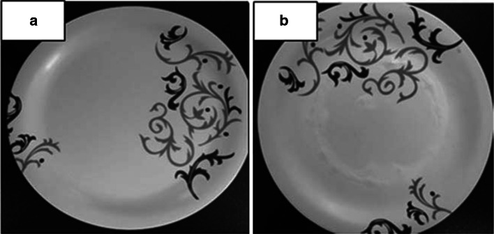
After 2.47 min cracking of melamine ware exemplified by a bursting noise was noticed from the microwave oven. The oven was immediately turned off. The plates were found to be cracked. The cracked melamine plate (Fig. 1b) was highly lachrymatory in nature. This may be due to the degradation of melamine–formaldehyde resin on heating the plates. The collected water samples were recorded in FT-IR. No other food stimulant was subjected to microwave in melamine wares as melamine wares are getting damaged. The surprising fact is no warning indication was given on the melamine plates for a common man to avoid use in a microwave oven.
Fig. 1.
a Melamine plate before subjected to microwave, b Melamine plate after subjected to microwave
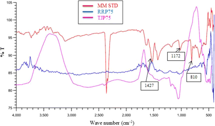
The fingerprint region in IR spectra gives valuable information and no two molecules can have the same spectral bands in the fingerprint region. The fingerprint region in Standard Melamine (Fig. 2) shows the peak at 763.81 cm−1 correspondings to side chain out of plane C–N bending, 810.10 cm−1 correspondings to C–N bending, 1172.72 cm−1 corresponding to NH2 rocking, 1427.32 cm−1 correspondings to NH2 wagging. The corresponding peaks in the fingerprint region are used for the determination of melamine leaching from the food samples. Figure 2 is the FT-IR spectra of the standard melamine, hot tomato soup in melamine plate (TJP75), hot rice porridge in melamine plate (RRP75). Most of the melamine ware in the market is melamine–formaldehyde (MF) resin (Bradley et al. 2011). In the MF resin formation mechanism; melamine a nucleophile by addition reaction adds to the electrophile formaldehyde. These reactions are favorable under almost all pH conditions; the rate of the reaction is slower at the neutral conditions. The melamine reacts with six formaldehyde groups to form two methylol groups on each exocyclic amine group (Ullah et al. 2014). The variation in the pH conditions is the reason for the melamine leaching. From FT-IR spectra it is clear that melamine leaching happens at 75 °C temperature when food stimulants which are in contact with melamine plate and also the microwaved water sample. In all the spectra the peak at 1149 cm−1 corresponds to NH2 rocking. Also, there is a difference in the group frequency region of the food samples incubated in melamine tablewares at a higher temperatures and the standard melamine spectra. It is because upon heating, melamine in melamine tablewares will be degraded and this is the reason for the difference in the group frequency region. From Fig. 3 it is clear that at lower temperatures there is no melamine leeching. This clearly portrays that the hot food items in used melamine wares are not safe for consumer use as it leaches melamine. Also there is no leaching of melamine from new melamine bowl and also at lower temperature there is no leaching of melamine from used melamine plates.
Fig. 2.
FTIR spectra of the food samples treated in used Melamine plates compared to Standard Melamine
Fig. 3.
FTIR spectra of the food samples treated in New Melamine bowl compared to standard Melamine
Determination of DNA damage using the comet assay
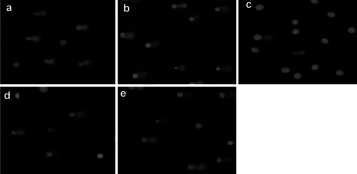
The food stimulants treated in used melamine plates under 75 °C showed the leaching of melamine. Those samples were subjected to comet assay by treating them with ct-DNA. The optical density 1.8 at absorbance 260 ensured the DNA is pure. In presence of an electric field negatively charged DNA fragments will be worn out into the agarose gel. Several factors decide DNA migration which happens with DNA damage in the cell. DNA migration increases when DNA strand breaks after treatment with alkali or in combination with certain enzymes DNA migration, whereas migration retards when DNA–DNA and DNA–protein cross-links. Ethidium bromide (EB) is the dye used to stain DNA on the slides. The ratio of the dye binding to the DNA depends on the amount of DNA present also the amount of light emitted after excitation with ultraviolet light of appropriate wavelength. Very low concentrations of the dyes are usually employed as higher concentrations will saturate the system. Visual scoring technique one of the methods used to interpret the comet assay results. This method is rapid, simple, and economical without depending on the software packages (Kumaravel et al. 2009). The visual scoring is employed for the present study (Fig. 4).
Fig. 4.
Comet tails for the samples aTJP75, b RRP75, c MMPR75, d UMMPW, e MMSTD
By visual scoring, it is clear that all the samples, damage the DNA as the cell shows the comet tail. Figure 4 shows the comet tail appearing for the samples treated with DNA. The Standard melamine solution treated with ct-DNA (e) is used for reference. Comparing the tail formed RRP75(c) has the least tail compared to other samples. The tail formed for the other samples portrays a severe DNA damage. The DNA movement to the anode during the electrophoresis might be due to the presence of melamine. By visual classification, the extent of DNA damage can be classified into 0–3(Bruschweiler et al. 2016) corresponding to the tail size. If there is no tail the score = 0(no DNA damage), beginning of tail indicates score = 1(low damage), if the tail is visible the score = 2(medium damage) and if the tail is long the score = 3(high damage). From Fig. 4 it is clear that all the samples fall under the score 3 and the DNA are highly damaged. Sun et al. (2010) have reported the toxic interaction of melamine with Herring sperm DNA and comparison between double-stranded ct-DNA. The mode of binding was electrostatic in nature. The electrostatic binding of melamine with the DNA phosphates is possible and this would be the reason for DNA damage in the present study. This is an alarming sign for melamine ware users. However, the quantitation of melamine in the samples has to be carried out to ensure the extent of DNA damage due to melamine.
Conclusion
This is the first report from India on Melamine leaching from Melamine table wares marketed in India with common Indian food cuisines. FT-IR is used as an analytical tool for the qualitative determination of melamine leaching from melamine tablewares. The study reveals melamine wares to be microwave unsafe and melamine leaching with hot food materials. Also, the study portrays used melamine dine wares to be unsafe than the new dine wares. Hot food stimulants in melamine ware cause DNA damage as evidenced by the comet assay results.
Acknowledgements
The authors sincerely acknowledge the Avinashilingam Institute for Home Science and Higher Education for Women, Coimbatore for providing facilities to carry out the research work. The consumables were procured under the DST-CURIE fund. The small financial assistance (Rs.3000/month) in the form of fellowship by GURUKADAKSHAM TRUST is gratefully acknowledged.
Footnotes
Publisher's Note
Springer Nature remains neutral with regard to jurisdictional claims in published maps and institutional affiliations.
References
- Bradley EL, Boughtflower V, Smith TL, Speck DR, Castle L. Survey of the migration of melamine and formaldehyde from melamine food contact articles available on the UK market. Food Addit Contam. 2005;22(6):597–606. doi: 10.1080/02652030500135243. [DOI] [PubMed] [Google Scholar]
- Bradley EL, Castle L, Day JS, Leak J. Migration of melamine from can coatings cross-linked with melamine-based resins, into food simulants and foods. Food Addict Contam. 2011;28(2):243–250. doi: 10.1080/19440049.2010.536167. [DOI] [PubMed] [Google Scholar]
- Bruschweiler ED, Wild P, Khanh C, Huynh A, Savova-Bianchi D, Danuser B, Nancy BH. DNA damage among wood workers assessed with the Comet assay. Environ Health Insights. 2016;10:105–112. doi: 10.4137/EHI.S38344. [DOI] [PMC free article] [PubMed] [Google Scholar]
- Firdhouse JM, Lalitha P. Binding properties of biosynthesized gold nanoparticles with calf-thymus DNA in vitro. Int J Biol Chem. 2015;9(4):188–197. doi: 10.3923/ijbc.2015.188.197. [DOI] [Google Scholar]
- https://www.who.int/mediacentre/news/release/2008/pr48/en/
- Ingelfinger Julie R. Melamine and the global implications of food contamination. N Engl J Med. 2008;359:2745–2748. doi: 10.1056/NEJMp0808410. [DOI] [PubMed] [Google Scholar]
- Kumaravel TS, Barbara V, Stephen PF, Awadhesh NJ. Comet Assay measurements: a perspective. Cell Biol Toxicol. 2009;25(1):53–64. doi: 10.1007/s10565-007-9043-9. [DOI] [PubMed] [Google Scholar]
- Partanen H, Vähäkangas K, Woo CSJ, Auriola S, Veid J, Chen Y, Myllynen P, Nezami Eli P. Transplacental transfer of melamine. Placenta. 2012;33:60–66. doi: 10.1016/j.placenta.2011.10.010. [DOI] [PubMed] [Google Scholar]
- Sun Y, Rutao L, Zhenxing C, Pengfei Q, Xiaoyan F, Yue M. Spectroscopic investigation on the toxic interaction of melamine with herring sperm DNA. J Biochem Mol Toxicol. 2010;24(5):323–329. doi: 10.1002/jbt.20341. [DOI] [PubMed] [Google Scholar]
- Tice RR, Agurell E, Anderson D, Burlinson B, Hartmann A, Kobayashi H, Miyamae Y, Rojas E, Ryu JC, Sasaki YF. Single cell gel/comet assay: guidelines for in vitro and in vivo genetic toxicology testing. Environ Mol Mutagen. 2000;35:206–221. doi: 10.1002/(SICI)1098-2280(2000)35:3<206::AID-EM8>3.0.CO;2-J. [DOI] [PubMed] [Google Scholar]
- Ullah S, Bustam MA, Naz MN, Tan MY, W.L. Synthesis and thermal degradation studies of melamine formaldehyde resins. Sci World J. 2014;2014:1–6. doi: 10.1155/2014/940502. [DOI] [PMC free article] [PubMed] [Google Scholar]
- Wiwanitkit V. Melamine: problems in obstetrics. Arch Gynecol Obstet. 2009;280:345–346. doi: 10.1007/s00404-008-0926-7. [DOI] [PubMed] [Google Scholar]
- Xiang D, Guoping Z, Kun Z, Li L, Zhike X. Determination of melamine in milk powder based on the fluorescence enhancement of Au nanoparticles. Analyst. 2011;136:2837–2844. doi: 10.1039/c1an00013f. [DOI] [PubMed] [Google Scholar]
- Yang VL, Batlle D. Acute renal failure from adulteration of milk with melamine. Sci Worl J. 2008;8:974–975. doi: 10.1100/tsw.2008.137. [DOI] [PMC free article] [PubMed] [Google Scholar]
- Yang T, Huangfu WG, Wu YL. Melamine residues in eggs of laying hens exposed to melamine-contaminated feed. Poult Sci. 2011;90:701–704. doi: 10.3382/ps.2010-00775. [DOI] [PubMed] [Google Scholar]
- Yurdakok B, Filazi A, Ekici H, Celikm TH, Sireli UT. Melamine in breast milk. Toxicol Res. 2014;3:242–246. doi: 10.1039/C3TX50095K. [DOI] [Google Scholar]
- Zhang Q, Yang G, Li J, Li W, Zhang B, Zhu W. Melamine induces sperm DNA damage and abnormality, but not genetic toxicity. Regul Toxicol Pharmacol. 2011;60:144–150. doi: 10.1016/j.yrtph.2011.03.004. [DOI] [PubMed] [Google Scholar]
- Zhou W, Yang H, Jiang Y, Shi H. The Characteristics of immune system changes in children who ingested melamine-contaminated powdered formula in China. Int J Environ Health Res. 2010;20(4):289–297. doi: 10.1080/09603121003663479. [DOI] [PubMed] [Google Scholar]
- Zhu H, Kannan K (2018) Distribution profiles of melamine and its derivatives in indoor dust from 12 countries and the implications for human exposure. Environ Sci Technol 52, 12801−12808. [DOI] [PubMed]


