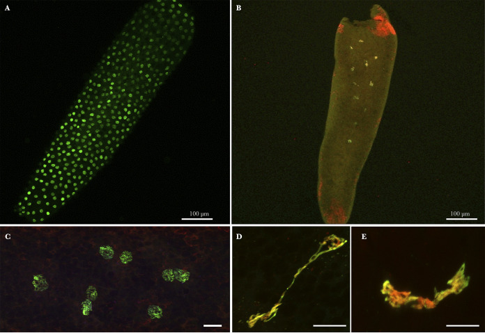
FIG 4.
Culex pipiens embryos 5 h postoviposition in INTER-INTER crosses. Green/yellow (acetylated histone H4 labeling) and red (propidium iodide labeling). (A) Global view of a normal C. pipiens embryo having reached the expected syncytial stage. (B) Global view of an abnormal C. pipiens embryo exhibiting only few (less than 15) nuclei 5 h postoviposition. (C) Normal nuclei in a syncytial embryo. (D and E) Atypical mitotic features observed in abnormal embryos. Confocal stacks were obtained on embryos from several INTER-INTER crosses. Red dots (especially visible at the embryo’s poles in panel B) are propidium iodide-labeled Wolbachia in the embryo’s cytoplasm. Scale bar represents 10 μm.

