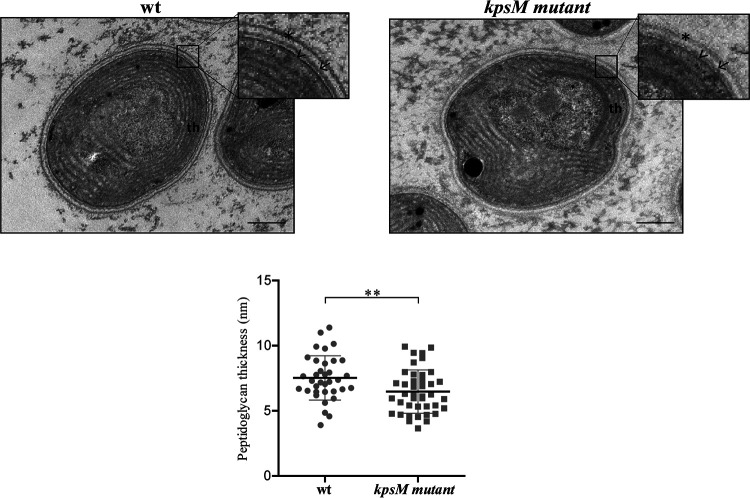
FIG 10.
Ultrastructure of Synechocystis sp. PCC 6803 wild-type (wt) and kpsM mutant cells. The inset panels show details of the cell walls. Asterisk, S layer; arrow, outer membrane; arrowhead, peptidoglycan; th, thylakoids. Bars, 100 nm. The measurements of peptidoglycan thickness are presented below the micrographs (**, P value of ≤0.01).

