Abstract
Background and Aims:
A wide variety of neurological diseases result in clinical and/or radiological enlargement of nerves, roots and plexuses. With the advancement in techniques and use of magnetic resonance neurography (MRN), aided by electrophysiology, proximal segments of the lower motor neuron (LMN) can be well studied. The relative merits of investigative modalities have not been well defined and comprehensive information on this subject is sparse.
Methods:
This retrospective study included data from January 2010 to June 2018. Patients having clinical and/or radiological enlargements of lower motor neuron were included. Clinical and laboratory work up, electrophysiology, MRN and biopsy studies were documented and analyzed.
Results:
133 patients fulfilled the inclusion criteria. The diagnostic categories were of leprosy (32%), immune neuropathies (27.8%), nerve infiltrations (8.2%), inherited neuropathies (9%), diabetic radiculopathies (9%) and others (12.7%). MRN was essential to diagnosis in 24.8% and supportive in 31.5% patients. Electrophysiology was essential in diagnosis in 70.6%, biopsy in 45.8% and genetic studies in 6.4% patients.
Conclusion:
The manuscript presents a large cohort of diseases causing enlargement of LMN with clinical and investigative aspects of 7 patients of the most unusual condition of chronic immune sensorimotor polyradiculopathy (CISMP) and details of 7 other patients with chronic mononeuropathies at non-entrapment sites. A table of comparative utility and an algorithm depicting the optimization of investigations has been presented.
Keywords: Causes of nerve enlargement, clinical nerve enlargement, investigations for nerve enlargement, radiological nerve enlargement
INTRODUCTION
Clinical enlargement of accessible nerves offers an important diagnostic clue to the etiology of neuropathies. Leprosy and inherited neuropathies are examples of such clinical enlargements of nerves. Deep seated and proximally located segments of the peripheral nervous system such as roots, plexuses and large nerve trunks are not accessible to clinical examination. These segments have to be studied with the help of electrophysiological assessments like the F waves, H reflex, somatosensory evoked potentials and inching technique, where the nerve is accessible for stimulation. These methods have the ability of providing accurate localization within various segments of peripheral nervous system. Once the lesion has been localized, further tests are directed to find out the extent of lesion, characterization of the lesion and to determine the site of biopsy, when needed. The development of magnetic resonance neurography (MRN) has helped to provide answers to some of these parameters. As more information becomes available the spectrum of disorders with enlargement of nerves, plexuses and roots widens further.
Enlargement of the lower motor neuron is encountered in a variety of conditions: infective neuropathies like leprosy; chronic inflammatory demyelinating polyneuropathy (CIDP); multifocal acquired demyelinating sensory and motor neuropathy (MADSAM); multifocal motor neuropathy (MMN); neoplastic/infiltrative, radiation related and immune plexopathies; and hereditary neuropathies. Besides these, lesser known forms of isolated proximal radiculopathies e.g. chronic immune sensory polyradiculopathy (CISP), chronic immune motor polyradiculopathy (CIMP) and the recently documented chronic immune sensorimotor polyradiculopathy (CISMP) which result in isolated proximal root enlargements, are being described.
While case studies and small series are available on the subject,[1] a systematic evaluation of enlargement of the lower motor neuron has not been presented. In particular, the optimization of investigations with their relative merits need to be studied. Hence this comprehensive study was undertaken.
METHODS
This retrospective observational study was carried out at a tertiary care teaching hospital. The departments of neurology, radiology, electrophysiology and histopathology participated in the study. The study period was January 2010 to June 2018. The institutional Ethics committee approval was obtained prior to commencement of the study.
Inclusion criteria
Patients with disorders of peripheral nervous system having clinical and/or radiological enlargement of peripheral nerves, plexuses and/or nerve roots.
-
Nerve enlargement:
The clinical examination and radiological assessment were performed by a single observer each (Faculty member of neurology and radiology departments).
Exclusion criteria
Patients with disorders of the peripheral nervous system who did not have enlargement of peripheral nerves or plexuses or roots
Entrapment neuropathies
Traumatic Neuropathies.
Historical information on the onset, duration and progression of symptoms, occupation, diet, family history, addictions, drug intake, toxin exposure and anaesthetic skin patches was documented. Neurological examination (motor system, sensory system, autonomic system, trophic changes and deep tendon reflexes) was documented. Clinical enlargement of supraorbital, infraorbital, greater auricular, ulnar, ulnar dorsal cutaneous, superficial radial, median, lateral popliteal, tibial, common and superficial peroneal nerves was charted. Investigations from available documents were recorded which included complete blood counts with RBC indices, erythrocyte sedimentation rate (ESR), serum B12, Human Immunodeficiency virus (HIV), Australia antigen (HbsAg) and anti-hepatitis C serology. Special investigations like Anti Nuclear Antibody (ANA), ANA blot, anti-neutrophil cytoplasmic antibody (ANCA), venereal disease research laboratory (VDRL), angiotensin converting enzyme (ACE) levels, cerebrospinal fluid (CSF) study, toxic/heavy metal screen, immunofixation electrophoresis, serum light chain assay and genetic results were noted when available.
Detailed nerve conduction studies were performed using a Natus synergy electromyograph. Studies included evaluation of F waves, H reflex and somatosensory evoked potentials as indicated.
Data of MRN performed on 3T Philips Achieva machine, acquiring T1 and T2 weighted images in axial and sagittal planes, short T1 inversion recovery (STIR) images in axial and coronal planes, diffusion-weighted imaging with background signal suppression (DWIBS) with coronal reconstruction and contrast enhanced T1 fat saturated axial, sagittal and coronal images were recorded.[2,5]
Biopsy of enlarged nerves was available in selected consenting patients in whom the diagnosis or therapy warranted tissue diagnosis. Available data on various stains including hematoxylin and eosin, Ziehl–Neelson, Fite Faraco and other special stains was recorded. Results from genetic studies (using the next generation sequencing and focused exome approach) were recorded.
The diagnosis of CIDP was made in patients fulfilling the European federation of neuromuscular societies (EFNS)/peripheral nerve society (PNS) criteria.[6] Inherited neuropathies were diagnosed when more than one family member was affected and/or pathogenic mutations were documented. Leprosy was diagnosed with positivity of skin or nerve biopsy findings.[7] MADSAM, MMN, CIMP and CISMP were diagnosed on clinical and electrophysiological criteria.[6] Diabetic lumbosacral radiculoplexoneuropathy[8] and idiopathic brachial neuritis[9,10] were diagnosed as per criteria. Nerve tumors, primary and secondary, were diagnosed on the basis of radiological and histological information.
RESULTS
133 patients fulfilled the inclusion criteria. There were 85 males and 48 females (M: F = 1.7:1) and the age ranged between 7 to 79 years. Clinical nerve thickening was seen in 55 patients (leprosy, n = 42, inherited neuropathies, n = 7 and CIDP, n = 6) and radiological thickening was seen in 95 patients, across all diagnostic categories. As per the diagnostic criteria mentioned in the material and methods, the distribution of disease categories was as follows [Table 1].
Table 1.
Disease categories
| Disease category | No. of patients (n=133) |
|---|---|
| Leprosy | 43 |
| Immune Neuropathies (CIDP, CISMP, MMN, MADSAM, CIMP) | 37 (23+7+4+2+1) |
| Inherited neuropathies | 12 |
| Tumor infiltrations | 11 |
| Diabetic lumbosacral radiculopathy | 11 |
| Unexplained Non compressive mononeuropathies (Sciatic 5, radial 1, ulnar 1) | 7 |
| Idiopathic brachial plexitis | 5 |
| Primary nerve tumors | 4 |
| Radiation plexopathy | 2 |
| Diabetic dorsal intercostal neuropathy | 1 |
| Total | 133 |
(CIDP: chronic inflammatory demyelinating neuropathy, CISMP: chronic immune sensorimotor polyradiculopathy, MMN: Multifocal motor neuropathy, MADSAM: multifocal acquired demyelinating sensory and motor neuropathy, CIMP: chronic immune motor polyradiculopathy)
42 out of the 43 patients with leprosy presented with sensory and motor mononeuritis multiplex and had clinical thickening of the distal nerves. Their skin (n = 34) or nerve (n = 9) biopsies confirmed leprosy. Biopsies revealed the following features: lepromatous (n = 8), borderline (n = 11) and tuberculoid leprosy (n = 14). The only patient, who did not have a clinical thickening of peripheral nerves, presented with distal tibial mononeuropathy. MRN showed thickening of the distal tibial nerve just proximal to the tarsal tunnel and the biopsy confirmed features of tuberculoid leprosy [Figure 1].
Figure 1.
Leprosy - (A1) T1 weighted sagittal image at the level of right distal tibia showing thickening of the right distal tibial nerve, (A2) T1 weighted axial image at the level of distal tibiofibular joint showing thickening of right distal tibial nerve just proximal to the tarsal tunnel (Arrow) as compared to the normal left tibial nerve (arrowhead), (B) Hematoxylin and eosin staining of biopsy of medial plantar nerve showing inflammation and granuloma (20×)
23 patients fulfilled the diagnostic criteria for CIDP. 6 of these had clinical thickening of nerves. All patients had non-length dependent sensorimotor neuropathies with slowed conduction velocities, conduction blocks and dispersion. Secondary CIDP was seen in 5 out of these 23 patients (multiple myeloma, n = 1); (solitary plasmacytoma, n = 1) and POEMS syndrome (Polyneuropathy, Organomegaly, Endocrinopathy, Monoclonal gammopathy, and Skin changes, n = 3).
7 patients were classified as CISMP. All of these patients had sensory ataxia, weakness and areflexia in the lower limbs. Their distal conductions in the sensory and motor nerves were normal, F waves were delayed and H reflex was absent in 6 and severely attenuated in the seventh patient. SSEPs showed normal N9, absent N22 and prolonged P40 responses in all patients. Electromyography showed chronic partial denervation in the lower limb musculature. Albuminocytological dissociation was seen in the CSF of all patients (protein 77-450 mg%).
Inherited neuropathies were diagnosed in 12 patients, 7 of whom had clinical thickening of nerves. Genetic diagnosis was established in 8 patients (PMP 22 n = 3, GJB1 n = 2, SH3TC2 n = 2, MPZ n = 1). 7 patients had affected family members.
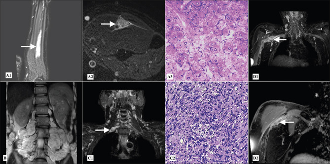
11 patients had infiltrations of nerves secondary to tumours (leukemias, lymphomas, carcinomas of breast, lung). Tumour infiltrations were much more common as compared to primary nerve tumors. The primary nerve tumours were perineuroma, schwannoma, neurofibromatosis and hemangioendothelioma [Figure 2].
Figure 2.
Peripheral nerve tumors - (A1) Postcontrast T1 weighted fat saturated coronal image of left forearm, (A2) STIR (short T1 inversion recovery) axial image at the level of left mid forearm showing fusiform enlargement with preserved fascicular pattern, intense STIR hyperintensity and post contrast enhancement of the left ulnar nerve (arrows) in a patient of perineuroma. (A3) Semithin section, toluidine blue stain of fascicular biopsy of left ulnar nerve showing pseudo-onion bulb appearance, which stained positive for epithelial membrane antigen and S-100 (not shown) suggestive of perineuroma (40×). (B) T2 weighted coronal image of the lumbosacral plexus showing plexiform neurofibromas along the lumbosacral plexuses. (C1) Reconstructed STIR MIP (maximum intensity projection) coronal image of the brachial plexus shows a small schwannoma along the post ganglionic right C7 root (arrow). (C2) Hematoxylin and eosin stain showing palisading appearance suggestive of schwannoma (40×). (D1) Reconstructed STIR MIP coronal and (D2) postcontrast T1 weighted fat saturated images of the right brachial plexus showing an ill-defined intensely enhancing lesion along the cords of right brachial plexus (arrows) which turned out to be hemangioendothelioma on histopathological examination
Diabetic lumbosacral radiculopathy was seen in 11 patients who presented with painful asymmetric lower limb weakness. Their MRIs showed asymmetric root enlargements and altered signal intensities. One patient had thoracic truncal neuropathy with enlargement of the intercostal nerve roots [Figure 3].
Figure 3.
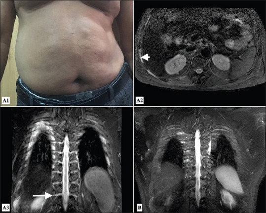
Diabetic truncal neuropathy - Clinical image (A1) showing protruded abdomen on right side due to weakness of abdominal wall muscles; MR images (A2) STIR axial at the level of D12 vertebra and (A3) STIR MIP coronal image of the dorsal spine showing thickening and hyperintense signal of the right sided dorsal nerve roots (arrow) and right intercostals nerves (arrowhead) in a patient with diabetic dorsal truncal neuropathy as opposed to (B) normal STIR MIP coronal image of the intercostal nerves for comparison
There were 7 unusual patients who had chronic progressive sensorimotor mononeuropathies (sciatic n = 5; radial n = 1; ulnar n = 1). In these patients, electrophysiology helped to focus the probable site of abnormality and MRN showed uniform signal changes (isointense on T1 weighted images, hyperintense on T2/STIR images with contrast enhancement) at non entrapment sites. 4 of the 5 patients with sciatic neuropathy underwent sciatic nerve fascicular biopsy. Nerve biopsy studies from these 5 patients showed axon loss and also helped to exclude etiologies like tumours, vasculitis and granulomas [Figure 4].
Figure 4.
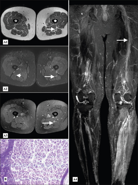
Sciatic mononeuropathy - MR Neurography (A1) T1weighted axial, (A2) STIR axial, (A3) postcontrast T1 weighted axial images at the level of mid-thigh and (A4) STIR MIP reconstructed coronal image of the lower limbs showing mild diffuse thickening, abnormal STIR hyperintense signal and postcontrast enhancement of the left sciatic nerve (arrows) as compared to normal appearing right sciatic nerve (arrowhead), (B) Semithin section, toluidine blue stain of fascicular biopsy of left sciatic nerve showing axon loss and regenerating fibers, preserved myelin and no evidence of vasculitis, granuloma or malignancy (40×)
Idiopathic brachial plexopathies were found in 5 patients, 4 other patients fulfilled the criteria for multifocal motor neuropathy and 2 patients each had MADSAM and radiation plexopathy.
MRN was essential to diagnosis in 24.8% including Immune neuropathies like CISMP (7), CIMP (1), tumor infiltrations (11), unexplained non-compressive mononeuropathies (7), primary nerve tumors (4), radiation plexopathy (2) and diabetic dorsal intercostal neuropathy (1). MRN was supportive in 31.5% patients diagnosing CIDP (7), MADSAM (2), MMN (4), inherited neuropathies (12), Diabetic LSPRN (11), Idiopathic brachial plexitis (5) and leprosy (1). Electrophysiology was essential for diagnosis in 70.6% including leprosy (43), immune neuropathies (37), inherited neuropathies (12), unexplained non-compressive mononeuropathies (7), idiopathic brachial plexitis (5). Biopsy provided results in 45.8% patients including leprosy (43), tumour infiltration (10), unexplained non-compressive mononeuropathies (4), primary nerve tumors (4), and genetic studies yielded diagnosis in 6.7% patients of inherited neuropathies (8) and primary nerve tumor in a case of neurofibromatosis.
DISCUSSION
This study evaluates large data on enlargement of the peripheral nerves, plexuses and roots. As can be seen from Table 1, the diagnostic spectrum included almost all conditions known to give rise to nerve enlargements,[1] the common groups being leprosy, dysimmune neuropathies, infiltrative conditions, hereditary neuropathies and plexopathies. We now discuss various diagnostic categories, highlighting the importance of investigative modalities in them.
Leprosy
This diagnosis was suspected clinically as patients had sensory mononeuritis multiplex with clinical thickening of the peripheral nerves. Patients in whom there were no skin manifestations (pure neuritic leprosy), thickening of multiple distal nerves provided a lead to the diagnosis and also helped to select the site for the nerve biopsy. The biopsies of skin or nerve were necessary to confirm the diagnosis. MRN was required in only one patient with single nerve involvement as described in results [Figure 1 A1-A3]. Interestingly, this patient was thought to have a tarsal tunnel syndrome, but the MRN highlighted the nerve thickening and biopsy confirmed leprosy.
Immune neuropathies
In this category, typical CIDP was most common followed by CISMP, MMN, MADSAM and CIMP. In typical CIDP, MADSAM and MMN, the diagnosis was clinical and electrophysiological. Electrophysiology was pivotal in determining the multifocal nature of the acquired demyelinating process and the involvements of sensory and motor components, thus enabling the categorizations e.g. MMN, MADSAM. The role of MRN was supportive, e.g. to help differentiate MMN from ALS.
However, MRN and electrophysiology (study of F waves, H reflexes and somatosensory evoked potentials) was essential to diagnose the rare isolated proximal conditions like CISMP and CIMP.[11,12]
CISMP
These 7 patients presented with proximal weakness, sensory ataxia and areflexia and had normal CMAPs and SNAPs, suggesting normalcy of the distal nerve segments. However, F-waves were absent in nerves of lower limbs, H-reflexes were delayed or absent and SSEPs showed delayed conduction at lumbar spine, highly suggestive of localization to the lumbosacral nerve roots. In this set of patients, MRN of lumbosacral nerve roots showed a diffuse thickening of lumbosacral nerve roots within the thecal sac leading to obliteration of surrounding CSF spaces. Contrast study showed diffuse enhancement of these roots without any leptomeningeal enhancement [Figure 5]. Thus, these 7 patients could be diagnosed correctly using electrophysiology followed by MRN.
Figure 5.
CISMP - MRI lumbosacral spine (A1) T2 weighted sagittal of the lumbar spine, (A2) T1 weighted fat saturated postcontrast and (A3) T2 weighted axial images at the level of L3-L4 vertebrae showing thickening and clumping of the lumbosacral nerve roots within the thecal sac with abnormal postcontrast enhancement (arrows), (B) Normal T2 weighted axial image at the level of L4 vertebra showing normal lumbosacral roots within the thecal sac (arrowhead) for comparison
CISMPs as a group are extremely uncommon. Khadilkar et al. initially described 2 such patients and suggested the terminology.[13] Since then only 9 more patients with CISMP have been described by Katirji and colleagues.[14] Besides these two reports, no other documentations of CISMP exist in the literature and these 7 patients further add to the literature on this rare condition.
Inherited neuropathies
This diagnosis was often suspected clinically with the long duration of symptoms, skeletal abnormalities like pes cavus and kyphoscoliosis and a positive family history. Electrophysiology further characterized them in demyelinating and axonal categories. Electrophysiological studies were particularly important in patients with HNPP who present with recurrent mononeuropathies and in patients harboring GJB1 mutations. The GJB1 mutations or the X linked CMT cases were challenging, as their electrophysiology had features of acquired demyelination, by showing conduction blocks and dispersion. In these patients, final diagnosis was confirmed by genetic study. MRN assumed only a supportive role in the inherited neuropathies. As an adjunct test, MRN displayed some differentiating features in acquired and inherited neuropathies as discussed below.
Role of MRN in acquired and inherited neuropathies
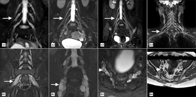
In acquired neuropathies, there was a diffuse symmetrical mild to moderate thickening of the extradural roots and proximal branches of the lumbosacral plexuses which showed abnormal hyperintense signal on T2 weighted images with preserved fascicles and abnormal post contrast enhancement depending on the stage of the disease [Figure 6]. In acute stage, there was only abnormal enhancement of the intra dural nerve roots.[5,15] In inherited neuropathies, there was thickening of the roots and proximal branches of the lumbosacral plexus with prominent preserved/demyelinating fascicles and increased fatty interfascicular epineurium giving multicystic appearance within the thickened nerves [Figure 6].[5,15]
Figure 6.
CIDP - MR Neurography (A1-A3) STIR MIP coronal reconstructed images of the lumbosacral plexuses in three different patients and (A4) of the brachial plexus, showing mild to moderate uniform thickening of the roots and branches of the lumbosacral plexuses (A1-A3) (arrows) and of the roots and trunks of brachial plexus (A4) (arrow) in acquired demyelinating neuropathies. CMT - MR Neurography (B1 and B2) Coronal STIR MIP reconstructed images of the lumbosacral plexus and (B3- B4) STIR axial and T1 axial images at the level of the lumbosacral plexus showing thickening of the roots and proximal branches of lumbosacral plexuses (arrows). There is increased inter-fascicular epineural fat with preserved fascicles (B3-B4) (arrowheads) giving multi cystic appearance of the thickened nerve roots in inherited neuropathies
Tumors
In tumoral infiltrations and primary nerve tumors, MRN and biopsies played the major role. MRN was extremely useful in arriving at the provisional diagnosis and choosing the site of biopsy.
In patients with diabetic lumbosacral radiculopathy, the diagnosis was made clinically and electrophysiologically. MRN supported the diagnosis by showing asymmetric enlargement of the lumbosacral roots or unilateral involvement of the lumbosacral plexus.
One patient presented with right sided intercostal pain and bulging of the right side of the abdomen, he was diagnosed with the intercostal diabetic neuropathy. In his MRN, the right intercostal nerves were found to be enlarged and showing abnormal T2 hyperintense signal, as compared to the normal side [Figure 3]. To the best of our knowledge, this finding has not been documented earlier and may be relevant to the pathophysiological basis of the condition.
The present cohort had only 5 patients with idiopathic brachial plexitis. The diagnosis was made by the clinical and electrophysiological features. Electrophysiologically, some had motor nerve involvement rather than the plexus itself. This is in keeping with the recent studies documenting the same[9,10] and others had involvement of the motor roots. MRN reinforced this observation by showing altered signals in affected segments. None of our patients exhibited the constrictions recently demonstrated on the MRN studies of patients with brachial plexitis but the number is too small to draw any conclusion.[16]
Unexplained non-compressive mononeuropathies
The combination of clinical, electrophysiological and MRN features identified 7 patients having sub-acute to chronic, progressive, sensorimotor mononeuropathies at non-entrapment sites. Over a mean observation period of 25.6 months, none of these patients showed involvement of contiguous or non-contiguous nerves beyond the nerve primarily affected (sciatic, radial or ulnar). Electrophysiology helped to accurately localize the site of lesion along the affected nerve and further guided the MRN studies. MRN was carried out to evaluate the extent and nature of the lesion and to rule out possible entrapment or compressions. All patients had uniform MRI features such asT2/STIR hyper intensity, diffuse nerve enlargement, homogenous contrast enhancement and disruption of perineural fat [Figure 4]. Biopsies were available in 4 of these patients, which showed axon loss without any demyelination or onion bulb formation. Biopsies excluded neoplastic or infiltrative processes. MR Neurography thus played a vital role in diagnosis of this group of mononeuropathies. 2 small series of chronic sciatic axonopathies have been documented in literature, but the pathophysiology, prognosis and therapy aspects of such mononeuropathies are as yet unknown.
Relative utility of workup
Considering the roles of MRN, electrophysiology, histopathology and genetic studies in this cohort, we now wish to propose the relative utility of clinical examination and various tests as summarized below in Table 2.
Table 2.
Comparative utility of examination and tests
| Clinical examination | Thickening of distal nerves |
| Leprosy (sensory mononeuritis multiplex) | |
| Inherited neuropathies (skeletal abnormalities) | |
| Some immune neuropathies | |
| Neurofibromatosis | |
| Electrophysiology | Diagnosis of CISMP |
| In patients with sensory ataxia, areflexia and weakness, when the distal segments were normal, study of the proximal segments with F wave and H reflex and SSEP studies assumes importance. | |
| Localization of mononeuropathies (Short segment or segmental study technique) | |
| Confirm presence or absence of other nerve involvements (e.g. HNPP, leprosy patients had mononeuritis multiplex) | |
| Information on conduction blocks and dispersion (e.g. MMN) | |
| To confirm demyelinating nature in cases of CIDP | |
| Differentiate between dysmyelinating (HMSN) and acquired demyelinating (e.g. CIDP). (Particularly relevant in the X linked Charcot Marie Tooth disease). | |
| Utility of the MR Neurography | For confirmation of lumbosacral plexus or proximal root involvement and to define the extent of involvement (e.g. CISMP) |
| Details of localization, extent of involvement, to rule out entrapment and to determine the site of biopsy | |
| Details of nerve tumors | |
| Differentiating MMN from amyotrophic lateral sclerosis | |
| As an adjunct tool in evaluation of demyelinating and dysmyelinating neuropathies | |
| Biopsy | Primary and secondary nerve tumors and infiltrations |
| Nerve infections like leprosy | |
| Unexplained neuropathies | |
| Genetic studies | Inherited neuropathies |
| Neurofibromatosis |
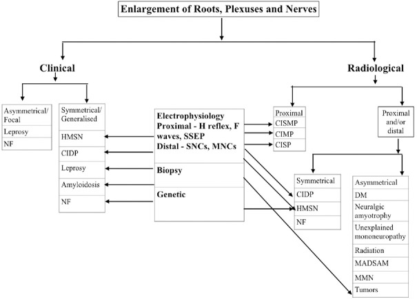
An algorithm has been proposed for optimisation of investigations for evaluation of this set of diseases resulting in enlargements of the lower motor neuron and is presented below [Figure 7]. While it contains conditions which we encountered in this study, amyloidosis has been additionally included for the sake of completeness.
Figure 7.
Algorithm for evaluation of enlargement of roots, plexuses and nerves
CONCLUSION
In this series of 133 patients, a large array of diagnostic categories was documented. Leprosy was the most common cause of clinical enlargement of nerves followed by immune neuropathies, tumour infiltrations and inherited neuropathies. Here, we wish to highlight our 7 cases of CISMP and 7 cases with non-entrapment mononeuropathies, five of which involved the sciatic nerves.
While clinical examination successfully detected enlargements of the distal nerve segments and skeletal abnormalities helped the diagnosis of conditions like leprosy and inherited neuropathies, proximal segments were largely inaccessible on routine clinical examination.
Electrophysiology contributed to the localization of disorders affecting the proximal segments, characterized the nature of these diseases, separated motor diseases like MMN form others; and helped the localization of various neuropathies. MRN was most useful to evaluate proximal conditions such as CISMP and sciatic neuropathies and characterization of nerve tumors.
The relative utility of available investigations for evaluation of enlargements of roots, plexuses and nerves has been presented and an algorithm for evaluation has been proposed.
Limitations
This is a retrospective analysis
Declaration of patient consent
The authors certify that they have obtained all appropriate patient consent forms. In the form, the patient(s) has/have given his/her/their consent for his/her/their images and other clinical information to be reported in the journal. The patients understand that their names and initials will not be published and due efforts will be made to conceal their identity, but anonymity cannot be guaranteed.
Financial support and sponsorship
Nil.
Conflicts of interest
There are no conflicts of interest.
REFERENCES
- 1.Khadilkar SV, Yadav RS, Soni G. A Practical approach to enlargement of nerves, plexuses and roots. Pract Neurol. 2015;15:105–15. doi: 10.1136/practneurol-2014-001004. [DOI] [PubMed] [Google Scholar]
- 2.Chhabra A, Andreisek G, Soldatos T, Wang KC, Flammang AJ, Belzberg AJ, et al. MR neurography: Past, present, and future. AJR. 2011;197:583–91. doi: 10.2214/AJR.10.6012. [DOI] [PubMed] [Google Scholar]
- 3.Thawait SK, Chaudhry V, Thawait GK, Wang KC, Belzberg A, Carrino JA, et al. High-resolution MR neurography of diffuse peripheral nerve lesions. AJNR Am J Neuroradiol. 2011;32:1365–72. doi: 10.3174/ajnr.A2257. [DOI] [PMC free article] [PubMed] [Google Scholar]
- 4.Donaghy M. Enlarged peripheral nerves. Pract Neurol. 2003;3:40–5. [Google Scholar]
- 5.Chhabra A, Lee PP, Bizzell C, Soldatos T. 3 Tesla MR neurography—technique, interpretation, and pitfalls. Skeletal Radiol. 2011;40:1249–60. doi: 10.1007/s00256-011-1183-6. [DOI] [PubMed] [Google Scholar]
- 6.Van den Bergh PY, Hadden RD, Bouche P, Cornblath DR, Hahn A, Illa I, et al. European Federation of Neurological Societies/Peripheral Nerve Society guideline on management of chronic inflammatory demyelinating polyradiculoneuropathy: Report of a joint task force of the European Federation of Neurological Societies and the Peripheral Nerve Society-First revision. Eur J Neurol. 2010;17:356–63. doi: 10.1111/j.1468-1331.2009.02930.x. [DOI] [PubMed] [Google Scholar]
- 7.Massone C, Belachew WA, Schettini A. Histopathology of the lepromatous skin biopsy. Clin Dermatol. 2015;33:38–45. doi: 10.1016/j.clindermatol.2014.10.003. [DOI] [PubMed] [Google Scholar]
- 8.Bhanushali MJ, Muley SA. Diabetic and non- diabetic lumbosacral radiculoplexus neuropathy. Neurol India. 2008;56:420–5. doi: 10.4103/0028-3886.44814. [DOI] [PubMed] [Google Scholar]
- 9.van Alfen N. Clinical and pathophysiological concepts of neuralgic amyotrophy. Nat Rev Neurol. 2011;7:315–22. doi: 10.1038/nrneurol.2011.62. [DOI] [PubMed] [Google Scholar]
- 10.Van Eijk JJ, Groothuis JT, Van Alfen N. Neuralgic amyotrophy: An update on diagnosis, pathophysiology, and treatment. Muscle Nerve. 2016;53:337–50. doi: 10.1002/mus.25008. [DOI] [PubMed] [Google Scholar]
- 11.Devic P, Petiot P, Mauguiere F. Diagnostic utility of somatosensory evoked potentials in chronic polyradiculopathy without electrodiagnostic signs of peripheral demyelination. Muscle Nerve. 2016;53:78–83. doi: 10.1002/mus.24693. [DOI] [PubMed] [Google Scholar]
- 12.Yiannikas C, Vucic S. Utility of somatosensory evoked potentials in chronic acquired demyelinating neuropathy. Muscle Nerve. 2008;38:1447–54. doi: 10.1002/mus.21078. [DOI] [PubMed] [Google Scholar]
- 13.Khadilkar S, Patel B, Mansukhani KA, Jaggi S. Two cases of chronic immune sensorimotor polyradiculopathy: Expanding the spectrum of chronic immune polyradiculopathies. Muscle Nerve. 2017;55:135–7. doi: 10.1002/mus.25360. [DOI] [PubMed] [Google Scholar]
- 14.Thammongkolchai T, Suhaib O, Termsarasab P, Li Y, Katirji B. Chronic immune sensorimotor polyradiculopathy: Report of a case series. Muscle Nerve. 2019;59:658–64. doi: 10.1002/mus.26436. [DOI] [PubMed] [Google Scholar]
- 15.Soldatos T, Andreisek G, Thawait GK, Guggenberger R, Williams EH, Carrino JA. High-resolution 3-T MR neurography of the lumbosacral plexus. Radiographics. 2013;33:96787. doi: 10.1148/rg.334115761. [DOI] [PubMed] [Google Scholar]
- 16.Sneag DB, Rancy SK, Wolfe SW, Lee SC, Kalia V, Lee SK, et al. Brachial plexitis or neuritis.MRI features of lesion distribution in Parsonage-Turner syndrome? Muscle Nerve. 2018;58:359–66. doi: 10.1002/mus.26108. [DOI] [PubMed] [Google Scholar]


