Abstract

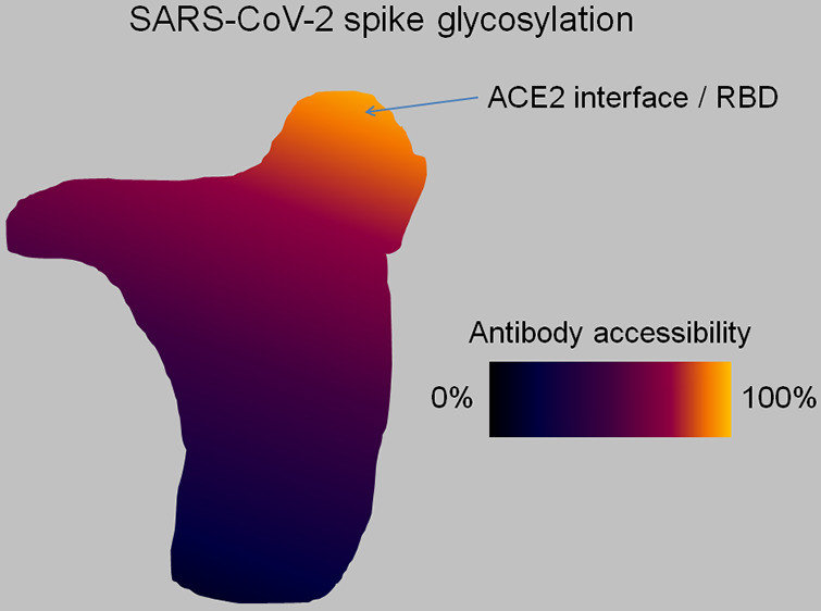
There is a need to assess evolutionary outcomes for SARS-CoV-2 in the postvaccination phase. The role of virus glycosylation in deterring the development of vaccine resistance is weighed against the epitopes of extant vaccines and the modulation of induced immune surveillance on antigens containing glycosylation sites.
Keywords: SARS-CoV-2, COVID-19 vaccine, protein glycosylation, evolutionary change, structural biology, vaccine resistance
SARS-CoV-2 Glycosylation and the Modulation of Immune Surveillance
Glycans result from linear or branched condensation of monosaccharides. The post-translational or cotranslational attachment of glycans to specific protein side chains is known as glycosylation. This chemical process is believed to provide a locus destination code for the protein within the cell. This code has not yet been fully elucidated. From a chemical perspective, glycosylation may influence the protein folding process, be it by altering the outcome structure or the expediency of the process—or probably both—due to the high solubility and high conformational entropy of the glycans attached to specific residues along the chain.1
Glycosylation is important to virologists because glycan attachment to capside proteins in viruses is often extensive and serves as a form of camouflage to immune surveillance. Many pathogenic agents, including SARS-CoV-2, resort to glycosylation to disguise their antigenicity and most likely exploit glycosylation to modulate the immune response.2 To enable their biosynthesis within the host cell, viruses hijack the host glycosylation machinery, yet differences from typical host glycosylation patterns, glycan density, and composition are known to occur. Thus, some level of glycan-based immunogenicity may be attained when the glycan types and glycosylation patterns are significantly different from those typically encountered in proteins from the host and thus become immunologically targeted.
The glycosylation camouflage may be rationalized from a chemical perspective: the association of an antibody to a glycosylated antigen is unlikely to have the high affinities expected from antibody recognition of protein loci due to the significant conformational entropy loss (−ΔS ≫ R, R = gas constant) associated with the constraining of glycans that are implicated in the recognition event. Thus, besides shielding the underlying protein from antibody recognition, the glycans attenuate the ability of the host immune system to recruit antibodies against glycosylated epitopes.
Glycosylation also serves as camouflage vis-à-vis the T cell mediated adaptive immune response. Peptides from the pathogen are placed on antigen-presenting cells by the major histocompatibility complex II (MHC II). This complex has preferred peptide motifs, and hence it is possible to predict which antigenic regions from a pathogenic protein are likely to elicit an adaptive immune response. However, when the purportedly antigenic peptide is glycosylated, its incorporation within the MHC may be abrogated due to the steric hindrance brought about by the glycan dynamics.
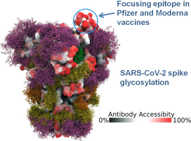
In the case of SARS-CoV-2, it has been estimated that glycosylation covers about 40% of the surface of the spike (S) protein trimer (Figure 1).2 As expected, the ACE2 receptor-binding domain (RBD) does not get glycosylated, representing the largest antigenic region exposed to host immune surveillance. Because SARS-CoV-2 relies on ACE2 recognition for host-cell entry, the virus simply cannot afford a glycan shielding of the RBD from the host immune response without substantially reducing its fitness. The requirement that the virus maintain proteic integrity at the ACE2 RBD to maintain fitness, i.e. capability of anchoring to the host cell, suggests that all extant vaccines which include this epitope may retain efficacy despite antigenic drift brought about by evolutionary changes to the glycosylation pattern. This success trend will likely prevail as long as the virus continues to target the same host receptor.
Figure 1.

Glycosylation camouflage on the spike protein of SARS-CoV-2 covering 40% of the spike surface and modulating immune surveillance. The exposed ACE2-RBD (circled) is the largest antigenic region with maximum antibody accessibility, constituting the focusing epitope of the Pfizer and Moderna vaccines. The “moss colors” indicate the different glycan types. Adapted from ref (2) under Creative Commons CC BY license.
Capside Glycosylation Would Likely Influence the Evolutionary Outcome of SARS-CoV-2 in the Postvaccination Scenario
The evolutionary pathway to vaccine resistance expected to be launched by SARS-CoV-2 is currently unknown, with speculation largely outpacing evidence.3 This is because we are not in a position to assess a priori the selection pressure to be exerted on the virus.4 The selection forces that will trigger vaccine-evasion pathways will become operative only in a postvaccination phase, during the endemic phase, with the evolutionary outcome ultimately resulting in an attenuated virus prevalence or extinction.4
The extensive glycosylation of the S protein trimer funnels the vaccine-induced immune surveillance toward epitopes that are deprived of the glycosylation camouflage, i.e. toward the ACE2 RBD. In this regard, the vaccines that only contain this antigen,5 like the ones from Pfizer/BioNTech (BNT162b2) or Moderna/NIAID (mRNA-1273), are better positioned to launch an effective adaptive immune attack than those that also include antigens that become glycosylated in the virus surface. The introduction of the latter antigens may in fact distract the immune system without enabling the induced antibodies or elicited T cell response to effectively target the virus. Furthermore, even if some participation from glycosylated antigen recognition can be factored into the immune response, the vaccines from Pfizer/BioNTech and Moderna/NIAID are likely to be better positioned. This is so because the antigens get administered in mRNA formulation,5 which essentially implies that the vaccines may co-opt the host glycosylation machinery to modify the vaccine-encoded protein co- or post-translationally to reproduce the glycosylation pattern of the virus in the specific underlying antigen.
This discussion leads to the conclusion that the evolutionary strategies launched by SARS-CoV-2 to evade the vaccine-induced immune response are most likely to play out by focusing on the ACE2 RBD region. This region is significantly constrained in the sense that most mutations are expected to be deleterious because they may compromise fitness. However, there are a substantial number of mutations in that region and even at the ACE2 interface that are well-tolerated by the structure of the spike protein and may even enhance the stability of the ACE2 association.6 Such mutations are likely to impair immune response since the induced antibodies will become less competitive in terms of displacing ACE2 as they bind to the epitope.
There is no evidence that such ACE2 affinity-enhancing mutations have been selected so far during the Covid-19 pandemic.6 But this is not surprising because the virus will only be under severe selection pressure during the postvaccination phase, not during the pandemic, where only social distancing has exerted a mild selection pressure.4
In this way, the role of SARS-CoV-2 glycosylation in deterring the development of vaccine resistance has been weighed against the epitopes of extant Covid-19 vaccines and the modulation of the induced immune surveillance on antigens susceptible to glycosylation.
The author declares no competing financial interest.
References
- Walsh C. (2006) Posttranslational Modification of Proteins: Expanding Nature’s Inventory; Roberts and Co. Publishers, Englewood, CO. [Google Scholar]
- Grant O. C.; Montgomery D.; Ito K.; Woods R. J. (2020) Analysis of the SARS-CoV-2 spike protein glycan shield reveals implications for immune recognition. Sci. Rep. 10, 14991. 10.1038/s41598-020-71748-7. [DOI] [PMC free article] [PubMed] [Google Scholar]
- Kennedy D. A.; Read A. F. (2020) Monitor for COVID-19 vaccine resistance evolution during clinical trials. PLoS Biol. 18, e3001000 10.1371/journal.pbio.3001000. [DOI] [PMC free article] [PubMed] [Google Scholar]
- Fernández A. (2020) COVID-19 Evolution in the Post-Vaccination Phase: Endemic or Extinct?. ACS Pharm. Transl. Sci. 10.1021/acsptsci.0c00220. [DOI] [PMC free article] [PubMed] [Google Scholar]
- World Health Organization . Draft landscape of COVID-19 candidate vaccines. December 17, 2020. https://www.who.int/publications/m/item/draft-landscape-of-covid-19-candidate-vaccines (accessed January 8, 2021). [Google Scholar]
- Starr T. N.; Greaney A. J.; Hilton S. K.; Ellis D.; Crawford K. H.D.; Dingens A. S.; Navarro M. J.; Bowen J. E.; Tortorici M. A.; Walls A. C.; King N. P.; Veesler D.; Bloom J. D. (2020) Deep Mutational Scanning of SARS-CoV-2 Receptor Binding Domain Reveals Constraints on Folding and ACE2 Binding. Cell 182, 1295–1310.e20. 10.1016/j.cell.2020.08.012. [DOI] [PMC free article] [PubMed] [Google Scholar]


