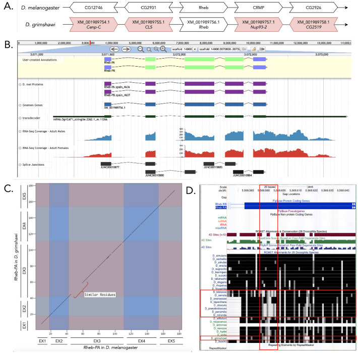
Figure 1.

(A) Synteny of genomic neighborhood of Rheb in both D. melanogaster as well as D. grimshawi. (B) Gene Model in Apollo: A screenshot of the Apollo instance housing the gene model, containing student annotations, Dmel proteins, Gnomon annotations, transdecoder genes, RNA-Seq tracks and splice junctions; (C) Dot Plot of gene in D. melanogaster (x-axis) vs. the gene in D. grimshawi (y-axis); (D) Low sequence conservation near the start of the third coding exon, which is more obvious in other species, but still existent in D. grimshawi.
Description
Introduction
The Ras homolog enriched in brain (Rheb) encodes a Ras homolog (Karassek et al. 2010). Ras is family of genes that make proteins involved in cell signaling pathways that control cell growth and cell death (Banerjee and Resat 2016).The gene model reported here (dgri_Rheb) was developed for the May 2011 assembly of D. grimshawi Agencourt dgri_caf1/DgriCAF1 (GCA_000005155.1) to describe the ortholog to D. melanogaster Rheb(FBgn0041191) at the locus previously annotated as XM_001989756.1. The general protocol and datasets for the genome browser tracks used to establish this reported gene model are reported in Rele et al. 2020. Additional tools and resources used in generating and confirming this model include HISAT (Kim et al. 2015), BEDTools (Quinlan et al. 2010), and the Sequence Read Archive (trace.ncbi.nlm.nih.gov/Traces/sra/?study=SRP073087).
Synteny
The Rheb gene on chromosome 3R in D. melanogaster is surrounded by the genes CG12746, CG2931, CRMP, and CG2926. Upon a tblastn search, the Rheb ortholog gene in D. grimshawi, on scaffold 14906, is surrounded by the genes XM_001989754.1, XM_001989755.1, XM_001989575.1, and XM_001989576.1 (orthologous to Cenp-C , CLS, Nup93-2, and CG2519) in D. melanogaster, Figure 1A). Though none of these flanking genes appear to be orthologous between the two species, we determined this region to contain the ortholog for Rheb in D. grimshawi because this location had a substantially better blastp hit to the Rheb protein sequence than to the second-best hit.
Gene Model
This gene model contains two isoforms of the Rheb protein in D. grimshawi, Rheb-PA and Rheb-PB (Figure 1B). Each of these isoforms contains five identical coding exons. The model in D. melanogaster and D. grimshawi have the same length and number of exons, and are similar in peptide sequence. However, there is a dissimilarity at the start of the third exon; but the peptides replacing the ones in D. melanogaster have similar properties. The coordinates of the corrected gene model can be found in NCBI at GenBank/BankIt using the accession BK014396 and archived in CaltechData here.
Special Characters of Gene Model
Improper alignment at start of coding exon three: As shown in Figure 1D, there is a stretch of amino acids (Figure 1D) that are not identical (black color), but similar (gray/white), between D. melanogaster and D. grimshawi. This pattern is also evident in a few other species as compared to D. melanogaster (Figure 1D). Though this is more obvious in D. kikkawai, D. serrata, D. obscura, D. pseudoobscura, D. persimilis, and D. miranda (Figure 1D) as compared to D. grimshawi (Figure 1D), it nonetheless exists.
Changes in the splice donor site for coding exon four: Rheb-PA in D. grimshawi has a canonical GT splice donor site at the end of coding exon four. In contrast, the end of coding exon four of Rheb-PA in D. melanogaster, and in most of the other Drosophila species across the genus Sophophora, use a non-canonical GC splice donor site.
Extended data:
Fasta (.faa and .fna) and GFF files are archived and available on CaltechData:D_grimshawi_Rheb_geneModel_gff_faa_fna. This file contains
Peptide Sequence of Rheb in D. grimshawi
Nucleotide Sequence of Rheb in D. grimshawi
GFF of Rheb in D. grimshawi
Acknowledgments
Acknowledgments
We would like to acknowledge Katie M. Sandlin, Gary Williams, and Terence Murphy for their constructive criticism of this article.
Funding
This material is based upon work supported by the National Science Foundation under Grant No. IUSE-1915544 to LKR and the National Institute of General Medical Sciences of the National Institute of Health Award R25GM130517 to LKR. The Genomics Education Partnership is fully financed by Federal moneys. The content is solely the responsibility of the authors and does not necessarily represent the official views of the National Institutes of Health.
References
- Aspuria P. J., and F. Tamanoi, 2004. The Rheb family of GTP-binding proteins. Cell. Signal. Volume 16, Issue 10: 1105-1112. [DOI] [PubMed]
- Banerjee K. , Resat H. 2016. Constitutive activation of STAT3 in breast cancer cells: A review. Int. J. Cancer 138: 2570–2578 [DOI] [PMC free article] [PubMed]
- Karassek S., C. Berghaus, M. Schwarten, C. G. Goemans, N. Ohse, et al. 2010. Ras homolog enriched in brain (Rheb) enhances apoptotic signaling. J. Biol. Chem. 285: 33979–91 [DOI] [PMC free article] [PubMed]
- Kim D, Langmead B, Salzberg SL. HISAT: a fast spliced aligner with low memory requirements. Nat Methods. 2015 Mar 01;12(4):357–360. doi: 10.1038/nmeth.3317. [DOI] [PMC free article] [PubMed] [Google Scholar]
- NCBI Expression Archives “Sequence Read Archive : NCBI/NLM/NIH.” National Center for Biotechnology Information, U.S. National Library of Medicine, trace.ncbi.nlm.nih.gov/Traces/sra/?study=SRP073087
- Quinlan AR, Hall IM. BEDTools: a flexible suite of utilities for comparing genomic features. Bioinformatics. 2010 Jan 28;26(6):841–842. doi: 10.1093/bioinformatics/btq033. [DOI] [PMC free article] [PubMed] [Google Scholar]
- Rele CP, Sandlin KM, Leung W, Reed LK. 2020. Manual annotation of genes within drosophila species: the genomics education partnership protocol. bioRxiv: 2020.12.10.420521.


