Abstract
Background
Autophagy, meaning ‘self-eating’, is required for the degradation and recycling of cytoplasmic constituents under stressful and non-stressful conditions, which helps to maintain cellular homeostasis and delay aging and longevity in eukaryotes. To date, the functions of autophagy have been heavily studied in yeast, mammals and model plants, but few studies have focused on economically important crops, especially tea plants (Camellia sinensis). The roles played by autophagy in coping with various environmental stimuli have not been fully elucidated to date. Therefore, investigating the functions of autophagy-related genes in tea plants may help to elucidate the mechanism governing autophagy in response to stresses in woody plants.
Results
In this study, we identified 35 C. sinensis autophagy-related genes (CsARGs). Each CsARG is highly conserved with its homologues from other plant species, except for CsATG14. Tissue-specific expression analysis demonstrated that the abundances of CsARGs varied across different tissues, but CsATG8c/i showed a degree of tissue specificity. Under hormone and abiotic stress conditions, most CsARGs were upregulated at different time points during the treatment. In addition, the expression levels of 10 CsARGs were higher in the cold-resistant cultivar ‘Longjing43’ than in the cold-susceptible cultivar ‘Damianbai’ during the CA period; however, the expression of CsATG101 showed the opposite tendency.
Conclusions
We performed a comprehensive bioinformatic and physiological analysis of CsARGs in tea plants, and these results may help to establish a foundation for further research investigating the molecular mechanisms governing autophagy in tea plant growth, development and response to stress. Meanwhile, some CsARGs could serve as putative molecular markers for the breeding of cold-resistant tea plants in future research.
Supplementary Information
The online version contains supplementary material available at 10.1186/s12864-021-07419-2.
Keywords: Autophagy, Camellia sinensis, Expression, Hormone, Abiotic stress, Cold acclimation
Background
Autophagy (ATG) is an evolutionarily conserved eukaryotic system that involves the degradation of various cytoplasmic components, including many biological macromolecules (such as proteins and protein aggregates), and entire organelles in vacuoles or lysosomes [1]. Normally, autophagy occurs at basal levels in eukaryotic cells but induces autophagic flux by specific developmental processes or stressful environments to degrade oxidative damaged proteins, damaged organelles, and other toxic compounds. Autophagy ensures that the degradation products can be recycled and the cells remodeled in cells to sustain their survival. Autophagy occurs through at least four pathways, classified as microautophagy [2], macroautophagy [3], chaperone-mediated autophagy [4], and selective autophagy [5]. Among these pathways, macroautophagy, which is manipulated by a special organelle, known as the autophagosome, is the most thoroughly characterized and is commonly referred to as autophagy. In this report, macroautophagy is referred to as autophagy. Accumulating evidence indicates that macroautophagy is derived from the formation of cup-shaped double membranes named phagophores (or isolation membranes), which engulf cytoplasmic material and then obturate to generate autophagosomes. Next, the outer membrane of the autophagosome fuses with the tonoplast, and the rest of the autophagosome forms an autophagic body, which is degraded in the vacuolar lumen to release its cargo for recycling [6].
Autophagy is primarily mediated by a collection of ATG genes. To date, more than 30 ATG genes have been identified in yeast and Arabidopsis [7–11]. Among these genes, ATG1–10, − 12-14, − 16-18, − 29, and − 31 serve as key modulators that participate in autophagy initiation, nucleation, elongation, maturation, and fusion with vacuoles [12–14]. In yeast and Arabidopsis, the initiation of autophagy is mediated by the target of rapamycin (TOR) kinase via association with ATG1/13 [15]. Next, the PI3K complex, containing Vps34, Vps15, ATG6/Vps30 and ATG14, is activated to promote vesicle nucleation. This step is followed by the expansion and enclosure of autophagy through the ATG5-12-16 and ATG8-PE conjugation systems. Later, the autophagosome is docked and fused to the tonoplast, which employs a vesicle trafficking system, known as the v-SNARE complex. Finally, the autophagic body in the vacuole is digested by a series of hydrolases, including the lipase ATG15 and proteinases A (PEP4) and B (PRB1) [16]. Following the identification of the ATG protein families in yeast and Arabidopsis, numerous orthologs have been identified in various plant and animal genomes, indicating that the core autophagic systems controlled by these proteins are conserved during evolution. In recent years, ATG genes have been identified in a number of plant species, such as 33 OsATGs in rice [17], 30 NtATGs in tobacco [18], 24 SlATGs in tomato [19], 30 VaARGs in grapevine [20], 32 MaATGs in banana [21], 37 SiATGs in foxtail millet [22], and 29 CaATGs in pepper [23]. Based on the alignment of multiple ATG amino acid sequences, many of the ATGs showed remarkable overall conservation in various plants, strongly suggesting that autophagic processes are mechanistically identical in different plants.
Along with the identification of ATG genes across the eukaryotic kingdoms, accumulating evidence indicates that the mechanism governing autophagy is involved in the whole life cycle of the plant, ranging from vegetative and reproductive development to environmental stress responses. Recently, reverse genetics approaches have effectively accelerated the functional analyses of ATG genes in plants, especially mutation and overexpression techniques. Many research findings have indicated that most ATG-mutated Arabidopsis lines exhibit premature senility phenotypes, although they still have intact life cycles, indicating that autophagy contributes to leaf longevity during senescence [9, 11, 24, 25]. Autophagy addresses organ senescence and nutrient starvation (e.g., carbon and nitrogen starvation, sucrose deficiency, and lack of light) by degrading damaged or unwanted proteins and organelle compounds to promote the recycling and remobilization of nutrients [1, 26–29]. It has been demonstrated that the biomass production and nitrogen remobilization efficiency in ATG-mutated Arabidopsis and rice were notably lower than those in the wild-type plants, indicating that autophagy contributes to efficient nitrogen remobilization [29–31]. In addition, the synthesis of amino acids was reduced in autophagy mutants during carbon starvation, indicating that the autophagy machinery controls cellular homeostasis [32]. However, increased autophagic activity can promote yield and nitrogen use efficiency in plants. For example, overexpression of OsATG8a was observed to strongly improve the level of autophagy and significantly improved nitrogen uptake efficiency in transgenic rice under suboptimal N conditions [26]. Apart from responding to nutrient deficiency, autophagy can also be induced by various abiotic stresses, and the kinases SnRK1 and TOR may be the central regulators of these processes [27]. Under high salt and osmotic stress conditions, the expression of an autophagy-related gene, AtATG18a, was upregulated in Arabidopsis, and the AtATG18a mutants were more sensitive to salt and drought conditions than were the wild-type plants [33]. However, overexpression of ATG5 or ATG7 promoted Atg8 lipidation, autophagosome formation and autophagic flux, thereby increasing the resistance of necrotrophic pathogens and oxidative stress and delaying senescence and improving growth, seed set, and seed oil content [34]. Moreover, autophagy can shape plant innate immune responses by a variety of means, but there are three main ways to induce autophagy, including virulent or related pathogen-induced programmed cell death, salicylic acid and jasmonic acid, and virus-induced RNA silencing [35]. Overall, autophagy is involved in the regulation of the whole life cycle of plants during different growth phases under different growing conditions.
As a special type of evergreen plant, the tea plant (Camellia sinensis) requires specific growing conditions, such as acidic soil, high humidity, and ordinary temperature; thus, this plant is primarily distributed in tropical and subtropical areas in Asia. However, tea plants are also regularly exposed to pathogen attack and herbivory, nutrient deficiency, and various types of abiotic challenges, such as extreme temperature, drought, salt, ozone, and ion toxicity. Under these adverse conditions, the morphology, physiology, and metabolism of tea plants have changed to survive. Accordingly, numerous studies on the stress resistance of tea plants in response to various stimuli have been performed in recent years. For example, multiple omic techniques, including transcriptomic, proteomic and metabonomic techniques, have been widely employed to explore the dynamic changes in genes, proteins and metabolites under different stress conditions [36–40]. In addition, many genes that respond to various stimuli have been identified and analyzed using draft genome sequences [41–46], and functional studies of some differentially expressed genes (DEGs), such as the Basic leucine zipper (bZIP) gene (CsbZIP6) [47], SWEET transporter gene (CsSWEET16) [48], vacuolar invertase gene (CsINV5) [49], and 12-Oxophytodienoate reductase gene (CsOPR3) [50], have been widely performed. However, no one autophagy-related gene (ARG) has been comprehensively analyzed in tea plants, and the roles played by autophagy in coping with different environmental stimuli have also not been fully elucidated to date in tea plants. In this study, the in vivo roles played by autophagy and the mechanisms governing the expression of ARG genes in tea plants were investigated through the genome-wide identification, characterization, and expression analysis of CsARGs. The results of this study may facilitate a deeper understanding of the diverse roles played by autophagy in response to different growth phases or environmental stress conditions in tea plants.
Results
Identification of CsARGs in tea plants
Based on four different identification methods, a total of 35 CsARGs were identified from the two published tea plant genomes (‘ShuChaZao’ and ‘YunKang10’). Among these genes, four genes (CsATG1s, CsATG8s, CsATG18s and CsVTI13s) were determined to have isoforms (Table 1). In this study, we identified a UV radiation resistance protein/autophagy-related protein 14, known as CsATG14, which has not been fully studied in plants to date and may be deficient in Arabidopsis. In addition, as there is no single strict criterion to identify all the paralogs of these four genes, and as the genome assembly and gene annotation of the two reported tea plant genomes have not been fully completed to date compared to the Arabidopsis, rice and tobacco genomes, the products of the partial paralogs of these four genes, as mentioned in Arabidopsis, rice, and tobacco, were not determined in the tea plant genomes. Bioinformatic analysis results showed that as a type of biological macromolecule, the CsARG ORF lengths varied from 285 to 7410 bp, the corresponding numbers of deduced amino acids ranged from 94 to 2469 aa, and the molecular weights ranged from 10.51 to 276.87 kD. The theoretical isoelectric points (pIs) were predicted to range from 4.52 to 9.41. The prediction of subcellular location results suggested that most CsARGs were located in the nucleus, and some of them were also predicted to be located in the cytoplasm, chloroplasts and mitochondria. The results of signal peptide prediction showed that none of these CsARGs contained signal peptides. In addition, CsATG9 was predicted to contain 5 TMHs, CsATG18b, and three transport vesicle-soluble NSF attachment receptor (v-SNARE) proteins, namely, CsVTI12, CsVTI13a and CsVTI13b, were predicted to contain 1 TMH.
Table 1.
Basic information of CsARGs
| Gene name | Accession number | ORF (bp) | AA | MW (kDa) | pI | instability index | Aliphatic index | Loc | SignalP | TMHs |
|---|---|---|---|---|---|---|---|---|---|---|
| CsATG1c | XP_028071137.1 | 2193 | 730 | 80.78 | 6.69 | unstable | 83.37 | Nucleus | NO | NO |
| CsATG1t | XM_028254805.1 | 852 | 283 | 31.70 | 6.72 | unstable | 100.21 | Cytoplasm | NO | NO |
| CsATG2 | XM_028214951.1 | 6039 | 2012 | 220.59 | 5.69 | unstable | 87.78 | Nucleus | NO | NO |
| CsATG3 | XM_028269562.1 | 945 | 314 | 35.72 | 4.72 | unstable | 78.82 | Cytoplasm | NO | NO |
| CsATG4 | XM_028205167.1 | 1473 | 490 | 54.16 | 5.54 | unstable | 73.45 | Nucleus | NO | NO |
| CsATG5 | XM_028239072.1 | 1047 | 367 | 41.29 | 4.77 | unstable | 98.26 | Cytoplasm | NO | NO |
| CsATG6 | XM_028209545.1 | 1581 | 526 | 59.43 | 5.87 | unstable | 71.77 | Nucleus | NO | NO |
| CsATG7 | XM_028207488.1 | 2121 | 706 | 77.95 | 5.63 | unstable | 91.20 | Cytoplasm | NO | NO |
| CsATG8a | XM_028204481.1 | 354 | 117 | 13.65 | 6.60 | unstable | 84.19 | Cytoplasm | NO | NO |
| CsATG8c | XM_028257959.1 | 360 | 119 | 13.65 | 8.78 | unstable | 83.61 | Nucleus | NO | NO |
| CsATG8f | XM_028237334.1 | 369 | 122 | 14.00 | 8.75 | stable | 95.08 | Nucleus | NO | NO |
| CsATG8g | XM_028213593.1 | 354 | 117 | 13.64 | 8.73 | stable | 86.67 | Nucleus | NO | NO |
| CsATG8i | XM_028202806.1 | 393 | 130 | 14.96 | 7.58 | unstable | 64.38 | Nucleus | NO | NO |
| CsATG9 | XM_028219288.1 | 2610 | 869 | 99.93 | 6.25 | unstable | 79.55 | plasmid | NO | 5 |
| CsATG10 | XM_028214294.1 | 717 | 238 | 27.19 | 4.96 | unstable | 82.73 | Nucleus | NO | NO |
| CsATG11 | XM_028237709.1 | 3471 | 1156 | 129.98 | 5.57 | unstable | 82.95 | Nucleus | NO | NO |
| CsATG12 | XM_028206145.1 | 285 | 94 | 10.51 | 9.41 | unstable | 88.19 | Cytoplasm | NO | NO |
| CsATG13 | XM_028260289.1 | 1863 | 620 | 68.75 | 8.90 | unstable | 65.27 | Nucleus | NO | NO |
| CsATG14 | XM_028265144.1 | 1440 | 479 | 53.78 | 8.85 | unstable | 77.37 | Chloroplast | NO | NO |
| CsATG16 | XM_028206301.1 | 1527 | 508 | 55.74 | 6.08 | unstable | 91.44 | Cytoplasm | NO | NO |
| CsATG18a | XM_028253213.1 | 1296 | 431 | 47.63 | 6.62 | stable | 77.82 | Nucleus | NO | NO |
| CsATG18b | XP_028071781.1 | 1107 | 368 | 40.18 | 7.15 | unstable | 96.49 | Cytoplasm | NO | 1 |
| CsATG18c | XM_028196882.1 | 1257 | 418 | 46.36 | 8.01 | unstable | 84.40 | Chloroplast | NO | NO |
| CsATG18f | XM_028238387.1 | 2697 | 898 | 97.13 | 8.47 | unstable | 75.46 | Mitochondria | NO | NO |
| CsATG18g | XM_028202982.1 | 2988 | 995 | 108.70 | 5.78 | unstable | 80.87 | Chloroplast | NO | NO |
| CsATG18h | XM_028252480.1 | 3069 | 1022 | 111.90 | 5.79 | unstable | 76.91 | Chloroplast | NO | NO |
| CsATG20 | XM_028265532.1 | 1206 | 401 | 46.15 | 8.20 | unstable | 83.47 | Chloroplast | NO | NO |
| CsATG101 | XM_028236970.1 | 657 | 218 | 25.43 | 6.46 | stable | 88.03 | Nucleus | NO | NO |
| CsATI | XP_028079241.1 | 948 | 315 | 35.00 | 4.52 | unstable | 64.13 | Nucleus | NO | 1 |
| CsVTI12 | CSA033576 | 669 | 222 | 25.26 | 9.22 | unstable | 105.81 | Nucleus | NO | 1 |
| CsVTI13a | XM_028240825.1 | 666 | 221 | 25.12 | 9.30 | unstable | 102.81 | Cytoplasm | NO | 1 |
| CsVTI13b | XM_028226760.1 | 666 | 221 | 24.89 | 9.41 | unstable | 101.95 | Cytoplasm | NO | 1 |
| CsVPS15 | XM_028202873.1 | 4632 | 1543 | 172.13 | 6.19 | unstable | 87.43 | Nucleus | NO | NO |
| CsVPS34 | XM_028243914.1 | 2361 | 814 | 93.36 | 6.39 | unstable | 92.46 | Cytoplasm | NO | NO |
| CsTOR | XM_028205854.1 | 7410 | 2469 | 276.87 | 6.22 | unstable | 102.06 | Cytoplasm | NO | NO |
ORF opening reading fame, AA the numbers of amino acid residues, pI Theoretical isoelectric point, MW Molecule weight, Loc Subcellular location, TMHs Transmembrane helices
Phylogenetic analysis of CsARGs in tea plant
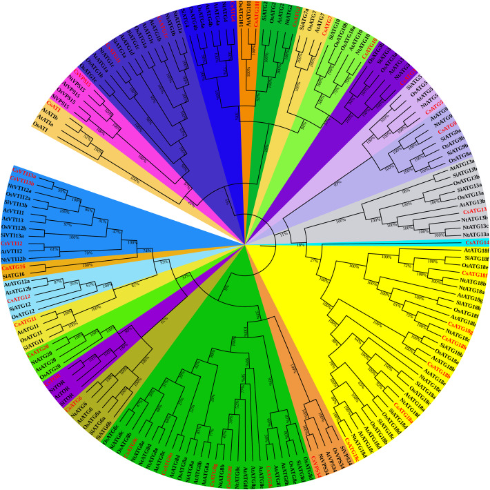
To explore the evolutionary relationships and classification of CsARGs in tea plant, a total of 177 ARG proteins from tea plants, Arabidopsis, Setaria italic, Oryza sativa, and Nicotiana tabacum were aligned to construct a phylogenetic tree. As shown in Fig. 1, except for CsATG14, which had only been identified in tea plants, all of the CsARG proteins were highly clustered together with the homologous proteins derived from the other four species, and almost all of the CsARGs showed the closest relationship with NtARGs. Meanwhile, we observed that the bootstrap values among the different ARG proteins in each subtree were nearly 100%, except for the ATG8 subfamily, which suggests that ARG protein sequences are highly conserved and may exhibit similar functions among different species.
Fig. 1.
Phylogenetic analysis of CsARGs and known ARGs in Arabidopsis, Setaria italic, Oryza sativa and Nicotiana tabacum. A total of 177 ARG protein sequences were used to construct phylogenetic tree throughout the neighbor-joining method with 1000 repeated bootstrap tests, p-distance, and pairwise deletion in MEGA 5.0 software. CsARGs are highlighted with red color, and different ARG subfamilies were covered with different colors
Gene structure, protein domain distribution and cis-acting element analysis
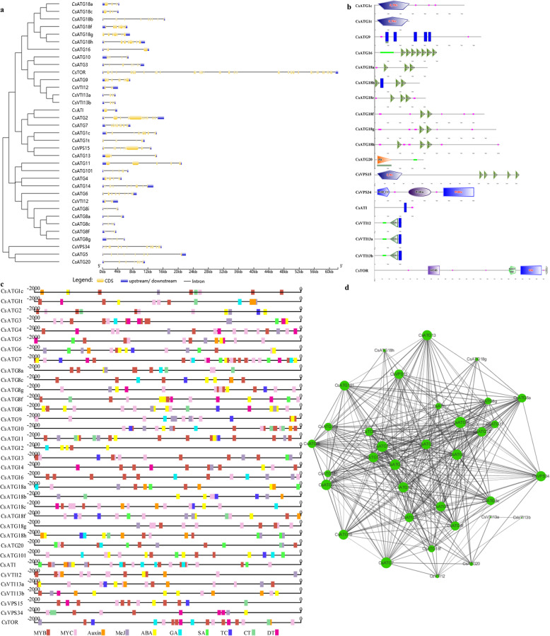
Understanding the exon-intron structure is beneficial for exploring the evolution of multiple gene families [51]. To investigate how the differences in exon-intron structure were generated, both the genomic and ORF sequences of CsARGs were uploaded into GSDS v2.0 to predict the exon-intron structure. As shown in Fig. 2a, the numbers of exons in the CsARG family varied, with members within the CsATG8s or CsVTI13s subfamilies exhibiting similar exon-intron structures.
Fig. 2.
The exon-intron structures, protein domains, cis-acting elements and protein-protein interaction networks of CsARGs. a Exon-intron structure of CsATG genes. The coding sequence and the corresponding genomic sequence of each CsARG were compared by using the Gene Structure Display Server (GSDS) program. Blue boxes represent untranslated upstream/downstream regions, yellow boxes represent exons, and lines indicate introns. b Protein domains of CsARGs. c The cis-acting regulatory elements of CsARGs. 2000-bp upstream noncoding region sequences of each CsARG gene were used to predict cis-acting elements, and different colored blocks represent different elements. d Protein-protein interaction networks of CsARGs. Thirty five orthologs of CsARGs were obtained from Inparanoid web server, and those 35 orthologs formed 360 protein-protein association patterns
To further dissect the functions of CsARG proteins, the protein domains of each CsARG were analyzed by the SMART program. As shown in Fig. 2b, CsATG1s encode serine/threonine protein kinases, which contain catalytic domains involved in protein phosphorylation occurring during the progression of autophagy [52]. CsATG9 contains 5 transmembrane helix regions (88–110, 155–177, 320–342, 403–425 and 438–457), as detected by the TMHMM v2.0 program, which play a unique role in autophagosome formation derived from the endoplasmic reticulum (ER) in plants [53]. CsATG16 contains a coiled coil region and 7 WD40 domains, which form a conserved Atg12-Atg5-Atg16 complex during the autophagy process. Each member of the CsATG18s subfamily is largely a β-propeller and is formed by 2 or 3 WD40 domains. CsATG20, also known as Snx42, contains a PX domain, which plays a central role in efficiently inducing nonselective autophagy. CsVPS15 encodes a serine/threonine protein kinase, which is formed by 5 WD40 domains and is regulated by a phosphoinositide 3-kinase (PI3K), CsVPS34. At present, CsVPS34 is characterized as a central regulator in mediating vesicular trafficking and cellular homeostasis [54]. As an ATG8-interacting protein, CsATI contains a transmembrane region that may help the protein complex to move to the ER network and reach the lytic vacuole. CsVTI1s, including CsVTI12, CsVTI13a and CsVTI13b, all contain a coiled coil region, a t-SNARE domain and a transmembrane region, and these domains may be involved in the trafficking of cargoes to the vacuole. CsTOR, as a conserved phosphatidylinositol kinase-related protein kinase, contains a specific rapamycin-binding domain, a P13Kc-catalyzing domain and a FATC domain, which suggests that it is involved in mediating redox-dependent structural and cellular stability.
To elucidate the regulatory mechanisms governing CsARGs in response to growth and development, stress defenses, and hormone signaling, a 2000-bp 5′-upstream noncoding region sequence of each CsARG was isolated to predict cis-elements. As Fig. 2c shows, the distributions, numbers and types of cis-elements vary among the promoter sequences. Nevertheless, most of the promoters contain a number of MYB- and MYC-binding sites, except for the promoter of CsATG12, which lacks MYC-binding sites. In addition, most of the promoters of CsARGs contain ABA- and MeJA-responsive elements, and some of them contain GA, SA, auxin, cold, drought, defense and stress (TC-rich repeats)-responsive elements. In addition, all of the promoters of CsARGs contain numerous light-responsive elements, including G-boxes, MREs, Box-4, and AE-boxes (not shown in Fig. 2c). Overall, each CsARG may play a vital role in responding to circadian variation, hormones, and biotic and abiotic stresses.
Protein-protein interaction networks of CsARGs
To investigate the interactions among CsARGs in tea plants, the ortholog groups of CsARGs, which originated from Arabidopsis, were used to construct PPINs. As a result, 35 orthologs of CsARGs were obtained from Inparanoid web server, and those 35 orthologs formed 360 protein-protein association patterns. The ARGs were determined to be closely related to one another, except for ATG14, which has not been fully studied in Arabidopsis. Among these proteins, 24 ARGs, including ATG9, ATG7, and ATG1c, were reported or predicted to interact with more than 20 ARGs, suggesting that the occurrence of autophagy requires interactions among numerous ARG proteins.
Conserved domain and motif distribution analysis of CsATG8s
As members of the UBQ superfamily, ATG8s coupled with their conjugation system are key components for autophagy. In our study, to clearly understand the regulatory mechanisms governing these proteins, the bioinformatic characteristics of CsATG8 subfamily proteins were further explored. As Fig. 3 shows, a total of 5 CsATG8s were identified in tea plants based on homologous alignment analysis. Phylogenetic analysis results showed that CsATG8s were subdivided into 3 clades. Among these proteins, CsATG8a/c and CsATG8g/f were clustered into one group, and CsATG8i was aligned closely with MdATG8i (Fig. 3). Motif distribution analysis results showed that CsATG8s contain motifs 1–4, and CsATG8i contains an additional motif 7 at the C-terminus (Additional file 2). All of these CsATG8s proteins contain conserved GABARAP domains, four putative tubulin binding sites, three ATG7 binding sites, and a conserved glycine (G) residue. In addition, we found that the conserved G residues in both CsATG8a and CsATG8g were directly exposed at the C-terminus (Fig. 3), suggesting that the functions of CsATG8a and CsATG8g involved in autophagy may be distinct from those of the other three proteins. These proteins may not require the cysteine protease Atg4 to cleave the C-terminus but may directly bind to the E1-like enzyme Atg7.
Fig. 3.
Conserved domains analysis of CsATG8s. a Phylogenetic analysis of CsATG8s and known ATG8s in Arabidopsis, Oryza sativa, Malus domestica, Saccharomyces cerevisiae and Humans. CsATG8s were highlighted with red boxes. b Amino acids alignment analysis of CsATG8s and known ATG8s in Arabidopsis, Oryza sativa and Malus domestica. Three putative ATG7 binding sites were contained in the red boxes respectively, four putative tubulin binding sites were contained in the pink boxes, and a conserved glycine (G) residue was framed in the bright blue box
Expression profiles of CsARGs in various tea plant tissues
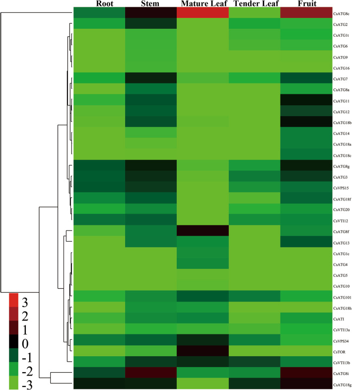
To confirm the tissue specificity of CsARGs, the roots, stems, mature leaves, tender leaves, and seeds of tea plants were obtained for qRT-PCR analysis. The results showed that the transcription of all CsARGs was detected among the above mentioned tissues, although the mRNA level of each CsARG varied across the various tissues (Fig. 4). In addition, we found that most CsARGs exhibited higher transcription abundances in stems and seeds, suggesting that autophagy plays important roles in the development of stems and seeds in tea plants. Moreover, we found that the CsATG3/7/101, CsVPS15/34, CsATI, CsVTI12/13b, CsATG8s and CsATG18s subfamily genes were highly expressed in different tissues. Notably, CsATG8c was significantly expressed in mature leaves and seeds, and CsATG8i was dramatically expressed in stems and seeds. In brief, our results found that the expression patterns of the CsARGs varied across different tissues, but some of them showed a degree of tissue specificity, suggesting that CsARGs mediated the growth and development of tea plants.
Fig. 4.
Expression profiles of CsARGs in different tea plant tissues. The transcription abundances of CsARGs in different tissues were monitored by using qRT-PCR, and the results were calculated by using 2–ΔCt method. CsPTB was chose as actin gene. The heat map was generated by using Cluster 3.0 software. The colorbar was displayed on the lower-left of the heat map, red and green colors represent higher and lower expression levels respectively
Differential expression of CsARGs in response to hormone treatments
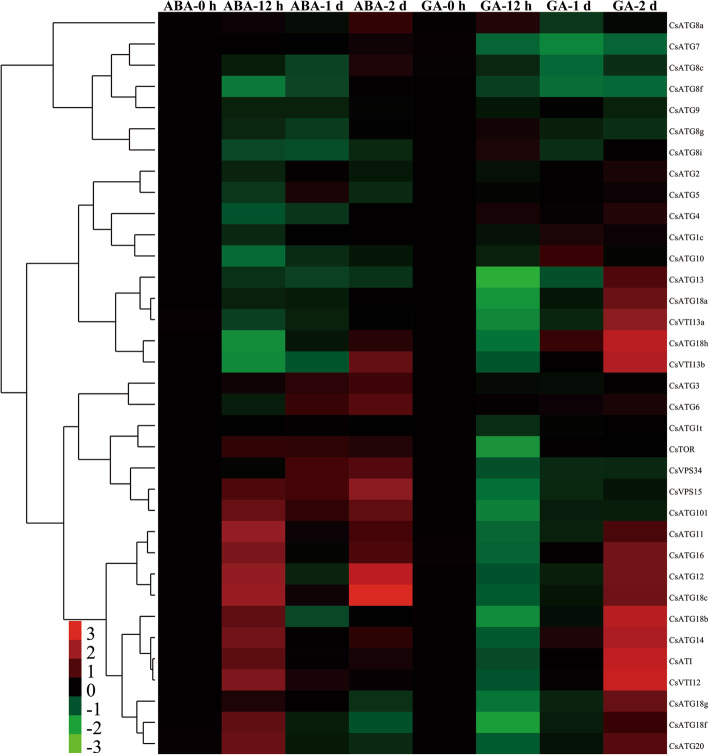
To elucidate the comprehensive roles of CsARGs under ABA and GA treatment conditions, we analyzed the expression pattern of each CsARG. Under ABA treatment conditions, we observed that multiple CsARGs were highly induced after 12 h and/or 2 d of ABA treatment. Among these genes, the expression levels of CsATG6/11/12/14/16/18b/18c/18f/20/101 and CsVTI12/VTI13b were induced more than 2-fold for at least at one time point. Meanwhile, some genes, such as CsATG3/11/18c/101/VPS15/TOR, were directly upregulated during the entire ABA treatment period (Fig. 5). In contrast, CsARGs exhibited contrary expression profiles under GA stress compared to ABA stress. Most CsARGs initially decreased but significantly increased after 2 d of GA treatment. Among these genes, the expression levels of 13 genes, including CsATG12/14/16/18a/18b/18c/18 g/18 h/20/ATI/VTI12/VTI13a/VTI13b, were increased more than 2-fold after 2 d of GA treatment. Furthermore, 6 genes, CsATG7/8c/8f/101/VPS15/VPS34, were negatively downregulated throughout GA stress periods. These results indicated that CsARGs are required for the response to hormone treatments in tea plants.
Fig. 5.
Temporal-spatial expression patterns of CsARGs in response to hormone treatments. The expressions of each CsARG gene within 2 d of ABA and GA treatments were performed by using qRT-PCR technique respectively. CsPTB was chose as the actin gene. The final results were calculated with 2–ΔΔCt method, and the samples that collected at 0 h were set as control. The heat map was generated by using Cluster 3.0 software. The colorbar was displayed on the lower-left of the heat map, red and green colors represent higher and lower expression levels, respectively
Expression patterns of CsARGs in response to different abiotic stresses
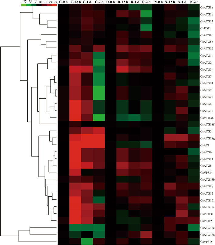
Similarly, to explore the temporal expression patterns of CsARGs under abiotic stress conditions, the related expression level of each CsARG was determined by qRT-PCR. As shown in Fig. 6, the expression levels of all CsARGs were regulated to different degrees under various abiotic stress conditions.
Fig. 6.
Temporal-spatial expression patterns of CsARGs in tea plant under various abiotic stresses. The expressions of each CsARG gene within 2 d of cold, drought, NaCl treatments were performed by using qRT-PCR technique respectively. CsPTB was chose as actin gene. The final results were calculated with 2–ΔΔCt method, and the samples that collected at 0 h were set as control. The heat map was generated by using Cluster 3.0 software. The colorbar was displayed on the upper- left of the heat map, red and green colors represent higher and lower expression levels, respectively
During the CT period, the expression levels of almost all CsARGs were upregulated at different processing time points, except for 2 genes, CsATG18c/h, which were downregulated throughout the CT period. In addition, 20 induced CsARGs showed the highest expression levels after 12 h of CT. Specifically, the expression levels of CsATG3/4/6/8i/10/11/18a/18 g/101/VTI12/VTI13a/VTI13b/VPS34 were more than 5-fold higher than those at 0 h of CT. In addition, CsATG5/ATG18g/ATI expression was gradually induced within 2 d of CT. Under DT conditions, 15 CsARGs were induced at different DT time points, and most of these genes showed the highest expression levels after 12 h of DT. The expression levels of CsATG2/3/6/8i/11/16/VPS34 were more than 2-fold higher than those at 0 h of DT. Moreover, CsATG18h and CsATG101 were gradually upregulated as the DT time extended. Conversely, the expression of the remaining CsARGs, such as CsATG4/7/8a/10/14, was slightly deduced or not affected by DT. Within NT periods, many CsARGs were also induced to different degrees. In contrast to CT, most of the induced CsARGs showed the highest expression levels after 1 d of NT but were deduced following the extended treatment time. In particular, the expression levels of CsATG9/10/18a/18 g/101/VTI13 were increased more than 2-fold after 1 d of NT compared to the control. However, CsATG8g/12/16/ATI was significantly upregulated after 12 h of NT. Taken together, these results demonstrated that CsARGs play central roles in responding to abiotic stress in tea plants.
Differential expression of CsARGs in different tea plant cultivars during CA periods
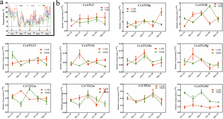
The differential expression patterns of CsARGs were compared in different tea plant cultivars (the cold-resistant cultivar ‘Longjing43’ and the cold-susceptible cultivar ‘DaMianBai’) within CA periods. Eleven CsARGs, which were confirmed to be notably induced under CT conditions, as shown in Fig. 7, were selected for analysis by qRT-PCR. The results showed that these 11 genes presented different expression patterns in ‘Longjing43’ and ‘DaMianBai’ during the CA period in 2018–2019. Specifically, these 11 genes showed contrary expression patterns from November 14th to December 13th in ‘Longjing43’ and ‘DaMianBai’. With the exception of CsVPS34, the other CsARGs were continuously upregulated in ‘Longjing43’, but all of them were gradually reduced in ‘DaMianBai’ from November 14th to December 13th. In contrast, the transcription levels of these 11 genes were all increased in ‘DaMianBai’ from Dec 13th to Jan 17th, but many CsARGs, such as CsATG16/18 g/101/VTI12, were decreased in ‘Longjing43’. Notably, we observed that the transcription level of CsATG101 was lower in ‘LongJing43’ than in ‘DaMianBai’ throughout the CA period. These results indicated that these CsARGs play important roles in the response of tea plants to cold resistance, but their regulatory mechanisms may vary among different cultivars.
Fig. 7.
Expression analysis of CsARGs during CA periods in 2018–2019. a Changes in air temperature from November 2018 to March 2019. The maximum (Max), average (Avg) and minimum (Min) daily temperatures were respectively indicated by red, blue and green colored lines. The red arrows represent sampling days. b The relative expression levels of 11 CsARGs in two tea cultivars during CA periods in 2018–2019. The expression profiles of CsARGs in two tea cultivars were displayed with different colored lines. The results were calculated by using the 2–ΔCt method with CsPTB as the actin gene. Data are shown as the means ± SE (n = 3)
Discussion
CsARGs are involved in different stages of autophagy in tea plants
Autophagy is a catabolic degradation pathway that is essential for degrading long-lived proteins, protein aggregates, and damaged organelles [55]. It has been proven that autophagy is highly conserved from yeast to humans and is the result of the interactions of many proteins. To date, more than 30 ATG-related genes have been identified in many eukaryotes, and those ATGs encode many core proteins involved in the entire process of autophagy, extending from induction to the degradation, recovery and recycling of autophagosomes. As the tea plant is a type of evergreen wooden plant, the recycling of some broken or discarded macromolecular substances plays an important role in the special development period or in the resistance to stresses in this plant. In the present study, a total of 35 CsARGs were identified in the tea plant genome. Some CsARGs, such as CsATG8 and CsATG18, exhibit multiple copies in the tea plant genome, and similar results were also observed in many other species, such as Oryza sativa [17], Nicotiana tabacum [18], Vitis vinifera [20], Musa acuminate [21], and Setaria italic [22]. In addition, the results of phylogenetic and protein domain analysis further confirmed that ATGs are highly homologous among different plant species. In yeast, ATG proteins are divided into four functional groups based on their roles in the autophagy process [56]. Similarly, the identified CsARGs also constitute a relatively complete autophagic machinery in which they function in forming the ATG1 kinase complex (CsATG1s/13, CsTOR), Class III PI3K complex (CsATG6/14, CsVPS15/34), ATG9 recycling complex (CsATG2/9/18 s), Atg8-lipidation system (CsATG3/4/7/8 s) and Atg12-conjugation system (CsATG5/7/10/12/16).
To date, there have been numerous reports on the functional analysis of ATG14 in mammals but few in plants. In mammals, the human ATG14 homolog, hAtg14/Barkor/Atg14L, has been shown to be the sole specific subunit in the phosphatidylinositol 3-kinase (PI3-kinase) complex. Atg14L was observed to interact with Beclin-1/2 through their coiled-coil domains and proved to be the targeting factor for PI3KC3 to the autophagosome membrane [57, 58]. Similarly, ATG14 is only integrated into phosphatidylinositol 3-kinase complex I to direct the association of complex I with the pre-autophagosomal structure (PAS) in yeast [59]. In this study, we identified an ATG14 homologous protein, referred to as CsATG14, in tea plants. Bioinformatic analysis results predicted that CsATG14 is a hydrophilic protein and contains a coiled-coil motif at the N-terminus region from 10 to 367 aa, suggesting that CsATG14 may interact with Beclin-1 (designated CsATG6 in our study) to serve as a scaffold for recruiting class III phosphatidylinositol-3-kinase (PIK3C3). However, this possibility requires further verification by using the yeast double hybrid (Y2H) method.
As a highly conserved ubiquitin-like protein, ATG8 is activated by conjugation to lipid phosphatidylethanolamine (PE) to form the ATG8-PE adduct, thereby participating in autophagosome formation and phagophore expansion [60, 61]. During conjugation, the C-terminus of ATG8 must be cleaved by a cysteine protease, Atg4, to expose a glycine residue [62, 63]. In this study, five ATG8 isoforms were identified, and they displayed high sequence similarity to AtATG8s, NtATG8s and OsATG8s. However, a conserved glycine residue for lipidation was directly exposed at the C-terminus of CsATG8a/g, and similar phenomena were also observed in MdATG8g/i, AtATG8h/i and OsATG8e, suggesting that ATG4 may not be necessary for the conjugation of ATG8-PE. In addition, ATG8 can also interact with its specific substrates or receptors via an Atg8 interacting motif (AIM) in the target proteins during selective autophagy. Recently, two plant-specific proteins in Arabidopsis, termed ATI1 and ATI2, were identified and proven to interact with AtATG8f and AtATG8h [64]. In our study, however, only one unique ATI homologous gene, CsATI, was identified. Sequence alignment analysis found that CsATI also contains two putative AIMs (17–20, 267–270) and a predicted transmembrane domain (242–259) (Additional file 2), suggesting that CsATI can also bind one of five CsATG8 isoforms in tea plants. To clarify whether CsATI interacts with CsATG8 isoforms, Y2H could be performed by using a CsATG8 isoform recombinant plasmid as bait and a CsATI recombinant plasmid as prey.
During macroautophagy, a v-SNARE complex, including v-SNARE VTI1, Rab-like GTP-binding protein (YKT6), and syntaxin (VAM3), contributes to the maturation and fusion of autophagosomes to lysosomes/vacuoles [65]. In plants, three VTI1-type SNARE members (VTI11, VTI12, VTI13) have been identified [66]. Sanmartin et al. (2007) suggested that VTI12 and VTI11 might be involved in trafficking to storage and lytic vacuoles in vegetative and seed tissues in Arabidopsis, respectively [67]. Moreover, VTI13 participates in the trafficking of cargo to the vacuole within root hairs and plays an essential role in the maintenance of cell wall organization in Arabidopsis [68]. In our study, CsVTI12 and CsVTI13a/b were all predicted to contain typical v-SNARE domains (Fig. 2b), and phylogenetic analysis showed a close relationship with other VTI1s (Fig. 1), suggesting that CsVTI1s are essential for autophagy and may mediate various protein transport pathways.
CsARGs mediate the growth and development of tea plants
Under normal growth conditions, autophagy serves as a housekeeping process to degrade unwanted proteins, organelles and damaged cytoplasmic contents. Recently, numerous studies observed that autophagy mediates plant senescence [69]. Indeed, autophagy is also necessary for anther and seed development [26, 30, 70], root elongation [71], and chloroplast recycling [72]. In the present study, we found that the transcription abundances of most CsARGs were higher in stems and seeds than in other tissues, which indicates that autophagy may mediate nutrient allocation or recycling from source tissues to sink tissues in tea plants. Specifically, CsATG8s subfamily genes showed high transcription levels in all detected tissues, which demonstrates that they play a notable role in modulating tea plant growth and development. A similar result was also observed in Arabidopsis, where AtATG8s were distinctly expressed throughout the plant [73]. As core ATG proteins, ATG8s have been used as very convenient markers to monitor autophagic activity and play vital roles in regulating nitrogen remobilization efficiency and grain quality in plants [74]. For instance, overexpression of OsATG8b increased the nitrogen recycling efficiency to grains in transgenic plants while reducing nitrogen recycling efficiency and grain quality in OsATG8b-RNAi transgenic plants [75]. Similarly, ATG8a, ATG8e, ATG8f and ATG8g overexpressed in Arabidopsis could promote autophagic activity and improve nitrogen remobilization efficiency and grain filling in transgenic plants [76]. In our study, both CsATG8c and CsATG8i were strongly expressed in tea seeds, suggesting that the high mRNA levels of these two genes may promote nitrogen remobilization efficiency in tea seeds. From this perspective, selecting tea plant germplasms with higher CsATG8s transcription abundances may be employed to improve tea seed quality and thus guarantee the seedling emergence rate and survival rate.
Chloroplasts are specific energy converters of higher plants and photoautotrophs, and nearly 80% of the total leaf nitrogen is stored in chloroplasts in C3 plants [77]. More recent findings determined that chloroplasts could be degraded by autophagy to serve as a principal nitrogen source for recycling and remobilization during leaf senescence. Indeed, it has been reported that autophagy also contributes to leaf starch degradation [78]. Tea plants are evergreen and C3 plants, and the numbers of chloroplasts in leaves gradually increase from tender leaves to mature leaves and subsequently decrease with leaf senescence. In the present study, we analyzed CsARG expression in both mature and tender leaves, and the results showed that 11 CsARGs exhibited more than 3-fold higher expression levels in mature leaves than in tender leaves, indicating that autophagic activity is changed following the maturation of leaves. The higher autophagic activity in mature leaves may be attributed to prolonged leaf longevity, which maintains the evergreen nature of tea plants for an extended period of time. However, to illustrate this assumption, the expression of CsARGs in various tissues at different growth stages in different tea plant cultivars should be determined.
CsARGs improved abiotic stress tolerance in tea plant
In addition to mediate plant growth and development, autophagy also plays a critical role in plant resistance to various stresses, such as nutrient deficiency, oxidation stress, cold, drought, salt, wounding, heavy metal, and pathogen attack. Currently, overexpression and mutation methods have been widely employed to explore the functions of ATG genes in different species. For example, overexpression of an ATG gene, MdATG18a, in apple plants could enhance drought resistance, probably by inducing greater autophagosome production and higher autophagic activity [79]. In addition, overexpression of MdATG18a also regulated the expression of many genes involved in anthocyanin biosynthesis, sugar metabolism, and nitrate uptake and assimilation and finally promoted soluble sugar and anthocyanin accumulation, starch degradation and nitrate utilization improvement in response to N depletion [80]. Aside from ATG18a, ATG3/5/7/8 s/10 has also been reported to play critical roles in addressing different stresses. Overexpression of MdATG3s enhanced tolerance to multiple abiotic stresses in both transgenic Arabidopsis and apple plants [81]. Overexpression of AtATG5 or AtATG7 in transgenic Arabidopsis activated AtAtg8-PE conjugation, autophagosome formation, and autophagic flux, thereby increasing the tolerance of necrotrophic pathogens and oxidative stress, retarding aging and improving growth and seed yields [34]. Indeed, MdATG8i was proven to interact with MdATG7a and MdATG7b, and overexpression of MdATG8i enhanced tolerance to nutrient starvation in both transgenic Arabidopsis and apple plants [22]. In the present study, the expression of CsATG3/5/7/8 g/8i/18a/18 g was quickly induced by cold, drought, and NaCl treatments. Furthermore, the promoters of these genes contain a series of cis-acting elements that may be involved in the response to environmental stresses or hormones, indicating that autophagy is induced in tea plants under adverse environmental conditions, and many CsATG genes participate in addressing different stresses.
Autophagy is also closely related to sugar signaling. The central energy-sensing SnRK1 acts as a positive regulator, which acts upstream of TOR on sugar-phosphate perception to activate autophagy, and TOR kinase acts as a negative factor to inhibit autophagy [82]. Accumulating evidence has proven that many genes involved in sugar metabolism, transport and signaling are differentially expressed in tea plants during abiotic stress conditions; for example, CsSnRK1.2 expression is induced, CsSnRK1.1 expression is not affected, and CsSnRK1.3 expression is notably reduced during CA periods in tea plants [83, 84]. Combined with our results, the expression profile of CsTOR did not show a strictly contrary tendency to CsSnRK1, whereas we found that the expression of CsTOR was slightly induced under cold conditions but was reduced under drought and NaCl conditions, suggesting that autophagy could be activated by TOR-independent pathways and that SnRK1 could also mediate autophagy through a TOR-independent mechanism in tea plants under certain stress conditions. Autophagy is also regulated by phytohormones. Under normal conditions, TOR kinase phosphorylates PYL receptors and represses ABA signaling, whereas ABA signaling represses TOR kinase activity through the phosphorylation of Raptor B mediated by SnRK2 under stress conditions [85]. In the present study, however, we found that the expression of CsTOR was slightly induced under ABA treatment conditions, which indicates that the inhibition of TOR is not simply affected by the transcription level but is primarily influenced at the posttranslational level during stress conditions. At present, few studies have explored the relationship between autophagy and GA. Kurusu et al. (2017) found that OsATG7 mutated in rice could reduce the endogenous level of active forms of gibberellins (GAs) in anthers of an autophagy-defective mutant, Osatg7–1, during the flowering stage, which suggested that autophagy mediated the biosynthesis of GAs in rice [86]. In the present study, we observed that two-thirds of CsARGs were induced after 2 d of GA treatment, which indicates that there is a close relationship between GA metabolism and autophagy, but the specific regulatory mechanism governing this phenomenon warrants further investigation.
Cold acclimation (CA) is an indispensable process to increase the cold tolerance of tea plants. During CA, higher ROS contents and lower SOD activity were observed in the cold-susceptible cultivar ‘DaMianBai’ than in the cold-resistant cultivar ‘Longjing43’ [87]. In addition, many genes related to ROS production and scavenging were induced and deduced in ‘DaMianBai’ under CA conditions, demonstrating that the stimulation of ROS-scavenging genes was a principal strategy for tea plants in response to cold stress. However, in addition to ROS-scavenging genes, autophagy could also inhibit ROS production under stress conditions [27]. In our study, we found that the expression of 10 CsATGs was higher in ‘Longjing43’ than in ‘DaMianBai’ during CA periods (from Dec. 13 to Jan. 17), which indicates that autophagic activity may be higher in the cold-resistant cultivar than in the cold-susceptible cultivar during CA periods. However, we found that another ATG gene, CsATG101, was more highly expressed in ‘DaMianBai’ than in ‘Longjing43’ throughout the CA period. At present, ATG101 is well-known as a component of the ULK1 complex, which serves as a stabilizer of ATG13 in cells. In mammals, ATG101 is required for maintaining tissue homeostasis in both adult brains and midguts, but the physiological role of ATG101 has not been fully elucidated in plants. Therefore, the specific regulatory mechanism governing the response of CsATG101 to CA needs to be further studied. In summary, based on the differential expression patterns in different cultivars, we believe that these 11 CsATG genes may serve as putative molecular markers for the breeding of cold-resistant tea plants in future research. However, to confirm the abovementioned claims, functional analyses of these CsARG genes, including autophagosome detection, subcellular analysis, promoter cloning and analysis, protein interactions and yeast complementation, and overexpression analysis in Arabidopsis or Nicotiana tabacum, should be performed in the future.
Conclusions
In the present study, a total of 35 CsARGs were identified, and each CsARG showed a close relationship to its homologue from other plant species. The transcriptional abundances of CsARGs vary in different tissues, but some of them show a certain degree of tissue specificity. Under various abiotic stress conditions, most CsARGs were induced at different treatment time points, which indicated that CsARGs play central roles in the response to abiotic stress in tea plants. In addition, 10 CsARGs were more highly expressed in the cold-resistant cultivar than in the cold-susceptible cultivar during CA periods; however, CsATG101 showed the opposite tendency, suggesting that these genes serve as putative molecular markers for cold-resistant breeding of tea plant. In this study, we performed a comprehensive bioinformatic and physiological analysis of CsARGs in tea plants, and these results may pave the way for further research on the molecular mechanism governing autophagy in tea plant growth, development and response to stress.
Methods
Plant materials and stress treatments
Three-year-old clonal potted seedlings of the ‘LongJing43’ cultivar, which were planted in the greenhouse of the Tea Research Institute of the Chinese Academy of Agricultural Sciences (TRI, CAAS, N30°10′, E120°5′), were used for tissue-specific analysis. Tissues including roots, stems, mature leaves, tender leaves, and seeds were sampled and quickly frozen in liquid nitrogen to store at − 80 °C. Three independent biological replicates were performed, and each replicate contained three seedlings with similar growth states.
One-year-old clonal potted seedlings of the ‘LongJing43’ cultivar, which were cultivated at the experimental base of Qingdao Agricultural University (QAU, N36°33′, E120°4′), were employed for cold, drought, salt, and hormones treatments. Before treatment, all seedlings were cultured in a growth chamber under the following growth conditions: temperature, 23 ± 0.5 °C; lighting time, 14 h/10 h (light/dark); and humidity, 75%. Cuttings with the same growth potential were used to process different treatments as described by Qian et al. (2016) [46] with some modifications. For cold treatment (CT), the temperature of the growth chamber was plummeted to 4 °C without changing any other growth conditions. PEG-6000 (10% (w/v)) and 250 mmol·L− 1 NaCl were used to imitate drought (DT) and salt treatment, respectively. To proceed hormone treatments, 100 μmol·L− 1 ABA and 100 μmol·L− 1 GA were sprayed onto the surfaces of tea leaves. During stress treatment periods, the other aspects of the growth conditions were maintained the same as the control. All of these treatments were carried out for 2 d, with samples of the third and/or fourth mature leaves from the terminal bud being taken at 0, 12, 24 and 48 h after treatment. For each stress treatment, the samples collected at the 0 h time point were taken as controls. All samples were quickly frozen in liquid nitrogen and stored at − 80 °C. Each stress treatment contained three biological replicates.
A cold-resistant cultivar, ‘LongJing43’, and a cold-susceptible cultivar, ‘DaMianBai’, as reported by Wang et al. (2019) [87], were used for natural cold acclimation (CA) analysis in 2018–2019. Both tea cultivars were 18 years old and cultivated at the Tea Research Institute of the Chinese Academy of Agricultural Sciences (TRI, CAAS, N30°10′, E120°5′). The sampling methods were performed as described by Qian et al. (2018) [49].
Identification of CsARGs in tea plant
To identify putative CsARGs in tea plant, four methods were used to search the homologous sequences of ARGs in published tea plant genomes and transcriptomes. First, ‘autophagy’ or ‘ATG’ as a keyword was searched in the Tea Plant Information Archive database (TPIA, http://tpia.teaplant.org/index.html) [88]. Second, ‘autophagy’ or ‘ATG’ as a keyword was searched in published transcriptome data [83]. Third, the nucleotide and protein sequences of ATG-related genes in Arabidopsis (44), rice (33), banana (31), grapevine (35), and tobacco (29) were retrieved from the Phytozome v12.1 database (JGI, https://phytozome.jgi.doe.gov/pz/portal.html), and then they were all used as references to perform BLASTn and BLASTx against the TPIA database. Fourth, the raw Hidden Markov Models (HMM) of ATG-related protein domains were also used for predicting and identifying ATG-related proteins from tea plant. Briefly, the ATG protein sequences of Arabidopsis were regarded as references to query the matched HMM from the Pfam database (http://pfam.xfam.org/), and then we used the HMMER 3.0 software to search the homologous sequences from the tea plant protein database through entering “hmmsearch --domtblout output_result.txt --cut_tc Pfam.hmm all.pep.fasta” codes into cmd.exe. After removing redundant sequences, all of the retained protein sequences were searched in the NCBI conserved domain database (https://www.ncbi.nlm.nih.gov/Structure/bwrpsb/bwrpsb.cgi) [89] to verify the presence of ATG-related domains and also performed BLASTp analysis for further guaranteeing whether they are belongs to ATG-related proteins. In addition, partial proteins that have been confirmed to interact with ATGs were also identified and analyzed using the same methods in this study.
Bioinformatics analysis of CsARGs in tea plant
The open reading frame (ORF) and potential amino acids were searched using ORF finder web (https://www.ncbi.nlm.nih.gov/orffinder/). Molecular weights and theoretical pI were calculated using the ProtParam tool (http://web.expasy.org/protparam/). Signal peptides and transmembrane regions were predicted using The SignalP 4.1 Server (http://www.cbs.dtu.dk/services/SignalP/) and the TMHMM Server v.2.0 (http://www.cbs.dtu.dk/services/TMHMM/), respectively. Plant-mPLoc (http://www.csbio.sjtu.edu.cn/bioinf/plant-multi/), WoLF PSORT (http://wolfpsort.org/), TargetP 1.1 Server (http://www.cbs.dtu.dk/services/TargetP/), MitoProt (https://ihg.gsf.de/ihg/mitoprot.html), and YLoc (http://abi.inf.unituebingen.de/Services/YLoc/webloc.cgi) were used to predict the subcellular locations of CsARGs.
Phylogenetic analysis
To explore the evolutionary relationships among the ARG proteins in various plant species, an unrooted phylogenetic tree of the ARGs identified in Arabidopsis, rice, tobacco, and foxtail millet was constructed. In brief, the amino acid sequences of AtARGs, OsARGs, NtARGs, and SiARGs were downloaded from Phytozome v12.1 database. Next, these ARG proteins were aligned together with CsARGs to proceed to complement alignment analysis by using ClustX2.1. Subsequently, the alignment results were imported into MEGA 5.0 and converted into meg. format. After that step, the phylogenetic tree was generated by using MEGA 5.0 with the following parameters: the neighbor-joining method, 1000 repeated bootstrap tests, p-distance, and pairwise deletion. A circle tree was formed with a scale length of 0.1 and a cutoff value for consensus tree of 50% and was finally managed using the ITOL website (https://itol.embl.de/).
Gene structure, protein domain distribution and cis-acting element analysis
The exon-intron structures of CsARGs were visualized by the GSDS 2.0 website (http://gsds.cbi.pku.edu.cn/) by comparing the coding sequences (CDSs) with their corresponding genomic sequences. Protein domains of CsARGs were determined using SMART online tools (http://smart.embl-heidelberg.de/). The 2000-bp upstream noncoding region sequences of each gene were used to predict cis-acting elements involved in responding to stresses and hormones by using PlantCARE software (http://bioinformatics.psb.ugent.be/webtools/plantcare/html/). To obtain the promoter sequences, the genome sequences of each CsARG gene were downloaded from the two published tea plant genomes (‘ShuChaZao’ and ‘YunKang10’) in the TPIA database. Then, a 2000-bp noncoding region sequence upstream of the translation initiation site (ATG) in each CsARG genome sequence was regarded as a promoter sequence for analyzing cis-acting element sites.
Conserved domains and motifs analysis
Multiple amino acids of ATG8s and ATI originating from different species were used to align conserved domains according to Clustlx2.0 software, and the results were exported using Genedoc software. The conserved domains of CsATG8s were identified using the MEME website (http://memesuite.org/) with optimum motifs ≥5 bp and ≤ 50 bp and a maximum number of motifs 15.
Construction of protein interaction networks
To construct the protein-protein interaction networks (PPINs) of CsARGs, the ortholog groups of CsARGs must be identified firstly from Arabidopsis due to there is no PPIs information of tea plant in the current PPI database. Here, the Blast search column in Inparanoid (http://inparanoid.sbc.su.se/cgi-bin/index.cgi) is used to determine the ortholog groups of CsARGs, and we only picked the orthologs originated from Arabidopsis to construct PPINs. Then, STRING (https://string-db.org, Ver10.5) was used to construct the PPINs of the ortholog groups. In brief, all geneID of obtained orthologs were uploaded into the ‘Multiple Proteins by Names/Identifiers’ column, and Arabidopsis thaliana was chosen as a reference for blast searching. Then, the matched STRING proteins were chosen to build PPINs. Finally, the result was uploaded into Cystoscape v3.7.2 to draw the interaction map.
qRT-PCR analysis
Total RNA isolation and first-strand cDNA synthesis of all samples were performed as described by Qian et al. (2018) [49]. For qRT-PCR analysis, 20 μL reaction volumes, including 10 μL SYBR Premix Ex Taq, 1.6 μL forward/reverse primers, 2 μL cDNA and 6.4 μL distilled water, were analyzed on a Roche 384 real-time PCR machine (Roche). The qRT-PCR program began with 95 °C for 10 min, followed by 45 cycles of 94 °C for 10 s, 60 °C for 15 s and 72 °C for 12 s; thus, a melting curve was added. CsPTB [90], as an actin gene, was used to quantify the relative expression levels of each CsARG according to the 2-ΔCt or 2−ΔΔCt methods [91]. Three replications were generated for each RNA sample for quantitative analysis, and the qRT-PCR primers are listed in Additional file 1.
Statistical analysis
The representative data of each CsARG are presented as the mean values ± standard error (± SE). In addition, heat maps were generated by Cluster 3.0 software, and the figures were plotted by GraphPad Prism 6.0 (GraphPad Software, Inc., LA Jolla, CA, USA).
Supplementary Information
Additional file 1: Table S1. Primers information used in qRT-PCR detection.
Additional file 2: Figure S1. Motif distribution in CsATG8s subfamily members of the tea plant, Arabidorpsis, Oryza sativa, Malus domestica, Saccharomyces cerevisiae and Humans. Different motifs are represented by various colors. Figure S2. Alignment analyses of CsATI with AtATIs and OsATI. The TM-HMM-predicted transmembrane helix of CsATI (242–259) are highlighted with a yellow box. The putative N-terminal and C-terminal AIMs are highlighted with red boxes.
Acknowledgements
Not applicable.
Abbreviations
- ATG
Autophagy
- ARG
Autophagy-related gene
- CA
Cold acclimation
- CT
Cold treatment
- DT
Drought treatment
- ORF
Open reading frame
- PI3K
Phosphoinositide 3-kinase
- qRT-PCR
Quantitative real-time PCR
- TOR
Target of rapamycin kinase
Authors’ contributions
QW and WX conceived and designed the experiments. QW and WH wrote the original draft. WH and GM performed the experiments. WL, HX, WY and DT sampled the materials. HJ and ZX analyzed the qRT-PCR results and performed figures. WY, DZ and YY reviewed and edited manuscript. All authors read and approved the final manuscript.
Funding
This work was supported by the National Natural Science Foundation of China (No. 31800588), the School Fund Project of Qingdao Agricultural University (No. 1118025), the Key R & D projects (Scientific and technological cooperation between shandong and Chongqing, No. 2019LYXZ009) in Shandong Province, and the Special Foundation for Distinguished Taishan Scholar of Shangdong Province (No. ts201712057).
Availability of data and materials
The datasets generated and/or analyzed during the current study are available in this article and the additional files. The nucleotide and protein sequences of ATG-related genes in Arabidopsis, rice, banana, grapevine, tobacco, and foxtail millet are available in Phytozome v12.1 database (JGI, https://phytozome.jgi.doe.gov/pz/portal.html).
Ethics approval and consent to participate
Not applicable.
Consent for publication
Not applicable.
Competing interests
The authors declare that they have no competing interests.
Footnotes
Publisher’s Note
Springer Nature remains neutral with regard to jurisdictional claims in published maps and institutional affiliations.
References
- 1.Bassham DC, Laporte M, Marty F, Moriyasu Y, Ohsumi Y, Olsen LJ, Yoshimoto K. Autophagy in development and stress responses of plants. Autophagy. 2006;2(1):2–11. doi: 10.4161/auto.2092. [DOI] [PubMed] [Google Scholar]
- 2.Dalibor M, Mark P, Devenish RJ. Microautophagy in mammalian cells: revisiting a 40-year-old conundrum. Autophagy. 2011;7(7):673–682. doi: 10.4161/auto.7.7.14733. [DOI] [PubMed] [Google Scholar]
- 3.Yang Z, Klionsky DJ. An overview of the molecular mechanism of autophagy. Curr Top Microbiol Immunol. 2009;335(1):1–32. doi: 10.1007/978-3-642-00302-8_1. [DOI] [PMC free article] [PubMed] [Google Scholar]
- 4.Orenstein SJ, Cuervo AM. Chaperone-mediated autophagy: molecular mechanisms and physiological relevance. Semin Cell Dev Biol. 2010;21(7):719–726. doi: 10.1016/j.semcdb.2010.02.005. [DOI] [PMC free article] [PubMed] [Google Scholar]
- 5.Reumann S, Voitsekhovskaja O, Lillo C. From signal transduction to autophagy of plant cell organelles: lessons from yeast and mammals and plant-specific features. Protoplasma. 2010;247(3–4):233–256. doi: 10.1007/s00709-010-0190-0. [DOI] [PubMed] [Google Scholar]
- 6.Li F, Vierstra RD. Autophagy: a multifaceted intracellular system for bulk and selective recycling. Trends Plant Sci. 2012;17(9):526–537. doi: 10.1016/j.tplants.2012.05.006. [DOI] [PubMed] [Google Scholar]
- 7.Barth H, Meiling-Wesse K, Epple UD, Thumm M. Autophagy and the cytoplasm to vacuole targeting pathway both require Aut10p. FEBS Lett. 2001;508(1):23–28. doi: 10.1016/s0014-5793(01)03016-2. [DOI] [PubMed] [Google Scholar]
- 8.Have M, Balliau T, Cottyn-Boitte B, Derond E, Cueff G, Soulay F, Lornac A, Reichman P, Dissmeyer N, Avice JC, et al. Increases in activity of proteasome and papain-like cysteine protease in Arabidopsis autophagy mutants: back-up compensatory effect or cell-death promoting effect? J Exp Bot. 2018;69(6):1369–1385. doi: 10.1093/jxb/erx482. [DOI] [PMC free article] [PubMed] [Google Scholar]
- 9.Xiong Y, Contento AL, Bassham DC. AtATG18a is required for the formation of autophagosomes during nutrient stress and senescence in Arabidopsis thaliana. Plant J. 2005;42(4):535–546. doi: 10.1111/j.1365-313X.2005.02397.x. [DOI] [PubMed] [Google Scholar]
- 10.Doelling JH, Walker JM, Friedman EM, Thompson AR, Vierstra RD. The APG8/12-activating enzyme APG7 is required for proper nutrient recycling and senescence in Arabidopsis thaliana. J Biol Chem. 2002;277(36):33105–33114. doi: 10.1074/jbc.M204630200. [DOI] [PubMed] [Google Scholar]
- 11.Hanaoka H, Noda T, Shirano Y, Kato T, Hayashi H, Shibata D, Tabata S, Ohsumi Y. Leaf senescence and starvation-induced chlorosis are accelerated by the disruption of an Arabidopsis autophagy gene. Plant Physiol. 2002;129(3):1181–1193. doi: 10.1104/pp.011024. [DOI] [PMC free article] [PubMed] [Google Scholar]
- 12.Nakatogawa H, Suzuki K, Kamada Y, Ohsumi Y. Dynamics and diversity in autophagy mechanisms: lessons from yeast. Nat Rev Mol Cell Biol. 2009;10(7):458–467. doi: 10.1038/nrm2708. [DOI] [PubMed] [Google Scholar]
- 13.Okamoto K, Kondo-Okamoto N, Ohsumi Y. Mitochondria-anchored receptor Atg32 mediates degradation of mitochondria via selective autophagy. Dev Cell. 2009;17(1):87–97. doi: 10.1016/j.devcel.2009.06.013. [DOI] [PubMed] [Google Scholar]
- 14.Kanki T, Wang K, Cao Y, Baba M, Klionsky DJ. Atg32 is a mitochondrial protein that confers selectivity during mitophagy. Dev Cell. 2009;17(1):98–109. doi: 10.1016/j.devcel.2009.06.014. [DOI] [PMC free article] [PubMed] [Google Scholar]
- 15.Boya P, Reggiori F, Codogno P. Emerging regulation and functions of autophagy. Nat Cell Biol. 2013;15(7):713–720. doi: 10.1038/ncb2788. [DOI] [PMC free article] [PubMed] [Google Scholar]
- 16.Teter SA, Eggerton KP, Scott SV, Kim J, Fischer AM, Klionsky DJ. Degradation of lipid vesicles in the yeast vacuole requires function of Cvt17, a putative lipase. J Biol Chem. 2001;276(3):2083–2087. doi: 10.1074/jbc.C000739200. [DOI] [PMC free article] [PubMed] [Google Scholar]
- 17.Xia K, Liu T, Ouyang J, Wang R, Fan T, Zhang M. Genome-wide identification, classification, and expression analysis of autophagy-associated gene homologues in Rice (Oryza sativa L.) DNA Res. 2011;18(5):363–377. doi: 10.1093/dnares/dsr024. [DOI] [PMC free article] [PubMed] [Google Scholar]
- 18.Zhou XM, Zhao P, Wang W, Zou J, Cheng TH, Peng XB, Sun MX. A comprehensive, genome-wide analysis of autophagy-related genes identified in tobacco suggests a central role of autophagy in plant response to various environmental cues. DNA Res. 2015;22(4):245–257. doi: 10.1093/dnares/dsv012. [DOI] [PMC free article] [PubMed] [Google Scholar]
- 19.Zhou J, Wang J, Yu JQ, Chen Z. Role and regulation of autophagy in heat stress responses of tomato plants. Front Plant Sci. 2014;5:174. doi: 10.3389/fpls.2014.00174. [DOI] [PMC free article] [PubMed] [Google Scholar]
- 20.Shangguan L, Fang X, Chen L, Cui L, Fang J. Genome-wide analysis of autophagy-related genes (ARGs) in grapevine and plant tolerance to copper stress. Planta. 2018;247(6):1449–1463. doi: 10.1007/s00425-018-2864-3. [DOI] [PubMed] [Google Scholar]
- 21.Wei Y, Liu W, Hu W, Liu G, Wu C, Liu W, Zeng H, He C, Shi H. Genome-wide analysis of autophagy-related genes in banana highlights MaATG8s in cell death and autophagy in immune response to Fusarium wilt. Plant Cell Rep. 2017;36(8):1237–1250. doi: 10.1007/s00299-017-2149-5. [DOI] [PubMed] [Google Scholar]
- 22.Li W, Chen M, Wang E, Hu L, Hawkesford MJ, Zhong L, Chen Z, Xu Z, Li L, Zhou Y, et al. Genome-wide analysis of autophagy-associated genes in foxtail millet (Setaria italica L.) and characterization of the function of SiATG8a in conferring tolerance to nitrogen starvation in rice. BMC Genomics. 2016;17(1):797. doi: 10.1186/s12864-016-3113-4. [DOI] [PMC free article] [PubMed] [Google Scholar]
- 23.Zhai Y, Guo M, Wang H, Lu J, Liu J, Zhang C, Gong Z, Lu M. Autophagy, a conserved mechanism for protein degradation, responds to heat, and other abiotic stresses in Capsicum annuum L. Front Plant Sci. 2016;7:131. doi: 10.3389/fpls.2016.00131. [DOI] [PMC free article] [PubMed] [Google Scholar]
- 24.Thompson AR, Vierstra RD. Autophagic recycling: lessons from yeast help define the process in plants. Curr Opin Plant Biol. 2005;8(2):165–173. doi: 10.1016/j.pbi.2005.01.013. [DOI] [PubMed] [Google Scholar]
- 25.Avila-Ospina L, Moison M, Yoshimoto K, Masclaux-Daubresse C. Autophagy, plant senescence, and nutrient recycling. J Exp Bot. 2014;65(14):3799–3811. doi: 10.1093/jxb/eru039. [DOI] [PubMed] [Google Scholar]
- 26.Yu J, Zhen X, Li X, Li N, Xu F. Increased autophagy of rice can increase yield and nitrogen use efficiency (NUE) Front Plant Sci. 2019;10:584. doi: 10.3389/fpls.2019.00584. [DOI] [PMC free article] [PubMed] [Google Scholar]
- 27.Signorelli S, Tarkowski LP, Van den Ende W, Bassham DC. Linking autophagy to abiotic and biotic stress responses. Trends Plant Sci. 2019;24(5):413–430. doi: 10.1016/j.tplants.2019.02.001. [DOI] [PMC free article] [PubMed] [Google Scholar]
- 28.Hirota T, Izumi M, Wada S, Makino A, Ishida H. Vacuolar protein degradation via autophagy provides substrates to amino acid catabolic pathways as an adaptive response sugar starvation in Arabidopsis thaliana. Plant Cell Physiol. 2018;59(7):1363–1376. doi: 10.1093/pcp/pcy005. [DOI] [PubMed] [Google Scholar]
- 29.Masclaux-Daubresse C, Chen Q, Have M. Regulation of nutrient recycling via autophagy. Curr Opin Plant Biol. 2017;39:8–17. doi: 10.1016/j.pbi.2017.05.001. [DOI] [PubMed] [Google Scholar]
- 30.Wada S, Hayashida Y, Izumi M, Kurusu T, Hanamata S, Kanno K, Kojima S, Yamaya T, Kuchitsu K, Makino A, et al. Autophagy supports biomass production and nitrogen use efficiency at the vegetative stage in rice. Plant Physiol. 2015;168(1):60–73. doi: 10.1104/pp.15.00242. [DOI] [PMC free article] [PubMed] [Google Scholar]
- 31.Guiboileau A, Yoshimoto K, Soulay F, Bataillé MP, Avice JC, Masclaux-daubresse C. Autophagy machinery controls nitrogen remobilization at the whole-plant level under both limiting and ample nitrate conditions in Arabidopsis. New Phytol. 2012;194(3):732–740. doi: 10.1111/j.1469-8137.2012.04084.x. [DOI] [PubMed] [Google Scholar]
- 32.Rose TL, Bonneau L, Der C, Marty-Mazars D, Marty F. Starvation-induced expression of autophagy-related genes in Arabidopsis. Biol Cell. 2006;98(1):53–67. doi: 10.1042/BC20040516. [DOI] [PubMed] [Google Scholar]
- 33.Liu Y, Xiong Y, Bassham DC. Autophagy is required for tolerance of drought and salt stress in plants. Autophagy. 2009;5(7):954–963. doi: 10.4161/auto.5.7.9290. [DOI] [PubMed] [Google Scholar]
- 34.Minina EA, Moschou PN, Vetukuri RR, Sanchez-Vera V, Cardoso C, Liu Q, Elander PH, Dalman K, Beganovic M, Lindberg Yilmaz J, et al. Transcriptional stimulation of rate-limiting components of the autophagic pathway improves plant fitness. J Exp Bot. 2018;69(6):1415–1432. doi: 10.1093/jxb/ery010. [DOI] [PMC free article] [PubMed] [Google Scholar]
- 35.Zhou J, Yu JQ, Chen Z. The perplexing role of autophagy in plant innate immune responses. Mol Plant Pathol. 2014;15(6):637–645. doi: 10.1111/mpp.12118. [DOI] [PMC free article] [PubMed] [Google Scholar]
- 36.Li Y, Wang X, Ban Q, Zhu X, Jiang C, Wei C, Bennetzen JL. Comparative transcriptomic analysis reveals gene expression associated with cold adaptation in the tea plant Camellia sinensis. BMC Genomics. 2019;20(1):624. doi: 10.1186/s12864-019-5988-3. [DOI] [PMC free article] [PubMed] [Google Scholar]
- 37.Wang Y, Fan K, Wang J, Ding ZT, Wang H, Bi CH, Zhang YW, Sun HW. Proteomic analysis of Camellia sinensis (L.) reveals a synergistic network in the response to drought stress and recovery. J Plant Physiol. 2017;219:91–99. doi: 10.1016/j.jplph.2017.10.001. [DOI] [PubMed] [Google Scholar]
- 38.Wang YX, Liu ZW, Wu ZJ, Li H, Zhuang J. Transcriptome-wide identification and expression analysis of the NAC gene family in tea plant [Camellia sinensis (L.) O. Kuntze] PloS one. 2016;11(11):e0166727. doi: 10.1371/journal.pone.0166727. [DOI] [PMC free article] [PubMed] [Google Scholar]
- 39.Zheng C, Zhao L, Wang Y, Shen JZ, Zhang Y, Jia SS, Li Y, Ding ZT. Integrated RNA-Seq and sRNA-Seq analysis identifies chilling and freezing responsive key molecular players and pathways in tea plant (Camellia sinensis) PLoS One. 2015;10(4):e0125031. doi: 10.1371/journal.pone.0125031. [DOI] [PMC free article] [PubMed] [Google Scholar]
- 40.Chen Q, Yang L, Ahmad P, Wan X, Hu X. Proteomic profiling and redox status alteration of recalcitrant tea (Camellia sinensis) seed in response to desiccation. Planta. 2011;233(3):583–592. doi: 10.1007/s00425-010-1322-7. [DOI] [PubMed] [Google Scholar]
- 41.Wei C, Yang H, Wang S, Zhao J, Liu C, Gao L, Xia E, Lu Y, Tai Y, She G, et al. Draft genome sequence of Camellia sinensis var. sinensis provides insights into the evolution of the tea genome and tea quality. Proc Natl Acad Sci U S A. 2018;115(18):4151–4158. doi: 10.1073/pnas.1719622115. [DOI] [PMC free article] [PubMed] [Google Scholar]
- 42.Xia EH, Zhang HB, Sheng J, Li K, Zhang QJ, Kim C, Zhang Y, Liu Y, Zhu T, Li W, et al. The tea tree genome provides insights into tea flavor and independent evolution of caffeine biosynthesis. Mol Plant. 2017;10(6):866–877. doi: 10.1016/j.molp.2017.04.002. [DOI] [PubMed] [Google Scholar]
- 43.Jin X, Cao D, Wang Z, Ma L, Tian K, Liu Y, Gong Z, Zhu X, Jiang C, Li Y. Genome-wide identification and expression analyses of the LEA protein gene family in tea plant reveal their involvement in seed development and abiotic stress responses. Sci Rep. 2019;9(1):14123. doi: 10.1038/s41598-019-50645-8. [DOI] [PMC free article] [PubMed] [Google Scholar]
- 44.Wang W, Gao T, Chen J, Yang J, Huang H, Yu Y. The late embryogenesis abundant gene family in tea plant (Camellia sinensis): Genome-wide characterization and expression analysis in response to cold and dehydration stress. Plant Physiol Biochem. 2019;135:277–286. doi: 10.1016/j.plaphy.2018.12.009. [DOI] [PubMed] [Google Scholar]
- 45.Zhou C, Zhu C, Fu H, Li X, Chen L, Lin Y, Lai Z, Guo Y. Genome-wide investigation of superoxide dismutase (SOD) gene family and their regulatory miRNAs reveal the involvement in abiotic stress and hormone response in tea plant (Camellia sinensis) PloS one. 2019;14(10):e0223609. doi: 10.1371/journal.pone.0223609. [DOI] [PMC free article] [PubMed] [Google Scholar]
- 46.Qian WJ, Yue C, Wang YC, Cao HL, Li NN, Wang L, Hao XY, Wang XC, Xiao B, Yang YJ. Identification of the invertase gene family (INVs) in tea plant and their expression analysis under abiotic stress. Plant Cell Rep. 2016;35(11):2269–2283. doi: 10.1007/s00299-016-2033-8. [DOI] [PubMed] [Google Scholar]
- 47.Wang L, Cao HL, Qian WJ, Yao LN, Hao XY, Li NN, Yang YJ, Wang XC. Identification of a novel bZIP transcription factor in Camellia sinensis as a negative regulator of freezing tolerance in transgenic Arabidopsis. Ann Bot. 2017;119(7):1195–1209. doi: 10.1093/aob/mcx011. [DOI] [PMC free article] [PubMed] [Google Scholar]
- 48.Wang L, Yao LN, Hao XY, Li NN, Qian WJ, Yue C, Ding CQ, Zeng JM, Yang YJ, Wang XC. Tea plant SWEET transporters: expression profiling, sugar transport, and the involvement of CsSWEET16 in modifying cold tolerance in Arabidopsis. Plant Mol Biol. 2018;96(6):577–592. doi: 10.1007/s11103-018-0716-y. [DOI] [PubMed] [Google Scholar]
- 49.Qian WJ, Xiao B, Wang L, Hao XY, Yue C, Cao HL, Wang YC, Li NN, Yu YB, Zeng JM, et al. CsINV5, a tea vacuolar invertase gene enhances cold tolerance in transgenic Arabidopsis. BMC Plant Biol. 2018;18(1):228. doi: 10.1186/s12870-018-1456-5. [DOI] [PMC free article] [PubMed] [Google Scholar]
- 50.Xin Z, Zhang J, Ge L, Lei S, Han J, Zhang X, Li X, Sun X. A putative 12-oxophytodienoate reductase gene CsOPR3 from Camellia sinensis. is involved in wound and herbivore infestation responses. Gene. 2017;615:18–24. doi: 10.1016/j.gene.2017.03.013. [DOI] [PubMed] [Google Scholar]
- 51.Xu G, Guo C, Shan H, Kong H. Divergence of duplicate genes in exon-intron structure. Proc Natl Acad Sci U S A. 2012;109(4):1187–1192. doi: 10.1073/pnas.1109047109. [DOI] [PMC free article] [PubMed] [Google Scholar]
- 52.Klionsky DJ, Cregg JM, Dunn WA, Emr SD, Sakai Y, Sandoval IV, Sibirny A, Subramani S, Thumm M, Veenhuis M, et al. A unified nomenclature for yeast autophagy-related genes. Dev Cell. 2003;5(4):539–545. doi: 10.1016/s1534-5807(03)00296-x. [DOI] [PubMed] [Google Scholar]
- 53.Zhuang X, Chung KP, Cui Y, Lin W, Gao C, Kang BH, Jiang L. ATG9 regulates autophagosome progression from the endoplasmic reticulum in Arabidopsis. Proc Natl Acad Sci U S A. 2017;114(3):426–435. doi: 10.1073/pnas.1616299114. [DOI] [PMC free article] [PubMed] [Google Scholar]
- 54.Thoresen SB, Pedersen NM, Liestøl K, Stenmark H. A phosphatidylinositol 3-kinase class III sub-complex containing VPS15, VPS34, Beclin 1, UVRAG and BIF-1 regulates cytokinesis and degradative endocytic traffic. Exp Cell Res. 2010;316(20):3368–3378. doi: 10.1016/j.yexcr.2010.07.008. [DOI] [PubMed] [Google Scholar]
- 55.Wold MS, Lim J, Lachance V, Deng Z, Yue Z. ULK1-mediated phosphorylation of ATG14 promotes autophagy and is impaired in Huntington's disease models. Mol Neurodegener. 2016;11(1):76. doi: 10.1186/s13024-016-0141-0. [DOI] [PMC free article] [PubMed] [Google Scholar]
- 56.Yoshimoto K. Beginning to understand autophagy, an intracellular self-degradation system in plants. Plant Cell Physiol. 2012;53(8):1355–1365. doi: 10.1093/pcp/pcs099. [DOI] [PubMed] [Google Scholar]
- 57.Fan W, Nassiri A, Zhong Q. Autophagosome targeting and membrane curvature sensing by Barkor/Atg14(L) Proc Natl Acad Sci U S A. 2011;108(19):7769–7774. doi: 10.1073/pnas.1016472108. [DOI] [PMC free article] [PubMed] [Google Scholar]
- 58.Su M, Li Y, Wyborny S, Neau D, Chakravarthy S, Levine B, Colbert CL, Sinha SC. BECN2 interacts with ATG14 through a metastable coiled-coil to mediate autophagy. Protein Sci. 2017;26(5):972–984. doi: 10.1002/pro.3140. [DOI] [PMC free article] [PubMed] [Google Scholar]
- 59.Obara K, Sekito T, Ohsumi Y. Assortment of phosphatidylinositol 3-kinase complexes-Atg14p directs association of complex I to the pre-autophagosomal structure in Saccharomyces cerevisiae. Mol Biol Cell. 2006;17(4):1527–1539. doi: 10.1091/mbc.e05-09-0841. [DOI] [PMC free article] [PubMed] [Google Scholar]
- 60.Nair U, Yen WL, Mari M, Cao Y, Xie Z, Baba M, Reggiori F, Klionsky DJ. A role for Atg8-PE deconjugation in autophagosome biogenesis. Autophagy. 2012;8(5):780–793. doi: 10.4161/auto.19385. [DOI] [PMC free article] [PubMed] [Google Scholar]
- 61.Xie Z, Nair U, Klionsky DJ. Atg8 controls phagophore expansion during autophagosome formation. Mol Biol Cell. 2008;19(8):3290–3298. doi: 10.1091/mbc.e07-12-1292. [DOI] [PMC free article] [PubMed] [Google Scholar]
- 62.Kim J, Huang WP, Klionsky DJ. Membrane recruitment of Aut7p in the autophagy and cytoplasm to vacuole targeting pathways requires Aut1p, Aut2p, and the autophagy conjugation complex. J Cell Biol. 2001;152(1):51–64. doi: 10.1083/jcb.152.1.51. [DOI] [PMC free article] [PubMed] [Google Scholar]
- 63.Kirisako T, Ichimura Y, Okada H, Kabeya Y, Mizushima N, Yoshimori T, Ohsumi M, Takao T, Noda T, Ohsumi Y. The reversible modification regulates the membrane-binding state of Apg8/Aut7 essential for autophagy and the cytoplasm to vacuole targeting pathway. J Cell Biol. 2000;151(2):263–276. doi: 10.1083/jcb.151.2.263. [DOI] [PMC free article] [PubMed] [Google Scholar]
- 64.Avin-Wittenberg T, Michaeli S, Honig A, Galili G. ATI1, a newly identified atg8-interacting protein, binds two different Atg8 homologs. Plant Signal Behav. 2012;7(6):685–687. doi: 10.4161/psb.20030. [DOI] [PMC free article] [PubMed] [Google Scholar]
- 65.Kuzuoglu-Ozturk D, Cebeci Yalcinkaya O, Akpinar BA, Mitou G, Korkmaz G, Gozuacik D, Budak H. Autophagy-related gene, TdAtg8, in wild emmer wheat plays a role in drought and osmotic stress response. Planta. 2012;236(4):1081–1092. doi: 10.1007/s00425-012-1657-3. [DOI] [PubMed] [Google Scholar]
- 66.Surpin M, Zheng H, Morita MT, Saito C, Avila E, Blakeslee JJ, Bandyopadhyay A, Kovaleva V, Carter D, Murphy A, Tasaka M, Raikhel N. The VTI family of SNARE proteins is necessary for plant viability and mediates different protein transport pathways. Plant Cell. 2003;15(12):2885–2899. doi: 10.1105/tpc.016121. [DOI] [PMC free article] [PubMed] [Google Scholar]
- 67.Sanmartín M, Ordóñez A, Sohn EJ, Robert S, Sánchez-Serrano JJ, Surpin MA, Raikhel NV, Rojo E. Divergent functions of VTI12 and VTI11 in trafficking to storage and lytic vacuoles in Arabidopsis. Proc Natl Acad Sci U S A. 2007;104(9):3645–3650. doi: 10.1073/pnas.0611147104. [DOI] [PMC free article] [PubMed] [Google Scholar]
- 68.Larson ER, Domozych DS, Tierney ML. SNARE VTI13 plays a unique role in endosomal trafficking pathways associated with the vacuole and is essential for cell wall organization and root hair growth in Arabidopsis. Ann Bot. 2014;114(6):1147–1159. doi: 10.1093/aob/mcu041. [DOI] [PMC free article] [PubMed] [Google Scholar]
- 69.Liu Y, Bassham DC. Autophagy: pathways for self-eating in plant cells. Annu Rev Plant Biol. 2012;63:215–237. doi: 10.1146/annurev-arplant-042811-105441. [DOI] [PubMed] [Google Scholar]
- 70.Kurusu T, Koyano T, Hanamata S, Kubo T, Noguchi Y, Yagi C, Nagata N, Yamamoto T, Ohnishi T, Okazaki Y. OsATG7 is required for autophagy-dependent lipid metabolism in rice postmeiotic anther development. Autophagy. 2014;10(5):878–888. doi: 10.4161/auto.28279. [DOI] [PMC free article] [PubMed] [Google Scholar]
- 71.Inoue Y, Suzuki T, Hattori M, Yoshimoto K, Ohsumi Y, Moriyasu Y. AtATG genes, homologs of yeast autophagy genes, are involved in constitutive autophagy in Arabidopsis root tip cells. Plant Cell Physiol. 2006;47(12):1641–1652. doi: 10.1093/pcp/pcl031. [DOI] [PubMed] [Google Scholar]
- 72.Ishida H, Izumi M, Wada S, Makino A. Roles of autophagy in chloroplast recycling. Biochim Biophys Acta. 2014;1837(4):512–521. doi: 10.1016/j.bbabio.2013.11.009. [DOI] [PubMed] [Google Scholar]
- 73.Slavikova S, Shy G, Yao Y, Glozman R, Levanony H, Pietrokovski S, Elazar Z, Galili G. The autophagy-associated Atg8 gene family operates both under favourable growth conditions and under starvation stresses in Arabidopsis plants. J Exp Bot. 2005;56(421):2839–2849. doi: 10.1093/jxb/eri276. [DOI] [PubMed] [Google Scholar]
- 74.Contento AL, Xiong Y, Bassham DC. Visualization of autophagy in Arabidopsis using the fluorescent dye monodansylcadaverine and a GFP-AtATG8e fusion protein. Plant J. 2005;42:598–608. doi: 10.1111/j.1365-313X.2005.02396.x. [DOI] [PubMed] [Google Scholar]
- 75.Fan T, Yang W, Zeng X, Xu X, Xu Y, Fan X, Luo M, Tian C, Xia K, Zhang M. A rice autophagy gene OsATG8b is involved in nitrogen remobilization and control of grain quality. Front Plant Sci. 2020;11(4):588. doi: 10.3389/fpls.2020.00588. [DOI] [PMC free article] [PubMed] [Google Scholar]
- 76.Chen Q, Soulay F, Saudemont B, Elmayan T, Marmagne A, Masclaux-daubresse CL. Overexpression of ATG8 in Arabidopsis stimulates autophagic activity and increases nitrogen remobilization efficiency and grain filling. Plant Cell Physiol. 2019;60(2):343–352. doi: 10.1093/pcp/pcy214. [DOI] [PubMed] [Google Scholar]
- 77.Makino A, Osmond B. Effects of nitrogen nutrition on nitrogen partitioning between chloroplasts and mitochondria in pea and wheat. Plant Physiol. 1991;96(2):355–362. doi: 10.1104/pp.96.2.355. [DOI] [PMC free article] [PubMed] [Google Scholar]
- 78.Wang Y, Liu Y. Autophagic degradation of leaf starch in plants. Autophagy. 2013;9(8):1247–1248. doi: 10.4161/auto.25176. [DOI] [PMC free article] [PubMed] [Google Scholar]
- 79.Sun X, Wang P, Jia X, Huo L, Che R, Ma FW. Improvement of drought tolerance by overexpressing MdATG18a is mediated by modified antioxidant system and activated autophagy in transgenic apple. Plant Biotech J. 2018;16(2):545–557. doi: 10.1111/pbi.12794. [DOI] [PMC free article] [PubMed] [Google Scholar]
- 80.Sun X, Jia X, Huo L, Che R, Gong X, Wang P, Ma FW. MdATG18a overexpression improves tolerance to nitrogen deficiency and regulates anthocyanin accumulation through increased autophagy in transgenic apple. Plant Cell Environ. 2018;41(2):469–480. doi: 10.1111/pce.13110. [DOI] [PubMed] [Google Scholar]
- 81.Wang P, Sun X, Jia X, Ma FW. Apple autophagy-related protein MdATG3s afford tolerance to multiple abiotic stresses. Plant Sci. 2017;256:53–64. doi: 10.1016/j.plantsci.2016.12.003. [DOI] [PubMed] [Google Scholar]
- 82.Janse van Rensburg HC, Van den Ende W, Signorelli S. Autophagy in plants: both a puppet and a puppet master of sugars. Front Plant Sci. 2019;10:14. doi: 10.3389/fpls.2019.00014. [DOI] [PMC free article] [PubMed] [Google Scholar]
- 83.Wang XC, Zhao QY, Ma CL, Zhang ZH, Cao HL, Kong YM, Yue C, Hao XY, Chen L, Ma JQ, et al. Global transcriptome profiles of Camellia sinensis during cold acclimation. BMC Genomics. 2013;14:415. doi: 10.1186/1471-2164-14-415. [DOI] [PMC free article] [PubMed] [Google Scholar]
- 84.Yue C, Cao HL, Wang L, Zhou YH, Huang YT, Hao XY, Wang YC, Wang B, Yang YJ, Wang XC. Effects of cold acclimation on sugar metabolism and sugar-related gene expression in tea plant during the winter season. Plant Mol Biol. 2015;88(6):591–608. doi: 10.1007/s11103-015-0345-7. [DOI] [PubMed] [Google Scholar]
- 85.Wang P, Zhao Y, Li Z, Hu CC, Liu X, Fu L, Hou YJ, Du Y, Xie S, Zhang C, et al. Reciprocal regulation of the TOR kinase and ABA receptor balances plant growth and stress response. Mol Cell. 2018;69(1):100–112. doi: 10.1016/j.molcel.2017.12.002. [DOI] [PMC free article] [PubMed] [Google Scholar]
- 86.Kurusu T, Koyano T, Kitahata N, Kojima M, Hanamata S, Sakakibara H, Kuchitsu K. Autophagy-mediated regulation of phytohormone metabolism during rice anther development. Plant Signal Behav. 2017;12(9):e1365211. doi: 10.1080/15592324.2017.1365211. [DOI] [PMC free article] [PubMed] [Google Scholar]
- 87.Wang L, Yao LN, Hao XY, Li NN, Wang YC, Ding CQ, Lei L, Qian WJ, Zeng JM, Yang YJ, et al. Transcriptional and physiological analyses reveal the association of ROS metabolism with cold tolerance in tea plant. Environ Exp Bot. 2019;160:45–58. doi: 10.1016/j.envexpbot.2018.11.011. [DOI] [Google Scholar]
- 88.Xia EH, Li FD, Tong W, Li PH, Wu Q, Zhao HJ, Ge RH, Li RP, Li YY, Zhang ZZ, et al. Tea plant information archive: a comprehensive genomics and bioinformatics platform for tea plant. Plant Biotech J. 2019;17(10):1938–1953. doi: 10.1111/pbi.13111. [DOI] [PMC free article] [PubMed] [Google Scholar]
- 89.Marchler-Bauer A, Derbyshire MK, Gonzales NR, Lu S, Chitsaz F, Geer LY, Geer RC, He J, Gwadz M, Hurwitz DI, et al. CDD: NCBI’s conserved domain database. Nucleic Acids Res. 2015;43:222–2226. doi: 10.1093/nar/gku1221. [DOI] [PMC free article] [PubMed] [Google Scholar]
- 90.Hao XY, Horvath DP, Chao WS, Yang YJ, Wang XC, Xiao B. Identification and evaluation of reliable reference genes for quantitative real-time PCR analysis in tea plant (Camellia sinensis (L.) O. Kuntze) Int J Mol Sci. 2014;15(12):22155–22172. doi: 10.3390/ijms151222155. [DOI] [PMC free article] [PubMed] [Google Scholar]
- 91.Livak KJ, Schmittgen TD. Analysis of relative gene expression data using real-time quantitative PCR and the 2(−Delta Delta C(T)) method. Methods. 2001;25(4):402–408. doi: 10.1006/meth.2001.1262. [DOI] [PubMed] [Google Scholar]
Associated Data
This section collects any data citations, data availability statements, or supplementary materials included in this article.
Supplementary Materials
Additional file 1: Table S1. Primers information used in qRT-PCR detection.
Additional file 2: Figure S1. Motif distribution in CsATG8s subfamily members of the tea plant, Arabidorpsis, Oryza sativa, Malus domestica, Saccharomyces cerevisiae and Humans. Different motifs are represented by various colors. Figure S2. Alignment analyses of CsATI with AtATIs and OsATI. The TM-HMM-predicted transmembrane helix of CsATI (242–259) are highlighted with a yellow box. The putative N-terminal and C-terminal AIMs are highlighted with red boxes.
Data Availability Statement
The datasets generated and/or analyzed during the current study are available in this article and the additional files. The nucleotide and protein sequences of ATG-related genes in Arabidopsis, rice, banana, grapevine, tobacco, and foxtail millet are available in Phytozome v12.1 database (JGI, https://phytozome.jgi.doe.gov/pz/portal.html).


