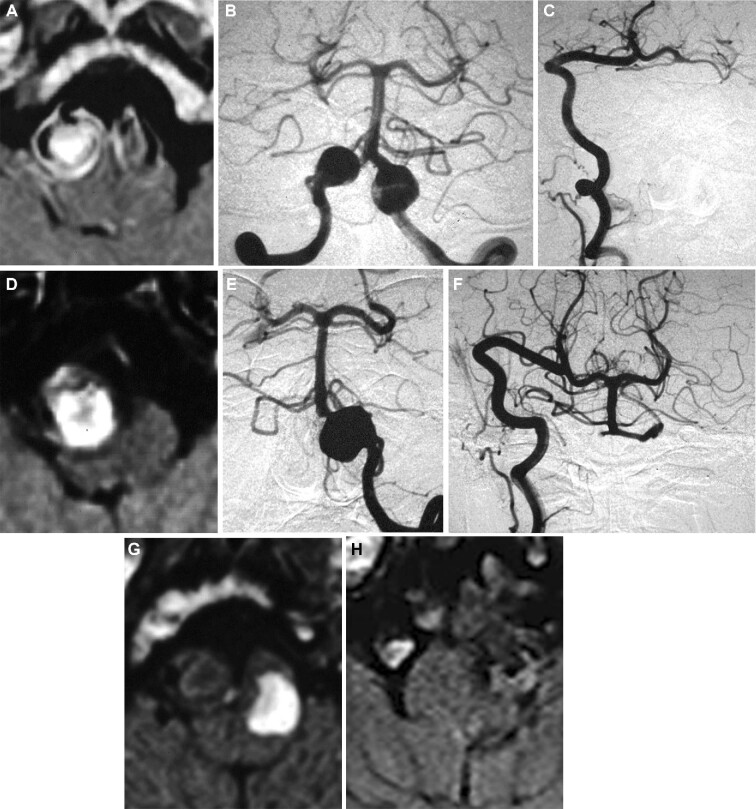
FIGURE 7.
Imaging studies of a case of remote proximal parent artery occlusion (RPO) with high-flow radial artery graft bypass from the extracranial vertebral artery (VA) to the posterior cerebral artery (PCA) (see Video, Supplemental Digital Content, first case). A 57-yr-old man presented with brainstem compression syndrome caused by bilateral large fusiform and dolichoectatic aneurysms of the vertebrobasilar junction (VBJ-BLFDA). A, Preoperative T1-weighted MR image with gadolinium demonstrated significant brainstem compression by the aneurysm. B, Preoperative bilateral vertebral angiogram revealing VBJ-BLFDA. C, Postoperative angiogram showing disappearance of the right VA aneurysm and patent extracranial VA to PCA bypass. D, Postoperative T1-weighted MR image showing complete thrombosis of the right VA aneurysm. Follow-up angiogram revealed slight enlargement of the left VA aneurysm E, so was occluded with a balloon at the C1-2 level 9 mo after initial treatment F. G, Postendovascular occlusion MR image revealed successful left VA aneurysm thrombosis. H, Recent T1-weighted MR image showed stable aneurysms on both sides at 184 mo after the treatment.

