Figure 3.

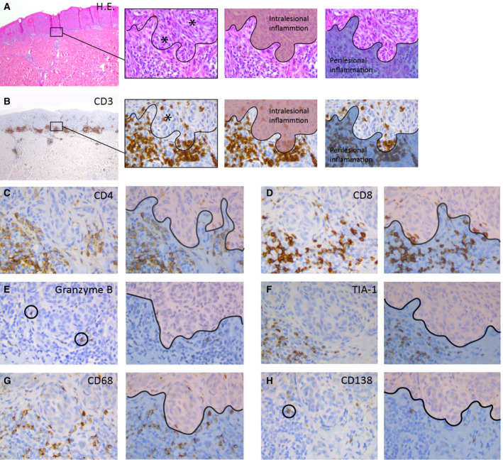
Immunohistochemical phenotypes in spitzoid melanocytic neoplasms. A, The left image shows a compound Spitz naevus (SN) used in this study with a symmetrical architecture and sharp lateral demarcation as well as circumscription towards the dermal depth. There is some intralesional inflammation as well as perilesional inflammatory aggregates at the base of the lesion. The second image from the left depicts a magnification from the left image and illustrates the two pathognomonic spitzoid cell types with epithelioid and spindle‐shaped melanocytes containing enlarged sharply demarcated nuclei with prominent nucleoli (black asterisks). The black curvilinear line demarcates the ‘grenz zone’ between intra‐ and perilesional inflammation. The two consecutive images accentuate the intralesional (highlighted in red) and perilesional (highlighted in blue) inflammatory areas. B, Left image with CD3 immunohistochemistry (IHC, brown staining) for the SN from (A). The three right images are magnifications from the left image and illustrate individual inflammatory CD3+ T cells in relation to the spitzoid epithelioid and spindle‐shaped melanocytes (blue‐stained nuclei). CD3+ T lymphocytes showed frequent peri‐ and intralesional presence (black asterisk, second image from the left). Intralesional inflammation is highlighted in red and perilesional areas are highlighted in blue in the two images from the right. The inflammatory pattern (IP) corresponds to an IP2 score (see also Figure 1A). C, The CD4 IHC staining depicts T helper lymphocytes and monocytes with a smudgy membranous accentuation (brown staining). CD4+ immune cells were frequently present in perilesional areas (highlighted with blue in the adjacent right image), but less often in intralesional zones (highlighted with red in the adjacent right image). D, There was a high amount of solitary infiltrating or clustering CD8+ T lymphocytes with circumscribed membranous staining (brown) with peri‐ (highlighted with blue in the adjacent right image) and intralesional localisation (highlighted with red in the adjacent right image). E, Rarely cytotoxic granzyme B+ T lymphocytes (encircled with black) were detected with granular cytoplasmic staining in perilesional (highlighted with blue in the adjacent right image) and intralesional (highlighted with red in the adjacent right image) localisation. F, TIA‐1 positive T lymphocytes with granular cytoplasmic staining (brown) were regularly present in the inflammatory infiltrate in perilesional areas (highlighted with blue in adjacent right image), but only rarely infiltrated within the individual spitzoid melanocytes (highlighted with red in adjacent right image). G, CD68+ histiocytes with smudgy cytoplasmic and membranous staining (brown) were presents in intralesional (highlighted with red in adjacent right image) and perilesional (highlighted with blue in adjacent right image) areas. H, Rarely a CD138+ plasma cell (encircled) with granular cytoplasmic staining and membranous accentuation (brown) was present in perilesional areas (highlighted with blue in adjacent right image) in the spitzoid melanocytic lesions. [Colour figure can be viewed at wileyonlinelibrary.com]
