

Abstract
Altered metabolism is a hallmark of cancer, and the reprogramming of energy metabolism has historically been considered a general phenomenon of tumors. It is well recognized that long noncoding RNAs (lncRNAs) regulate energy metabolism in cancer. However, lncRNA‐mediated posttranslational modifications and metabolic reprogramming are unclear at present. In this review, we summarized the current understanding of the interactions between the alterations in cancer‐associated energy metabolism and the lncRNA‐mediated posttranslational modifications of metabolic enzymes, transcription factors, and other proteins involved in metabolic pathways. In addition, we discuss the mechanisms through which these interactions contribute to tumor initiation and progression, and the key roles and clinical significance of functional lncRNAs. We believe that an in‐depth understanding of lncRNA‐mediated cancer metabolic reprogramming can help to identify cellular vulnerabilities that can be exploited for cancer diagnosis and therapy.
Keywords: cancer metabolism, enzyme, long noncoding RNA, metabolic reprogramming, posttranslational modification
Abbreviations
- 1,3‐BPG
1,3‐bisphosphoglycerate
- 3‐PG
3‐phosphoglycerate
- AGPG
actin gamma 1 pseudogene
- AML
acute myeloid leukaemia
- AMPK
adenosine monophosphate‐activated protein kinase
- ANRIL
antisense noncoding RNA in the INK4 locus
- ATG
autophagy‐related gene 5
- ATP
adenosine‐triphosphate
- BRK
breast tumor kinase
- CamK‐A
calcium‐dependent kinase activation
- CASC9
cancer susceptibility candidate 9
- CDK6
cyclin‐dependent kinase 6
- ceRNA
competing endogenous RNA
- circRNA
circular RNA
- CRC
colorectal cancer
- DHX9
DEAH (Asp‐Glu‐Ala‐ His) box helicase proteins 9
- DRAIC
downregulated RNA in cancer
- ENO1
enolase 1
- F‐2,6‐BP
fructose‐2,6‐bisphosphate
- F6P
fructose 6‐phosphate
- FEZF1‐AS1
Fez family zinc finger protein 1 antisense ribonucleic acid 1
- FGFR1
fibroblast growth factor receptor type 1
- FUS
fused in sarcoma
- G3BP1
Ras‐GTPase‐activating protein‐binding protein 1
- GBCDRlnc1
gallbladder cancer drug resistance‐associated lncRNA1
- GDH1
glutamate dehydrogenase 1
- GLCC1
glycolysis‐associated lncRNA of colorectal cancer 1
- GLS‐AS
antisense lncRNA of glutaminase
- GLUT1
glucose transporter isoform 1
- HCC
hepatocellular carcinoma
- HIF‐1α
hypoxia‐inducible factor‐1α
- HISLA
HIF‐1α‐stabilizing lncRNA
- HK2
hexokinase 2
- HNF4α
hepatocyte nuclear factor 4 alpha
- HSP90
heat shock protein 90
- HULC
highly up‐regulated in liver cancer
- IGF2BP2
insulin‐like growth factor 2 mRNA‐binding protein 2
- IKK
IκB kinase
- IκBα
inhibitor kappa B alpha
- KRT19P3
keratin 19 pseudogene 3
- LDHA
lactate dehydrogenase A
- lincRNA
long intergenic noncoding RNA
- LINK‐A
long intergenic noncoding RNA for kinase activation
- LINP1
lncRNA in nonhomologous end joining pathway 1
- LINRIS
long intergenic noncoding RNA for IGF2BP2 stability
- LKB1
liver kinase B1
- lncRNA
long noncoding RNA
- LRRK2
leucine‐rich repeat kinase 2
- m6A
N6‐methyladenosine
- MACC1
metastasis‐associated in colon cancer‐1
- MALAT1
metastasis‐associated lung adenocarcinoma transcript 1
- MDM2
murine double minute 2
- MEG3
maternally expressed gene 3
- miRNA
microRNA
- mTOR
mammalian target of rapamycin
- NADH
nicotinamide adenine dinucleotide
- NADPH
nicotinamide adenine dinucleotide phosphate
- NBR2
neighbour of BRCA1 gene 2
- NF‐κB
nuclear factor κB
- NSCLC
non‐small cell lung cancer
- OXPHOS
oxidative phosphorylation
- PFK1
phosphofructokinase‐1
- PFKFB3
6‐phosphofructo‐2‐kinase/fructose‐2,6‐bisphosphatase 3
- PGK1
phosphoglycerate kinase 1
- PHD2
proline hydroxylase domain 2
- PI3K
phosphoinositide‐3‐kinase
- PKM2
pyruvate kinase muscle isozyme M2
- PNCK
pregnancy upregulated non‐ubiquitous calmodulin kinase
- PRAL
p53 regulation‐associated lncRNA
- SIRT1
Sirtuin 1
- TCA
tricarboxylic acid
- VHL
von Hippel‐Lindau
- α‐KG
alpha‐ketoglutarate
1. BACKGROUND
Altered metabolism is a hallmark of cancer, and the reprogramming of energy metabolism has historically been considered a general phenomenon of tumors [1, 2, 3]. Specifically, tumor cells usually undergo energy metabolic reprogramming to satisfy the requirements for proliferation and survival in unfavourable environments, ultimately leading to cancer progression [4]. In addition to energy, tumor cells also require building blocks, such as the cofactors nicotinamide adenine dinucleotide phosphate (NADPH) and nicotinamide adenine dinucleotide (NADH), to support the housekeeping processes, growth, and proliferation in a dynamic microenvironment [5, 6]. Malignant tumor cells exhibit a highly regulated programme of metabolic plasticity [7]. Metabolic reprogramming and malignant transformation are complex and multistep processes that are associated with the accumulation of numerous molecular alterations [8]. On the one hand, altered metabolic signalling networks are driven by aberrant genetic alterations, including mutations, loss of heterozygosity, deletions, insertions, aneuploidy, etc., that lead to the activation of oncogenes and inactivation of tumor suppressors [9, 10]. On the other hand, epigenetic mechanisms, including DNA and histone methylation, noncoding RNA regulation, posttranslational modifications, etc., are also critical mediators of the functional changes that drive and maintain the malignant phenotypes and metabolic reprogramming [11, 12, 13].
Long noncoding RNAs (lncRNAs) are a heterogeneous group of non‐protein‐coding transcripts with the lengths of greater than 200 nucleotides [14, 15, 16]. LncRNAs are emerging regulators that are involved in the gene expression, diverse physiological and pathological processes [12, 17, 18]. Increasing evidence shows that lncRNAs play complex and precise regulatory roles in cancer initiation and progression by acting as oncogenes or tumor suppressors [19, 20]. They can regulate not only the proliferation, differentiation, invasion, and metastasis but also metabolic reprogramming of cancer cells [12, 21, 22]. Moreover, accumulating evidence suggests that lncRNAs play important functional roles in modulating the transcription and translation of metabolism‐related genes, serving as decoys, scaffolds, competing endogenous RNAs (ceRNAs), etc., ultimately leading to cancer metabolic reprogramming [23, 24]. In addition, recent studies by our group and other groups also suggested that lncRNAs promote energy metabolism and cancer progression through posttranslational modifications of key metabolism‐related proteins, including the ubiquitination, phosphorylation, and acetylation [25, 26, 27, 28, 29].
In this review, we discuss the significant roles of lncRNAs in the reprogramming of energy metabolism and the progression of cancers. In particular, we focus on lncRNA‐mediated posttranslational modifications of metabolic enzymes, metabolism‐related transcription factors, and other proteins in relevant signalling pathways, highlighting recent important insights into the mechanisms regulating cancer metabolism.
2. LncRNA‐MEDIATED POSTTRANSLATIONAL MODIFICATION OF METABOLIC ENZYMES
Tumor cell metabolism is reprogrammed from oxidative phosphorylation (OXPHOS) to aerobic glycolysis even when oxygen is sufficient, a phenomenon called the “Warburg effect” [30]. Therefore, the abnormal expression and regulation of related enzymes in the glycolytic pathway are vital for tumor development [31]. As shown in Figure 1 and Supplementary Table S1, we first summarize the available studies on lncRNA‐mediated posttranslational modifications of several metabolic enzymes in aerobic glycolysis. The relationship between lncRNAs and metabolic enzymes is complicated and flexible, because they can influence each other. The specific mechanisms of their actions and relationship deserve in‐depth studies in the future.
FIGURE 1.
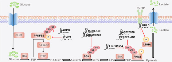
LncRNA‐mediated posttranslational modification of metabolic enzymes. LncRNA‐mediated posttranslational modifications directly affect metabolic enzymes to reprogram glucose metabolism. Examples are phosphorylation and ubiquitination of PFKFB3 by AGPG and YIYA; ubiquitination of PGK1 by MetaLnc9 and GBCDRlnc1; ubiquitination of PKM2 by AC020978, FEZF1‐AS1, and LINC01554; and phosphorylation of PKM2 and LDHA by HULC. LncRNA: long noncoding RNA; PFKFB3: 6‐phosphofructo‐2‐kinase/fructose‐2,6‐bisphosphatase 3; AGPG: actin gamma 1 pseudogene; PGK1: phosphoglycerate kinase 1; PKM2: pyruvate kinase muscle isozyme M2; FEZF1‐AS1: Fez family zinc finger protein 1 antisense ribonucleic acid 1; LDHA: lactate dehydrogenase A; HULC: highly up‐regulated in liver cancer; P, phosphorylation; Ub, ubiquitination; GLUT: glucose transporter isoform; HK2: hexokinase 2; F6P: fructose 6‐phosphate; F‐1,6‐BP: fructose‐1,6‐bisphosphate;F‐2,6‐BP: fructose‐2,6‐bisphosphate; 1,3‐BPG: 1,3‐bisphosphoglycerate; 3‐PG: 3‐phosphoglycerate; 2‐PG: 2‐phosphoglycerate; ENO: enolase; PEP: phosphoenolpyruvate
2.1. 6‐phosphofructo‐2‐kinase/fructose‐2,6‐bisphosphatase 3 (PFKFB3)
In aerobic glycolysis, PFKFB3 converts fructose 6‐phosphate (F6P) into fructose‐2,6‐bisphosphate (F‐2,6‐BP), which can act as an allosteric activator of the rate‐limiting enzyme phosphofructokinase‐1 (PFK1) [25]. Studies have identified that lncRNA‐mediated reprogramming of energy metabolism is closely related to the PFKFB3 ubiquitination and phosphorylation in cancer [25, 32]. Moreover, our recent study revealed that the lncRNA actin gamma 1 pseudogene (AGPG) specifically bound to the C‐terminal domain of PFKFB3, which stimulated the stable expression of PFKFB3 and activated the glycolytic flux by inhibiting the ubiquitination of PFKFB3 at Lys302 and its subsequent proteasome‐dependent degradation, leading to metabolic reprogramming in esophageal squamous cell carcinoma cells [25]. In addition, PFKFB3 could also be phosphorylated by the lncRNA YIYA (LINC00538), which was associated with cyclin‐dependent kinase 6 (CDK6) and facilitated its binding to cyclin D3. Thus, YIYA regulated the CDK6‐dependent phosphorylation of PFKFB3 in a cell cycle‐independent manner, which led to the enhanced conversion of F6P to F‐2,6‐BP and promoted the reprogramming of glucose metabolism and the growth of breast cancer [32].
2.2. Phosphoglycerate kinase 1 (PGK1)
PGK1 is the first crucial metabolic enzyme mediating adenosine‐triphosphate (ATP) production via glycolysis and catalysing the conversion of 1,3‐bisphosphoglycerate (1,3‐BPG) to 3‐phosphoglycerate (3‐PG) [33]. The activity of PGK1 is mediated by different posttranslational modifications. For example, PGK1 can frequently be phosphorylated, ubiquitinated, and acetylated at multiple sites to regulate tumor metabolism and cancer progression [34, 35, 36, 37]. A recent study showed that MetaLnc9 (LINC00963) played a dominant role in the posttranslational activity of PGK1 and stabilized PGK1 by blocking its ubiquitin‐mediated degradation, allowing it to exert its carcinogenic activity and accelerate the metastasis of non‐small cell lung cancer (NSCLC) cells via the phosphoinositide‐3‐kinase (PI3K)/AKT/mammalian target of rapamycin (mTOR) signalling [36]. Similarly, gallbladder cancer drug resistance‐associated lncRNA1 (GBCDRlnc1), which served as a “decoy molecule”, posttranslationally regulated the level of PGK1 by directly preventing its ubiquitin‐mediated degradation in gallbladder cancer. Expression of the autophagy initiator autophagy‐related gene 5 (ATG5)‐ATG12 conjugate was ultimately up‐regulated through the formation of the GBCDRlnc1/PGK1/ATG5‐ATG12 complex, which induced autophagy and drug resistance in gallbladder cancer cells [37].
2.3. Pyruvate kinase muscle isozyme M2 (PKM2)
PKM2 catalyses the conversion of phosphoenolpyruvate to pyruvate and generates ATP in the rate‐limiting step in glycolysis, which switches glucose metabolism from the normal respiratory chain to lactate production in tumor cells [38, 39]. Coincidentally, PKM2 activity is also regulated by posttranslational modifications, including O‐GlcNAcylation, acetylation, phosphorylation, ubiquitination, etc [40, 41, 42, 43, 44, 45]. The lncRNAs AC020978, Fez family zinc finger protein 1 antisense ribonucleic acid 1 (FEZF1‐AS1), and LINC01554 can reprogram tumor metabolism by regulating the stability of PKM2 via the ubiquitin‐mediated proteasome pathway. In detail, AC020978 specifically modulated the stability of the PKM2/hypoxia‐inducible factor‐1α (HIF‐1α) complex, furthered the action of HIF‐1α on its target genes, thus forming a positive feedback loop related to tumor‐specific metabolism and endowing NSCLC cells with an aggressive phenotype [40]. Additionally, FEZF1‐AS1 bound to and increased the stability of PKM2, increasing cytoplasmic and nuclear PKM2 levels. Thus, FEZF1‐AS1 promoted the aerobic glycolysis and accelerated the proliferation and metastasis of colorectal cancer (CRC) cells [41]. In contrast, the lncRNA LINC01554 accelerated ubiquitin‐mediated degradation of PKM2 to inhibit aerobic glycolysis in hepatocellular carcinoma (HCC) cells via the AKT/mTOR signalling pathway [42]. Moreover, the lncRNA highly up‐regulated in liver cancer (HULC) functioned as an adaptor molecule and enhanced the interactions of lactate dehydrogenase A (LDHA) and PKM2 with the intracellular domain of the upstream kinase fibroblast growth factor receptor type 1 (FGFR1), leading to the elevated phosphorylation of LDHA and PKM2, which consequently enhanced glycolysis and increased the proliferation of liver cancer cells [46].
3. LncRNA‐MEDIATED POSTTRANSLATIONAL MODIFICATION OF TRANSCRIPTION FACTORS
In addition to directly regulating energy metabolism via posttranslational modification of metabolic enzymes, lncRNAs can also regulate metabolism‐related transcription factors to reprogram energy metabolism since the regulation of transcription factors upon their targets is influenced by lncRNAs [47]. LncRNAs regulate the activity of these proteins through hydroxylation, phosphorylation, and ubiquitination, thus enhancing the expression of metabolism‐related enzymes and promoting the malignant progression of tumors (Figure 2 and Supplementary Table S1). Therefore, we next discuss the recently reported lncRNAs and the mechanisms by which they regulate representative factors, including HIF‐1α, c‐Myc, and nuclear factor κB (NF‐κB).
FIGURE 2.
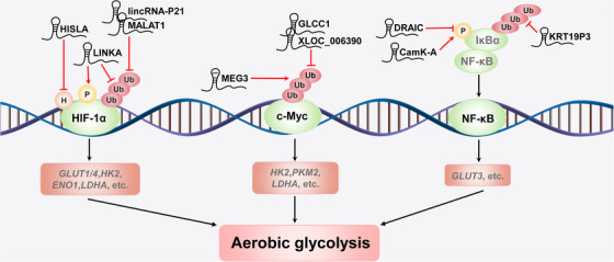
LncRNA‐mediated posttranslational modification of transcription factors. LncRNA‐mediated posttranslational modification of transcription factors regulates aerobic glycolysis‐related genes. Examples are hydroxylation, phosphorylation, and ubiquitination of HIF‐1α by HISLA, LINK‐A, lincRNA‐p21, and MALAT1, respectively; ubiquitination of c‐Myc by MEG3, GLCC1, and XLOC_006390; and phosphorylation and ubiquitination of NF‐κB by CamK‐A, KRT19P3, and DRAIC. LncRNA: long noncoding RNA; HIF‐1α: hypoxia‐inducible factor‐1α; HISLA: HIF‐1α‐stabilizing lncRNA; LINK‐A: long intergenic noncoding RNA for kinase activation; lincRNA: long intergenic noncoding RNA; MALAT1: metastasis‐associated lung adenocarcinoma transcript 1; MEG3: maternally expressed gene 3; GLCC1: glycolysis‐associated lncRNA of colorectal cancer 1; NF‐κB: nuclear factor κB; CamK‐A: lncRNA for calcium‐dependent kinase activation; KRT19P3: keratin 19 pseudogene 3; DRAIC: downregulated RNA in cancer; H: hydroxylation; P, phosphorylation; Ub, ubiquitination; GLUT: glucose transporter isoform; HK2: hexokinase 2; ENO1: enolase 1; LDHA: lactate dehydrogenase A; PKM2: pyruvate kinase muscle isozyme M2
3.1. HIF‐1α
HIF‐1α plays a key role in the reprogramming of cancer metabolism by activating the transcription of genes that encode glucose transporters and glycolytic enzymes [48, 49]. Accumulating evidence suggests that lncRNAs play important functional roles in regulating HIF‐1α activity and stability. For example, Su et al. [50] found that the lncRNA cancer susceptibility candidate 9 (CASC9) reprogramed glycolytic metabolism and stimulated the tumorigenic behaviour of nasopharyngeal cancer cells by interacting with HIF‐1α and increasing the abundance of HIF‐1α. Chen et al. [51] found that HIF‐1α‐stabilizing lncRNA (HISLA) inhibited the hydroxylation and degradation of HIF‐1α and promoted the aerobic glycolysis in breast cancer cells by blocking the interaction between proline hydroxylase domain 2 (PHD2) and HIF‐1α. More interestingly, by activating breast tumor kinase (BRK) phosphorylation at Tyr351, long intergenic noncoding RNA for kinase activation (LINK‐A) mediated HIF‐1α phosphorylation at Tyr565, which prevented the degradation of HIF‐1α under normoxic conditions; moreover, leucine‐rich repeat kinase 2 (LRRK2) recruited by LINK‐A could also phosphorylate HIF‐1α at Ser797, which led to the activation of HIF‐1α target genes. The above series of LINK‐A‐mediated posttranslational modifications promoted glycolytic reprogramming and tumorigenesis in triple‐negative breast cancer [52].
Under normoxic conditions, HIF‐1α is hydroxylated by PHD at Pro402 and/or Pro564 [53]. After hydroxylation, HIF‐1α binds to von Hippel‐Lindau (VHL) and undergoes VHL‐mediated ubiquitination and degradation via the proteasome pathway. However, the activity of PHD is suppressed under hypoxic conditions. A recent study reported that accumulation of HIF‐1α resulted in high expression of PHD, which rapidly induced transcription of long intergenic noncoding RNA (lincRNA)‐p21 [54]. Interestingly, highly expressed lincRNA‐p21 removed the polyubiquitin chain at Hyp564 installed by VHL and decreased HIF‐1α degradation via competitively binding with HIF‐1α. As discussed above, the formation of a positive HIF‐1α‐lincRNA‐p21‐HIF‐1α feedback loop ensured that HIF‐1α was highly induced in hypoxia, activating glucose transporter isoform 1 (GLUT1) and LDHA and enhancing the anaerobic glycolysis pathway, which promoted tumor growth [54]. Similarly, the lncRNA metastasis‐associated lung adenocarcinoma transcript 1 (MALAT1) increased the expression of core glycolytic enzymes, including hexokinase 2 (HK2), enolase 1 (ENO1), and GLUT4, by reducing the VHL‐mediated ubiquitination and subsequent degradation of HIF‐1α [20, 55].
3.2. c‐Myc
The c‐Myc protein acts as a “master regulator” of cellular metabolism and cancer progression by stimulating many metabolic changes that result in malignant transformation [56]. Additionally, lncRNAs have been reported to be involved in the regulation of cancer energy metabolism via the posttranslational modification of c‐Myc. Several lncRNAs are significantly overexpressed and function as oncogenes in CRC or pancreatic cancer, including glycolysis‐associated lncRNA of colorectal cancer 1 (GLCC1), long intergenic noncoding RNA for insulin‐like growth factor 2 mRNA‐binding protein 2 stability (LINRIS), and lncRNA XLOC_006390. Mechanistically, the lncRNA GLCC1 protected c‐Myc from ubiquitination by directly interacting with the chaperone heat shock protein 90 (HSP90) and further specified the transcriptional modification pattern of c‐Myc target genes such as LDHA, consequently reprogramming glycolytic metabolism for CRC cell proliferation [57]. The lncRNA LINRIS bound to insulin‐like growth factor 2 mRNA‐binding protein 2 (IGF2BP2), repressed its ubiquitination and prevented its degradation via the autophagy‐lysosome pathway. The LINRIS‐IGF2BP2 axis promoted the translation of its target gene c‐Myc and accelerated the malignant proliferation of CRC cells by up‐regulating aerobic glycolysis [26]. The lncRNA XLOC_006390 stablized c‐Myc by preventing its ubiquitination, increasing the expression of glutamate dehydrogenase 1 (GDH1), and subsequently stimulating the production of alpha‐ketoglutarate (α‐KG). Excess α‐KG supplied the tricarboxylic acid (TCA) cycle and facilitated glutamate metabolism, promoting pancreatic cancer growth [58].
In contrast, the lncRNA maternally expressed gene 3 (MEG3) and the lncRNA antisense lncRNA of glutaminase (GLS‐AS) were down‐regulated and functioned as tumor suppressors in cancer. Mechanistically, MEG3 overexpression enhanced ubiquitin‐dependent degradation of c‐Myc and repressed c‐Myc target genes involved in the glycolytic pathway, including LDHA, PKM2, and HK2 [59]. Similarly, the lncRNA GLS‐AS restricts glutaminase expression at the posttranscriptional level by binding with the glutaminase pre‐mRNA, resulting in the formation of a double‐stranded RNA. Furthermore, GLS‐AS reduce Myc expression by disrupting the glutaminase‐mediated stabilization of Myc. Conversely, in pancreatic cancer, Myc up‐regulated the expression of GLS‐AS under nutritional stress, thus forming a mutual feedback loop to inhibit metabolic reprogramming [60].
3.3. NF‐κB
NF‐κB is an inducible transcription factor that plays a core role in diverse biological processes, including cellular metabolism, epithelial‐to‐mesenchymal transition, invasion, angiogenesis, metastasis, therapeutic resistance, and other processes [61]. Aberrant NF‐κB signalling has been verified to be involved in the pathogenesis of most human cancers [62]. Direct regulation of cell metabolism via the NF‐κB pathway has been addressed by several recent studies [63, 64]. In particular, our recent study validated that lncRNA for calcium‐dependent kinase activation (CamK‐A) regulated Ca2+ signalling‐mediated remodelling of tumor microenvironment and reprogramming of energy metabolism through posttranslational modification and NF‐κB activation [27]. Mechanistically, CamK‐A bound both the calmodulin‐dependent kinase pregnancy up‐regulated non‐ubiquitous calmodulin kinase (PNCK) and the C‐terminal SH3‐like region of inhibitor kappa B alpha (IκBα). Thus, CamK‐A promotes PNCK‐induce phosphorylation of IκBα at Ser32 under hypoxic conditions, which further induce IκBα degradation and NF‐κB activation. In contrast, several NF‐κB‐related lncRNAs are down‐regulated and function as tumor suppressors during cancer progression. For example, Saha et al. [65] reported that the downregulated RNA in cancer (DRAIC) interacted with the subunits of the IκB kinase (IKK) complex and subsequently inhibited prostate cancer progression by repressing IκBα phosphorylation and NF‐κB activation. Additionally, the lncRNA keratin 19 pseudogene 3 (KRT19P3) inhibited gastric cancer cell growth and metastasis by modulating IκBα deubiquitination and inactivating NF‐κB signalling [66].
4. LncRNA‐MEDIATED POSTTRANSLATIONAL MODIFICATION OF OTHER PROTEINS INVOLVED IN METABOLIC PATHWAYS
Alterations in proteins involved in metabolism are accompanied by alterations in metabolic pathways, which will eventually become carcinogenic signalling pathways or be activated by carcinogenic factors [8]. Through these abnormal metabolic pathways, tumor cells can obtain the necessary materials and energy required for continuous proliferation [67]. In addition to directly regulating the posttranslational modification of key metabolic enzymes, lncRNAs can also indirectly modulate metabolic pathways through posttranslational modifications (Figure 3 and Supplementary Table S1). Among the observed effects, functional alterations in the adenosine monophosphate‐activated protein kinase (AMPK) pathway, PI3K/AKT pathway and p53 pathway are particularly prominent.
FIGURE 3.
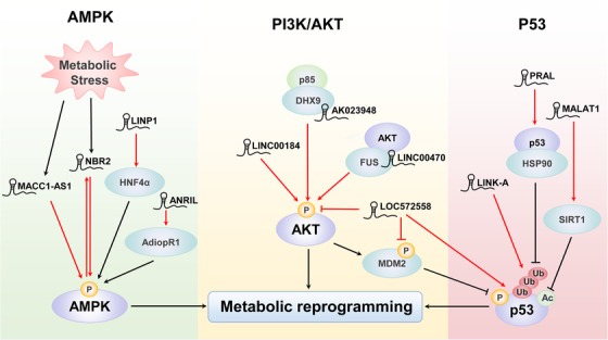
LncRNA‐mediated posttranslational modification of other proteins involved in metabolic pathways. LncRNA‐mediated posttranslational modifications of metabolic pathways include phosphorylation of AMPK signalling pathway components by MACC1‐AS1, NBR2, LINP1, and ANRIL; phosphorylation of PI3K/AKT signalling pathway components by LINC00184, AK023948, LINC00470, and LOC572558; and phosphorylation, ubiquitination, and acetylation of p53 signalling pathway components by LOC572558, LINK‐A, PRAL, and MALAT1. LncRNA: long noncoding RNA; AMPK: adenosine monophosphate‐activated protein kinase; MACC1: metastasis‐associated in colon cancer‐1; NBR2: neighbour of BRCA1 gene 2; LINP1: lncRNA in nonhomologous end joining pathway 1; ANRIL: antisense noncoding RNA in the INK4 locus; PI3K: phosphoinositide‐3‐kinase; LINK‐A: long intergenic noncoding RNA for kinase activation; PRAL: p53 regulation‐associated lncRNA; MALAT1: metastasis‐associated lung adenocarcinoma transcript 1; Ac, acetylation; P, phosphorylation; Ub, ubiquitination; HNF4α: hepatocyte nuclear factor 4 alpha; DHX9: DEAH (Asp‐Glu‐Ala‐ His) box helicase proteins 9; FUS: fused in sarcoma; MDM2: murine double minute 2; HSP90: heat shock protein 90; SIRT1: Sirtuin 1
4.1. AMPK signalling pathway
AMPK is a critical cellular energy sensor that mainly functions as a metabolic checkpoint to restore energy balance in response to different types of metabolic stress [68]. AMPK is activated by phosphorylation, which maintains energy balance, redox homeostasis and cell survival by regulating glycolysis, fatty acid metabolism, antioxidant reactions, and other processes [69, 70]. Recent studies have showed that AMPK phosphorylation is epigenetically regulated by different signalling pathways involving different lncRNAs, including metastasis‐associated in colon cancer‐1 (MACC1)‐AS1, antisense noncoding RNA in the INK4 locus (ANRIL), lncRNA in nonhomologous end joining pathway 1 (LINP1) and neighbour of BRCA1 gene 2 (NBR2). MACC1‐AS1 is the antisense lncRNA transcript of the transcriptional regulator MACC1, which is recognized as an oncogene in cancer. MACC1‐AS1 mediated metabolic plasticity by up‐regulating MACC1 and subsequently enhanced glycolysis and antioxidative capabilities via the phosphorylation of AMPK and the translocation of Lin28 (an evolutionarily conserved RNA‐binding protein), ultimately accelerating the growth of gastric cancer [71]. In addition, the lncRNA ANRIL promoted the survival of malignant cells and cellular glucose metabolism, accelerating acute myeloid leukaemia (AML) progression through AMPK phosphorylation and forming the AdipoR1/AMPK/Sirtuin 1 (SIRT1) pathway [72]. In addition, the lncRNA LINP1 regulated AML progression via increasing AMPK phosphorylation mediated through the hepatocyte nuclear factor 4 alpha (HNF4α)/AMPK/WNT5A signalling pathway [73]. In contrast, Liu et al. [74] proposed a feed‐forward model of NBR2‐AMPK regulation, in which the lncRNA NBR2 was induced by the liver kinase B1 (LKB1)‐AMPK pathway under energy stress, and in turn NBR2 interacted with AMPK and promoted its phosphorylation.
4.2. PI3K/AKT signalling pathway
PI3K/AKT plays a key role in diverse cellular processes in cancer, including cell survival, proliferation, angiogenesis, and energy metabolism [75, 76]. Evidence has revealed that numerous positive regulators, including growth factors, regulatory proteins, and microRNAs (miRNAs), promote the overactivation of AKT signalling [77]. Some lncRNAs, such as LOC572558, LINC00184, LINC00470 (also known as C18orf2) and AK023948, have been associated with AKT phosphorylation and energy metabolic reprogramming in diseases, especially in cancer. One study showed that LINC00184 modulated glycolysis and mitochondrial oxidative phosphorylation in oesophageal cancer cells through inducing AKT phosphorylation [78]. In addition, Liu et al. [79] demonstrated that LINC00470 was a positive regulator of AKT activation in glioblastoma cells. Mechanistically, LINC00470 bound to the RNA‐binding protein fused in sarcoma (FUS) and AKT, forming a ternary complex that anchors FUS in the cytoplasm and increases AKT phosphorylation. AKT activation by LINC00470 inhibited HK1 ubiquitination and promoted aerobic glycolysis and glioblastoma tumorigenesis. AK023948 is also reported to function as a positive regulator of AKT. AK023948 was required for the interaction between DEAH (Asp‐Glu‐Ala‐ His) box helicase proteins 9 (DHX9) and p85 to enhance p85 stability and promote AKT activity, which may contribute to breast tumor progression [80]. Overexpression of LOC572558, however, resulted in the dephosphorylation of AKT and murine double minute 2 (MDM2) while the phosphorylation of p53, which inhibited cell proliferation and tumor growth in bladder cancer through the AKT‐MDM2‐p53 axis [81].
4.3. p53 signalling pathway
In addition to the functions of the above basic metabolic pathways, the functions of p53‐related signalling pathways in metabolism cannot be ignored [82, 83]. Kruiswijk et al. [84] proposed that cell metabolism was a noncanonical p53‐controlled process, and this idea had attracted considerable attention. Importantly, activation of p53 reprograms tumor glucose metabolism by reversing the Warburg effect, which negatively affected the carcinogenic metabolism of cancer cells and prevented the development of aggressive tumor phenotypes [84, 85]. Recently, the importance of lncRNAs and p53 in cancer metabolic reprogramming has been confirmed [29, 86]. Mao et al. [87] confirmed that the interaction between the lncRNA P53RRA and Ras‐GTPase‐activating protein‐binding protein 1 (G3BP1) prevented p53 from forming the G3BP1 complex, which repressed the transcription of several metabolism‐related genes. In addition, various lncRNAs, such as LOC572558, p53 regulation‐associated lncRNA (PRAL), LINK‐A and MALAT1, also regulated p53 through the posttranslational modifications. In terms of ubiquitination, PRAL enhanced the stability of p53 by competitively binding with it and blocking its MDM2‐dependent degradation. The three stem‐loop motifs at the 5' end of PRAL promoted the interaction between p53 and HSP90 [88]. However, LINK‐A enhanced the K48‐polyubiquitination‐mediated degradation of p53 [89]. In addition, Chen et al. [90] suggested that MALAT1 suppressed cell apoptosis and promoted cell proliferation by enhancing the SIRT1‐mediated deacetylation of p53 and then down‐regulating its downstream target genes. Together, the above findings indicate that lncRNA‐mediated posttranslational modifications are crucial to the p53‐related metabolic pathways.
5. CONCLUSIONS
The discovery and functional studies of metabolism‐associated lncRNAs expand our understanding of the regulatory mechanisms in cancer. Importantly, the functions of these identified lncRNAs are critical to tumorigenesis, tumor progression, and metabolic reprogramming. As technological advances have flourished in the past decade, understanding the precise contributions of lncRNA‐mediated metabolic reprogramming has become easier. In this review, we highlight the flexible and complex energy metabolism network that is controlled by lncRNA‐mediated posttranslational modifications, one of the most rapid manners by which cells respond to external and internal stimuli, and is a hub of many cellular signalling pathways. Posttranslational modifications alter proteins by adding or removing different functional groups, thereby modulating the protein structure and function. In addition, the suite of epigenetic modifications mediated by lncRNAs is complex and diverse, and includes ubiquitination, phosphorylation and acetylation, all of which can lead to the reprogramming of energy metabolism. On the contrary, RNA epigenetic modification and its biological significance, such as N6‐methyladenosine (m6A), are emerging research frontiers in tumor biology and deserve extensive study. In general, the current research on this topic in cancer is limited, and the existing knowledge has not been fully translated into clinical applications. However, the current findings offer new scientific ideas and perspectives on cancer energy metabolism.
Recent studies of cancer metabolism have focused on assessing metabolic phenotypes in the tumor microenvironment, especially the tumor immune microenvironment. These studies have expanded the scope of metabolic dependencies beyond the classical pathways that dominate metabolism in cultured cells. Accumulating evidence from recent studies indicates that alterations in the tumor microenvironment can drive tumorigenesis. In addition, studies have indicated that cell metabolism influences T cell antitumor activity, and strategies to modulate metabolism can improve the tumor immune response. We believe that lncRNA‐mediated metabolic reprogramming also plays critical roles in remodelling of the tumor microenvironment and in tumor immune escape. In addition to lncRNA, the role of circular RNAs (circRNAs) in tumor metabolic remodelling and immune microenvironment has also attracted more attention. Thus, identification of these functional ncRNAs, and investigation of their biological roles and underlying regulation mechanisms in tumor microenvironment remodelling need to be further studied.
Taken together, in‐depth clarification of the common lncRNA‐mediated regulatory mechanisms in metabolic reprogramming will provide a deeper understanding of tumor development and cellular vulnerabilities. Moreover, the involved lncRNAs can also be exploited as reliable diagnostic biomarkers and therapeutic targets in cancer.
ETHICS APPROVAL AND CONSENT TO PARTICIPATE
Not applicable.
CONSENT FOR PUBLICATION
Not applicable.
AVAILABILITY OF DATA AND MATERIALS
Not applicable.
COMPETING INTERESTS
The authors declare that they have no competing interests.
FUNDING
This study was supported by the National Key R&D Program of China (2018YFC1313304 and 2018YFC1313300) and the National Natural Science Foundation of China (82073112, 82022052 and 81871951).
AUTHORS' CONTRIBUTIONS
Conceptualization: HQJ and RHX. Article writing and editing: YTT, JFL, TL, JJL and HQJ. Supervision: HQJand RHX. All authors read and approved the final manuscript.
Supporting information
Supporting information.
ACKNOWLEDGEMENTS
Not applicable.
Tan Y‐T, Lin J‐F, Li T, Li J‐J, Xu R‐H, Ju H‐Q. LncRNA‐mediated posttranslational modifications and reprogramming of energy metabolism in cancer. Cancer Commun. 2021;41:109–120. 10.1002/cac2.12108
Contributor Information
Rui‐Hua Xu, Email: xurh@sysucc.org.cn.
Huai‐Qiang Ju, Email: juhq@sysucc.org.cn.
REFERENCES
- 1. Faubert B, Solmonson A, DeBerardinis RJ. Metabolic reprogramming and cancer progression. Science. 2020;368(6487). 10.1126/science.aaw5473. [DOI] [PMC free article] [PubMed] [Google Scholar]
- 2. Chen PH, Cai L, Huffman K, Yang C, Kim J, Faubert B, et al. Metabolic diversity in human non‐small cell lung cancer cells. Mol Cell. 2019;76(5):838‐51 e5 10.1016/j.molcel.2019.08.028. [DOI] [PMC free article] [PubMed] [Google Scholar]
- 3. Wang Y, Xia Y, Lu Z. Metabolic features of cancer cells. Cancer Commun (Lond). 2018;38(1):65 10.1186/s40880-018-0335-7. [DOI] [PMC free article] [PubMed] [Google Scholar]
- 4. Tian Y, Yang B, Qiu W, Hao Y, Zhang Z, Yang B, et al. ER‐residential Nogo‐B accelerates NAFLD‐associated HCC mediated by metabolic reprogramming of oxLDL lipophagy. Nat Commun. 2019;10(1):3391 10.1038/s41467-019-11274-x. [DOI] [PMC free article] [PubMed] [Google Scholar]
- 5. Ju HQ, Ying H, Tian T, Ling J, Fu J, Lu Y, et al. Mutant Kras‐ and p16‐regulated NOX4 activation overcomes metabolic checkpoints in development of pancreatic ductal adenocarcinoma. Nat Commun. 2017;8:14437 10.1038/ncomms14437. [DOI] [PMC free article] [PubMed] [Google Scholar]
- 6. Ju HQ, Lu YX, Chen DL, Zuo ZX, Liu ZX, Wu QN, et al. Modulation of Redox Homeostasis by Inhibition of MTHFD2 in Colorectal Cancer: Mechanisms and Therapeutic Implications. J Natl Cancer Inst. 2019;111(6):584‐96. 10.1093/jnci/djy160. [DOI] [PMC free article] [PubMed] [Google Scholar]
- 7. Xiao Z, Dai Z, Locasale JW. Metabolic landscape of the tumor microenvironment at single cell resolution. Nat Commun. 2019;10(1):3763 10.1038/s41467-019-11738-0. [DOI] [PMC free article] [PubMed] [Google Scholar]
- 8. DeBerardinis RJ, Chandel NS. Fundamentals of cancer metabolism. Sci Adv. 2016;2(5):e1600200 10.1126/sciadv.1600200. [DOI] [PMC free article] [PubMed] [Google Scholar]
- 9. Pavlova NN, Thompson CB. The Emerging Hallmarks of Cancer Metabolism. Cell Metab. 2016;23(1):27‐47. 10.1016/j.cmet.2015.12.006. [DOI] [PMC free article] [PubMed] [Google Scholar]
- 10. Vogelstein B, Kinzler KW. The Path to Cancer –Three Strikes and You're Out. N Engl J Med. 2015;373(20):1895‐8. 10.1056/NEJMp1508811. [DOI] [PubMed] [Google Scholar]
- 11. Michalak EM, Burr ML, Bannister AJ, Dawson MA. The roles of DNA, RNA and histone methylation in ageing and cancer. Nat Rev Mol Cell Biol. 2019;20(10):573‐89. 10.1038/s41580-019-0143-1. [DOI] [PubMed] [Google Scholar]
- 12. Ransohoff JD, Wei Y, Khavari PA. The functions and unique features of long intergenic non‐coding RNA. Nat Rev Mol Cell Biol. 2018;19(3):143‐57. 10.1038/nrm.2017.104. [DOI] [PMC free article] [PubMed] [Google Scholar]
- 13. Wang YP, Lei QY. Metabolic recoding of epigenetics in cancer. Cancer Commun (Lond). 2018;38(1):25 10.1186/s40880-018-0302-3. [DOI] [PMC free article] [PubMed] [Google Scholar]
- 14. Nair L, Chung H, Basu U. Regulation of long non‐coding RNAs and genome dynamics by the RNA surveillance machinery. Nat Rev Mol Cell Biol. 2020;21(3):123‐36. 10.1038/s41580-019-0209-0. [DOI] [PMC free article] [PubMed] [Google Scholar]
- 15. Kopp F, Mendell JT. Functional classification and experimental dissection of long noncoding RNAs. Cell. 2018;172(3):393‐407. 10.1016/j.cell.2018.01.011. [DOI] [PMC free article] [PubMed] [Google Scholar]
- 16. Berger AC, Korkut A, Kanchi RS, Hegde AM, Lenoir W, Liu W, et al. A comprehensive pan‐cancer molecular study of gynecologic and breast cancers. Cancer Cell. 2018;33(4):690‐705 e9 10.1016/j.ccell.2018.03.014. [DOI] [PMC free article] [PubMed] [Google Scholar]
- 17. Li Y, Zhang J, Huo C, Ding N, Li J, Xiao J, et al. Dynamic organization of lncRNA and circular RNA regulators collectively controlled cardiac differentiation in humans. EBioMedicine. 2017;24:137‐46. 10.1016/j.ebiom.2017.09.015. [DOI] [PMC free article] [PubMed] [Google Scholar]
- 18. Li Y, Jiang T, Zhou W, Li J, Li X, Wang Q, et al. Pan‐cancer characterization of immune‐related lncRNAs identifies potential oncogenic biomarkers. Nat Commun. 2020;11(1):1000 10.1038/s41467-020-14802-2. [DOI] [PMC free article] [PubMed] [Google Scholar]
- 19. Esposito R, Bosch N, Lanzos A, Polidori T, Pulido‐Quetglas C, Johnson R. Hacking the cancer genome: profiling therapeutically actionable long non‐coding RNAs using CRISPR‐Cas9 screening. Cancer Cell. 2019;35(4):545‐57. 10.1016/j.ccell.2019.01.019. [DOI] [PubMed] [Google Scholar]
- 20. Kim J, Piao HL, Kim BJ, Yao F, Han Z, Wang Y, et al. Long noncoding RNA MALAT1 suppresses breast cancer metastasis. Nature genetics. 2018;50(12):1705‐15. 10.1038/s41588-018-0252-3. [DOI] [PMC free article] [PubMed] [Google Scholar]
- 21. Huarte M. The emerging role of lncRNAs in cancer. Nat Med. 2015;21(11):1253‐61. 10.1038/nm.3981. [DOI] [PubMed] [Google Scholar]
- 22. Luo J, Langer LF, Liu J. A novel role of LncRNA in regulating tumor metabolism and angiogenesis under hypoxia. Cancer Commun (Lond). 2019;39(1):2 10.1186/s40880-019-0348-x. [DOI] [PMC free article] [PubMed] [Google Scholar]
- 23. Thomson DW, Dinger ME. Endogenous microRNA sponges: evidence and controversy. Nat Rev Genet. 2016;17(5):272‐83. 10.1038/nrg.2016.20. [DOI] [PubMed] [Google Scholar]
- 24. Boque‐Sastre R, Guil S. A lncRNA Decoy Predicts Sensitivity to Cisplatin. Trends Mol Med. 2020;26(4):352‐4. 10.1016/j.molmed.2020.01.015. [DOI] [PubMed] [Google Scholar]
- 25. Liu J, Liu ZX, Wu QN, Lu YX, Wong CW, Miao L, et al. Long noncoding RNA AGPG regulates PFKFB3‐mediated tumor glycolytic reprogramming. Nat Commun. 2020;11(1):1507 10.1038/s41467-020-15112-3. [DOI] [PMC free article] [PubMed] [Google Scholar]
- 26. Wang Y, Lu JH, Wu QN, Jin Y, Wang DS, Chen YX, et al. LncRNA LINRIS stabilizes IGF2BP2 and promotes the aerobic glycolysis in colorectal cancer. Mol Cancer. 2019;18(1):174 10.1186/s12943-019-1105-0. [DOI] [PMC free article] [PubMed] [Google Scholar]
- 27. Sang LJ, Ju HQ, Liu GP, Tian T, Ma GL, Lu YX, et al. LncRNA CamK‐A Regulates Ca(2+)‐Signaling‐Mediated Tumor Microenvironment Remodeling. Mol Cell. 2018;72(1):71‐83 e7 10.1016/j.molcel.2018.08.014. [DOI] [PubMed] [Google Scholar]
- 28. Zheng X, Han H, Liu GP, Ma YX, Pan RL, Sang LJ, et al. LncRNA wires up Hippo and Hedgehog signaling to reprogramme glucose metabolism. EMBO J. 2017;36(22):3325‐35. doi: 10.15252/embj.201797609. [DOI] [PMC free article] [PubMed] [Google Scholar]
- 29. Liao M, Liao W, Xu N, Li B, Liu F, Zhang S, et al. LncRNA EPB41L4A‐AS1 regulates glycolysis and glutaminolysis by mediating nucleolar translocation of HDAC2. EBioMedicine. 2019;41:200‐13. 10.1016/j.ebiom.2019.01.035. [DOI] [PMC free article] [PubMed] [Google Scholar]
- 30. Baik SH, Kang S, Lee W, Choi H, Chung S, Kim JI, et al. A Breakdown in Metabolic Reprogramming Causes Microglia Dysfunction in Alzheimer's Disease. Cell Metab. 2019;30(3):493‐507 e6 10.1016/j.cmet.2019.06.005. [DOI] [PubMed] [Google Scholar]
- 31. Lu S, Wang Y. Nonmetabolic functions of metabolic enzymes in cancer development. Cancer Commun (Lond). 2018;38(1):63 10.1186/s40880-018-0336-6. [DOI] [PMC free article] [PubMed] [Google Scholar]
- 32. Xing Z, Zhang Y, Liang K, Yan L, Xiang Y, Li C, et al. Expression of Long Noncoding RNA YIYA Promotes Glycolysis in Breast Cancer. Cancer Res. 2018;78(16):4524‐32. 10.1158/0008-5472.CAN-17-0385. [DOI] [PMC free article] [PubMed] [Google Scholar]
- 33. Liang C, Shi S, Qin Y, Meng Q, Hua J, Hu Q, et al. Localisation of PGK1 determines metabolic phenotype to balance metastasis and proliferation in patients with SMAD4‐negative pancreatic cancer. Gut. 2020;69(5):888‐900. 10.1136/gutjnl-2018-317163. [DOI] [PubMed] [Google Scholar]
- 34. Qian X, Li X, Shi Z, Xia Y, Cai Q, Xu D, et al. PTEN Suppresses Glycolysis by Dephosphorylating and Inhibiting Autophosphorylated PGK1. Mol Cell. 2019;76(3):516‐27 e7 10.1016/j.molcel.2019.08.006. [DOI] [PubMed] [Google Scholar]
- 35. Hu H, Zhu W, Qin J, Chen M, Gong L, Li L, et al. Acetylation of PGK1 promotes liver cancer cell proliferation and tumorigenesis. Hepatology. 2017;65(2):515‐28. 10.1002/hep.28887. [DOI] [PubMed] [Google Scholar]
- 36. Yu T, Zhao Y, Hu Z, Li J, Chu D, Zhang J, et al. MetaLnc9 Facilitates Lung Cancer Metastasis via a PGK1‐Activated AKT/mTOR Pathway. Cancer Res. 2017;77(21):5782‐94. 10.1158/0008-5472.CAN-17-0671. [DOI] [PubMed] [Google Scholar]
- 37. Cai Q, Wang S, Jin L, Weng M, Zhou D, Wang J, et al. Long non‐coding RNA GBCDRlnc1 induces chemoresistance of gallbladder cancer cells by activating autophagy. Mol Cancer. 2019;18(1):82 10.1186/s12943-019-1016-0. [DOI] [PMC free article] [PubMed] [Google Scholar]
- 38. Hosios AM, Fiske BP, Gui DY. Vander Heiden MG. Lack of Evidence for PKM2 Protein Kinase Activity. Mol Cell. 2015;59(5):850‐7. 10.1016/j.molcel.2015.07.013. [DOI] [PMC free article] [PubMed] [Google Scholar]
- 39. Zahra K, Dey T, Ashish, Mishra SP, Pandey U. Pyruvate Kinase M2 and Cancer: The Role of PKM2 in Promoting Tumorigenesis. Front Oncol. 2020;10:159 10.3389/fonc.2020.00159. [DOI] [PMC free article] [PubMed] [Google Scholar]
- 40. Hua Q, Mi B, Xu F, Wen J, Zhao L, Liu J, et al. Hypoxia‐induced lncRNA‐AC020978 promotes proliferation and glycolytic metabolism of non‐small cell lung cancer by regulating PKM2/HIF‐1alpha axis. Theranostics. 2020;10(11):4762‐78. 10.7150/thno.43839. [DOI] [PMC free article] [PubMed] [Google Scholar]
- 41. Bian Z, Zhang J, Li M, Feng Y, Wang X, Zhang J, et al. LncRNA‐FEZF1‐AS1 Promotes Tumor Proliferation and Metastasis in Colorectal Cancer by Regulating PKM2 Signaling. Clin Cancer Res. 2018;24(19):4808‐19. 10.1158/1078-0432.CCR-17-2967. [DOI] [PubMed] [Google Scholar]
- 42. Zheng YL, Li L, Jia YX, Zhang BZ, Li JC, Zhu YH, et al. LINC01554‐Mediated Glucose Metabolism Reprogramming Suppresses Tumorigenicity in Hepatocellular Carcinoma via Downregulating PKM2 Expression and Inhibiting Akt/mTOR Signaling Pathway. Theranostics. 2019;9(3):796‐810. 10.7150/thno.28992. [DOI] [PMC free article] [PubMed] [Google Scholar]
- 43. Wang Y, Liu J, Jin X, Zhang D, Li D, Hao F, et al., O‐GlcNAcylation destabilizes the active tetrameric PKM2 to promote the Warburg effect. Proc Natl Acad Sci U S A. 2017;114(52):13732‐7. 10.1073/pnas.1704145115. [DOI] [PMC free article] [PubMed] [Google Scholar]
- 44. Liu F, Ma F, Wang Y, Hao L, Zeng H, Jia C, et al. PKM2 methylation by CARM1 activates aerobic glycolysis to promote tumorigenesis. Nat Cell Biol. 2017;19(11):1358‐70. 10.1038/ncb3630. [DOI] [PMC free article] [PubMed] [Google Scholar]
- 45. Liang J, Cao R, Zhang Y, Xia Y, Zheng Y, Li X, et al. PKM2 dephosphorylation by Cdc25A promotes the Warburg effect and tumorigenesis. Nat Commun. 2016;7:12431 10.1038/ncomms12431. [DOI] [PMC free article] [PubMed] [Google Scholar]
- 46. Wang C, Li Y, Yan S, Wang H, Shao X, Xiao M, et al. Interactome analysis reveals that lncRNA HULC promotes aerobic glycolysis through LDHA and PKM2. Nat Commun. 2020;11(1):3162 10.1038/s41467-020-16966-3. [DOI] [PMC free article] [PubMed] [Google Scholar]
- 47. Li Y, Li L, Wang Z, Pan T, Sahni N, Jin X, et al. LncMAP: Pan‐cancer atlas of long noncoding RNA‐mediated transcriptional network perturbations. Nucleic Acids Res. 2018;46(3):1113‐23. 10.1093/nar/gkx1311. [DOI] [PMC free article] [PubMed] [Google Scholar]
- 48. Kelly B, O'Neill LA. Metabolic reprogramming in macrophages and dendritic cells in innate immunity. Cell Res. 2015;25(7):771‐84. 10.1038/cr.2015.68. [DOI] [PMC free article] [PubMed] [Google Scholar]
- 49. Moldogazieva NT, Mokhosoev IM, Terentiev AA. Metabolic Heterogeneity of Cancer Cells: An Interplay between HIF‐1, GLUTs, and AMPK. Cancers (Basel). 2020;12(4). 10.3390/cancers12040862. [DOI] [PMC free article] [PubMed] [Google Scholar]
- 50. Su X, Li G, Liu W. The Long Noncoding RNA Cancer Susceptibility Candidate 9 Promotes Nasopharyngeal Carcinogenesis via Stabilizing HIF1alpha. DNA Cell Biol. 2017;36(5):394‐400. 10.1089/dna.2016.3615. [DOI] [PubMed] [Google Scholar]
- 51. Chen F, Chen J, Yang L, Liu J, Zhang X, Zhang Y, et al. Extracellular vesicle‐packaged HIF‐1alpha‐stabilizing lncRNA from tumour‐associated macrophages regulates aerobic glycolysis of breast cancer cells. Nat Cell Biol. 2019;21(4):498‐510. 10.1038/s41556-019-0299-0. [DOI] [PubMed] [Google Scholar]
- 52. Lin A, Li C, Xing Z, Hu Q, Liang K, Han L, et al. The LINK‐A lncRNA activates normoxic HIF1alpha signalling in triple‐negative breast cancer. Nat Cell Biol. 2016;18(2):213‐24. 10.1038/ncb3295. [DOI] [PMC free article] [PubMed] [Google Scholar]
- 53. Watts ER, Walmsley SR. Inflammation and Hypoxia: HIF and PHD Isoform Selectivity. Trends Mol Med. 2019;25(1):33‐46. 10.1016/j.molmed.2018.10.006. [DOI] [PubMed] [Google Scholar]
- 54. Yang F, Zhang H, Mei Y, Wu M. Reciprocal regulation of HIF‐1alpha and lincRNA‐p21 modulates the Warburg effect. Mol Cell. 2014;53(1):88‐100. 10.1016/j.molcel.2013.11.004. [DOI] [PubMed] [Google Scholar]
- 55. Luo F, Liu X, Ling M, Lu L, Shi L, Lu X, et al. The lncRNA MALAT1, acting through HIF‐1alpha stabilization, enhances arsenite‐induced glycolysis in human hepatic L‐02 cells. Biochimica et biophysica acta. 2016;1862(9):1685‐95. 10.1016/j.bbadis.2016.06.004. [DOI] [PubMed] [Google Scholar]
- 56. Bott AJ, Peng IC, Fan Y, Faubert B, Zhao L, Li J, et al. Oncogenic Myc Induces Expression of Glutamine Synthetase through Promoter Demethylation. Cell Metab. 2015;22(6):1068‐77. 10.1016/j.cmet.2015.09.025. [DOI] [PMC free article] [PubMed] [Google Scholar]
- 57. Tang J, Yan T, Bao Y, Shen C, Yu C, Zhu X, et al. LncRNA GLCC1 promotes colorectal carcinogenesis and glucose metabolism by stabilizing c‐Myc. Nat Commun. 2019;10(1):3499 10.1038/s41467-019-11447-8. [DOI] [PMC free article] [PubMed] [Google Scholar]
- 58. He J, Li F, Zhou Y, Hou X, Liu S, Li X, et al. LncRNA XLOC_006390 promotes pancreatic carcinogenesis and glutamate metabolism by stabilizing c‐Myc. Cancer Lett. 2020;469:419‐28. 10.1016/j.canlet.2019.11.021. [DOI] [PubMed] [Google Scholar]
- 59. Zuo S, Wu L, Wang Y, Yuan X. Long Non‐coding RNA MEG3 Activated by Vitamin D Suppresses Glycolysis in Colorectal Cancer via Promoting c‐Myc Degradation. Front Oncol. 2020;10:274 10.3389/fonc.2020.00274. [DOI] [PMC free article] [PubMed] [Google Scholar]
- 60. Deng SJ, Chen HY, Zeng Z, Deng S, Zhu S, Ye Z, et al. Nutrient Stress‐Dysregulated Antisense lncRNA GLS‐AS Impairs GLS‐Mediated Metabolism and Represses Pancreatic Cancer Progression. Cancer Res. 2019;79(7):1398‐412. 10.1158/0008-5472.CAN-18-0419. [DOI] [PubMed] [Google Scholar]
- 61. Taniguchi K, Karin M. NF‐kappaB, inflammation, immunity and cancer: coming of age. Nat Rev Immunol. 2018;18(5):309‐24. 10.1038/nri.2017.142. [DOI] [PubMed] [Google Scholar]
- 62. Choi MC, Jo J, Park J, Kang HK, Park Y. NF‐kappaB Signaling Pathways in Osteoarthritic Cartilage Destruction. Cells. 2019;8(7). 10.3390/cells8070734. [DOI] [PMC free article] [PubMed] [Google Scholar]
- 63. Fakhri S, Khodamorady M, Naseri M, Farzaei MH, Khan H. The ameliorating effects of anthocyanins on the cross‐linked signaling pathways of cancer dysregulated metabolism. Pharmacol Res. 2020;159:104895 10.1016/j.phrs.2020.104895. [DOI] [PubMed] [Google Scholar]
- 64. Cai J, Wang D, Liang S, Peng J, Zhao F, Liu J. Excessive supply of glucose elicits an NF‐kappaB2‐dependent glycolysis in lactating goat mammary glands. FASEB J. 2020. 10.1096/fj.201903088R. [DOI] [PubMed] [Google Scholar]
- 65. Saha S, Kiran M, Kuscu C, Chatrath A, Wotton D, Mayo MW, et al. Long Noncoding RNA DRAIC Inhibits Prostate Cancer Progression by Interacting with IKK to Inhibit NF‐kappaB Activation. Cancer Res. 2020;80(5):950‐63. 10.1158/0008-5472.CAN-19-3460. [DOI] [PMC free article] [PubMed] [Google Scholar]
- 66. Zheng J, Zhang H, Ma R, Liu H, Gao P. Long non‐coding RNA KRT19P3 suppresses proliferation and metastasis through COPS7A‐mediated NF‐kappaB pathway in gastric cancer. Oncogene. 2019;38(45):7073‐88. 10.1038/s41388-019-0934-z. [DOI] [PubMed] [Google Scholar]
- 67. Cui H, Han W, Zhang J, Zhang Z, Su X. Advances in the Regulatory Effects of Bioactive Peptides on Metabolic Signaling Pathways in Tumor Cells. J Cancer. 2019;10(11):2425‐33. 10.7150/jca.31359. [DOI] [PMC free article] [PubMed] [Google Scholar]
- 68. Hardie DG, Ross FA, Hawley SA. AMPK: a nutrient and energy sensor that maintains energy homeostasis. Nat Rev Mol Cell Biol. 2012;13(4):251‐62. 10.1038/nrm3311. [DOI] [PMC free article] [PubMed] [Google Scholar]
- 69. Lin SC, Hardie DG. AMPK: Sensing Glucose as well as Cellular Energy Status. Cell Metab. 2018;27(2):299‐313. 10.1016/j.cmet.2017.10.009. [DOI] [PubMed] [Google Scholar]
- 70. Herzig S, Shaw RJ. AMPK: guardian of metabolism and mitochondrial homeostasis. Nat Rev Mol Cell Biol. 2018;19(2):121‐35. 10.1038/nrm.2017.95. [DOI] [PMC free article] [PubMed] [Google Scholar]
- 71. Zhao Y, Liu Y, Lin L, Huang Q, He W, Zhang S, et al. The lncRNA MACC1‐AS1 promotes gastric cancer cell metabolic plasticity via AMPK/Lin28 mediated mRNA stability of MACC1. Mol Cancer. 2018;17(1):69 10.1186/s12943-018-0820-2. [DOI] [PMC free article] [PubMed] [Google Scholar]
- 72. Sun LY, Li XJ, Sun YM, Huang W, Fang K, Han C, et al. LncRNA ANRIL regulates AML development through modulating the glucose metabolism pathway of AdipoR1/AMPK/SIRT1. Mol Cancer. 2018;17(1):127 10.1186/s12943-018-0879-9. [DOI] [PMC free article] [PubMed] [Google Scholar]
- 73. Shi J, Dai R, Chen Y, Guo H, Han Y, Zhang Y. LncRNA LINP1 regulates acute myeloid leukemia progression via HNF4alpha/AMPK/WNT5A signaling pathway. Hematol Oncol. 2019;37(4):474‐82. 10.1002/hon.2651. [DOI] [PubMed] [Google Scholar]
- 74. Liu X, Xiao ZD, Han L, Zhang J, Lee SW, Wang W, et al. LncRNA NBR2 engages a metabolic checkpoint by regulating AMPK under energy stress. Nat Cell Biol. 2016;18(4):431‐42. 10.1038/ncb3328. [DOI] [PMC free article] [PubMed] [Google Scholar]
- 75. Janku F, Yap TA, Meric‐Bernstam F. Targeting the PI3K pathway in cancer: are we making headway? Nature Reviews Clinical Oncology. 2018;15(5):273‐91. 10.1038/nrclinonc.2018.28. [DOI] [PubMed] [Google Scholar]
- 76. Manning BD, Toker A. AKT/PKB Signaling: Navigating the Network. Cell. 2017;169(3):381‐405. 10.1016/j.cell.2017.04.001. [DOI] [PMC free article] [PubMed] [Google Scholar]
- 77. Hoxhaj G, Manning BD. The PI3K‐AKT network at the interface of oncogenic signalling and cancer metabolism. Nat Rev Cancer. 2020;20(2):74‐88. 10.1038/s41568-019-0216-7. [DOI] [PMC free article] [PubMed] [Google Scholar]
- 78. Li W, Huang K, Wen F, Cui G, Guo H, He Z, et al. LINC00184 silencing inhibits glycolysis and restores mitochondrial oxidative phosphorylation in esophageal cancer through demethylation of PTEN. EBioMedicine. 2019;44:298‐310. 10.1016/j.ebiom.2019.05.055. [DOI] [PMC free article] [PubMed] [Google Scholar] [Retracted]
- 79. Liu C, Zhang Y, She X, Fan L, Li P, Feng J, et al. A cytoplasmic long noncoding RNA LINC00470 as a new AKT activator to mediate glioblastoma cell autophagy. J Hematol Oncol. 2018;11(1):77 10.1186/s13045-018-0619-z. [DOI] [PMC free article] [PubMed] [Google Scholar]
- 80. Koirala P, Huang J, Ho TT, Wu F, Ding X, Mo YY. LncRNA AK023948 is a positive regulator of AKT. Nat Commun. 2017;8:14422 10.1038/ncomms14422. [DOI] [PMC free article] [PubMed] [Google Scholar]
- 81. Zhu Y, Dai B, Zhang H, Shi G, Shen Y, Ye D. Long non‐coding RNA LOC572558 inhibits bladder cancer cell proliferation and tumor growth by regulating the AKT‐MDM2‐p53 signaling axis. Cancer Lett. 2016;380(2):369‐74. 10.1016/j.canlet.2016.04.030. [DOI] [PubMed] [Google Scholar]
- 82. Levine AJ. p53: 800 million years of evolution and 40 years of discovery. Nat Rev Cancer. 2020;20:471‐80. 10.1038/s41568-020-0262-1. [DOI] [PubMed] [Google Scholar]
- 83. Kastenhuber ER, Lowe SW. Putting p53 in Context. Cell. 2017;170(6):1062‐78. 10.1016/j.cell.2017.08.028. [DOI] [PMC free article] [PubMed] [Google Scholar]
- 84. Kruiswijk F, Labuschagne CF, Vousden KH. p53 in survival, death and metabolic health: a lifeguard with a licence to kill. Nat Rev Mol Cell Biol. 2015;16(7):393‐405. 10.1038/nrm4007. [DOI] [PubMed] [Google Scholar]
- 85. Gomes AS, Ramos H, Soares J, Saraiva L. p53 and glucose metabolism: an orchestra to be directed in cancer therapy. Pharmacol Res. 2018;131:75‐86. 10.1016/j.phrs.2018.03.015. [DOI] [PubMed] [Google Scholar]
- 86. Fan C, Tang Y, Wang J, Xiong F, Guo C, Wang Y, et al. Role of long non‐coding RNAs in glucose metabolism in cancer. Mol Cancer. 2017;16(1):130 10.1186/s12943-017-0699-3. [DOI] [PMC free article] [PubMed] [Google Scholar]
- 87. Mao C, Wang X, Liu Y, Wang M, Yan B, Jiang Y, et al. A G3BP1‐Interacting lncRNA Promotes Ferroptosis and Apoptosis in Cancer via Nuclear Sequestration of p53. Cancer Res. 2018;78(13):3484‐96. 10.1158/0008-5472.CAN-17-3454. [DOI] [PMC free article] [PubMed] [Google Scholar]
- 88. Zhou CC, Yang F, Yuan SX, Ma JZ, Liu F, Yuan JH, et al. Systemic genome screening identifies the outcome associated focal loss of long noncoding RNA PRAL in hepatocellular carcinoma. Hepatology. 2016;63(3):850‐63. 10.1002/hep.28393. [DOI] [PubMed] [Google Scholar]
- 89. Hu Q, Ye Y, Chan LC, Li Y, Liang K, Lin A, et al. Oncogenic lncRNA downregulates cancer cell antigen presentation and intrinsic tumor suppression. Nat Immunol. 2019;20(7):835‐51. 10.1038/s41590-019-0400-7. [DOI] [PMC free article] [PubMed] [Google Scholar]
- 90. Chen R, Liu Y, Zhuang H, Yang B, Hei K, Xiao M, et al. Quantitative proteomics reveals that long non‐coding RNA MALAT1 interacts with DBC1 to regulate p53 acetylation. Nucleic Acids Res. 2017;45(17):9947‐59. 10.1093/nar/gkx600. [DOI] [PMC free article] [PubMed] [Google Scholar]
Associated Data
This section collects any data citations, data availability statements, or supplementary materials included in this article.
Supplementary Materials
Supporting information.
Data Availability Statement
Not applicable.