Abstract
Background
Colorectal cancer (CRC) is a deadly form of cancer worldwide. Heat shock protein 70 (Hsp70) belongs to the family of human HSPs and plays an essential role in multiple cellular developments and in responding to environmental changes. However, studies on the relationship between CRC and the Hsp70 family are rare.
Material/Methods
Data pertaining to 438 patients with CRC was downloaded from The Cancer Genome Atlas database. To investigate the prognostic significance of the Hsp70 genes, survival and joint-effect analyses were conducted. The correlation between prognosis-related Hsp70 genes and clinical factors in CRC was analyzed using a nomogram. Gene set enrichment analysis (GSEA) was performed to explore the complex enrichment pathway in CRC with the prognosis-related Hsp70 genes.
Results
According to multivariate Cox regression survival analysis, low expression levels of HSPA1A, HSPA1B, and HSPA1L were correlated with improved overall survival (OS). According to the joint-effects survival analysis, the joint low expression levels of HSPA1A, HSPA1B, and HSPA1L were related to improved OS. The 1-, 3-, 5-, and 10-year survival rates of patients with CRC were predicted by constructing a nomogram model based on HSPA1A, HSPA1B, HSPA1L, and tumor stage. The GSEA results indicated the biological roles of HSPA1A, HSPA1B, and HSPA1L in CRC.
Conclusions
Low expression levels of HSPA1A, HSPA1B, and HSPA1L were strongly correlated with improved prognosis in CRC and might serve as latent prognostic biomarkers in CRC.
Keywords: Colorectal Neoplasms, Hereditary Nonpolyposis; HSP70 Heat-Shock Proteins; Prognosis; Survival Analysis
Background
Colorectal cancer (CRC) causes many fatalities worldwide. However, timely diagnosis and therapy can slow the progression of CRC [1]. It has been found that during advanced stages of CRC, when metastasis has commenced, carcinoembryonic antigen (CEA) and carbohydrate antigen 199 (CA199) are elevated, and these antigen levels are being utilized in clinical practice with limited effectiveness [2].
Molecular chaperones aid in the dissolution of misfolded proteins. Consequently, they fulfill a crucial physiological role [3]. Heat shock protein 70 (Hsp70) is classified under the family of HSPs and has important functions in relation to different cellular processes that respond to environmental changes and survival [4]. Historically, Hsp70 has been regarded as an essential anti-stress defense system that keeps tumor cells alive. Hsp70 interacts at key points in cellular apoptotic pathways [5]. Elevated expression of extracellular Hsp70 is an indicator of a worse prognosis in the cancer process. Hsp70 inhibition leads to an anti-tumor system activation and apoptotic process in cancer [6]. The human Hsp70 is a multigene family consisting of 17 genes and 30 pseudogenes [7], and Hsp70 proteins are most likely related to the functional part of their differentiated C-terminal and N-terminal domains. The selection of the most effective and ideal molecule for anti-chaperone agents is based on the Hsp70 gene family [8].
Studies have demonstrated that Hsp70 is the worst independent prognostic factor in primary colon cancer [9], and the clinical value of Hsp70 overexpression in patients diagnosed with colon cancer has been summarized [10]. However, studies to date have not summarized the prognostic significance of all Hsp70 family genes in the context of CRC. Hence, this study is aimed at investigating the prognostic significance of Hsp70 family expression by using data from 438 patients with CRC obtained from The Cancer Genome Atlas (TCGA) database.
Material and Methods
Data Preparation
The TCGA dataset is a substantial network database for researchers (https://cancergenome.nih.gov/), which stores information on different genomes of primary tumors and matched normal tissues [11]. In this study, we analyzed data from 438 patients with CRC, which included Hsp70 gene family expression and clinical data. Scatter plots were generated for the Hsp70 gene family in CRC and matched normal tissues.
Interaction and Function Analysis of the Hsp70 Gene Family
The Pearson correlation coefficient was used for the correlation analysis of Hsp70 genes. The coexpression correlation of Hsp70 genes was performed in GeneMANIA (www.genemania.org) [12]. The functional bioinformatics analysis of Hsp70 genes was conducted using the online tool DAVID (david.ncifcrf.gov/tools.jsp) [13].
Survival and Joint-effect Analysis of the Hsp70 Gene Family
Univariate and multivariate Cox proportional hazard ratios (HRs) were used to determine the effects of all Hsp70 gene expressions on overall survival (OS). Adjustments included patient tumor-node-metastasis (TNM) stage, age, and sex. Following this, joint-effect analysis was conducted with the significant Hsp70 genes that exhibited prognostic value for CRC.
Nomogram
A nomogram was formulated for the prognosis-related Hsp70 genes and clinical factors in CRC. The 1-year, 3-year, 5-year, and 10-year survival rates in CRC patients were predicted using the nomogram [14].
Gene set Enrichment Analysis
Gene set enrichment analysis (GSEA) v.3.0 (http://software.broadinstitute.org/gsea/msigdb/index.jsp) was used to analyze the enrichment pathway in CRC with the prognosis-related Hsp70 genes [15]. The Kyoto Encyclopedia of Genes and Genomes (KEGG) and Gene Ontology (GO) datasets were used for analysis. Statistical significance was indicated by P<0.05 and a false discovery rate <0.25.
Statistical Analysis
SPSS version 25.0 (IBM, Chicago, IL, USA) was used for statistical analysis. The calculation of survival analysis was carried out with Cox proportional hazards regression and Kaplan-Meier analyses, which yielded log-rank P values, HRs, and 95% confidence intervals (CIs). Results were considered statistically significant when P<0.05.
Results
Clinical Characteristics
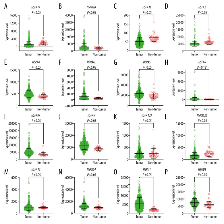
The clinical data from 438 patients with CRC was used in the analysis. Table 1 lists the correlations between clinical characteristics and OS in the patients with CRC [16]. The results showed that TNM stage was related with OS. Figure 1 illustrates each of the Hsp70 family gene levels in samples of CRC and normal colon tissue. HSPA1A, HSPA1B, HSPA1L, HSPA2, HSPA4, HSPA4L, HSPA5, HSPA8, HSPA9, HSPA12B, HSPA14, HSPH1, and HYOU1 gene levels were statistically significant.
Table 1.
The clinical data for 438 patients with colorectal cancer.
| Variables | Patients (n=438) | No. of events (%) | MST (days) | HR (95% CI) | Log-rank P |
|---|---|---|---|---|---|
| Age (years) | 0.398 | ||||
| <60 | 122 | 81.1 | 3039 | Ref. | |
| ≥60 | 316 | 76.3 | 2535 | 1.223 (0.766–1.952) | |
| Sex | 0.545 | ||||
| Female | 204 | 78.4 | 2990 | Ref. | |
| Male | 234 | 76.9 | 2320 | 1.131 (0.759–1.686) | |
| TNM stage | <0.001 | ||||
| I | 73 | 94.5 | 3234 | Ref. | |
| II | 167 | 83.8 | 2838 | 2.24 (0.781–6.421) | |
| III | 126 | 75.4 | 2856 | 4.068 (1.434–11.538) | |
| IV | 61 | 49.2 | 1114 | 11.291 (3.980–32.026) | |
| Missing | 11 | ||||
MST – median survival time; HR – hazard ratio; CI – confidence interval; TNM – tumor-node-metastasis.
Figure 1.
Expression levels of Hsp70 genes in colorectal cancer and normal colon tissue. (A) HSPA1A; (B) HSPA1B; (C) HSPA1L; (D) HSPA2; (E) HSPA4; (F) HSPA4L; (G) HSPA5; (H) HSPA6; (I) HSPA8; (J) HSPA9; (K) HSPA12A; (L) HSPA12B; (M) HSPA13; (N) HSPA14; (O) HSPH1; (P) HYOU1.
Interaction and Function Analysis of the Hsp70 Gene Family
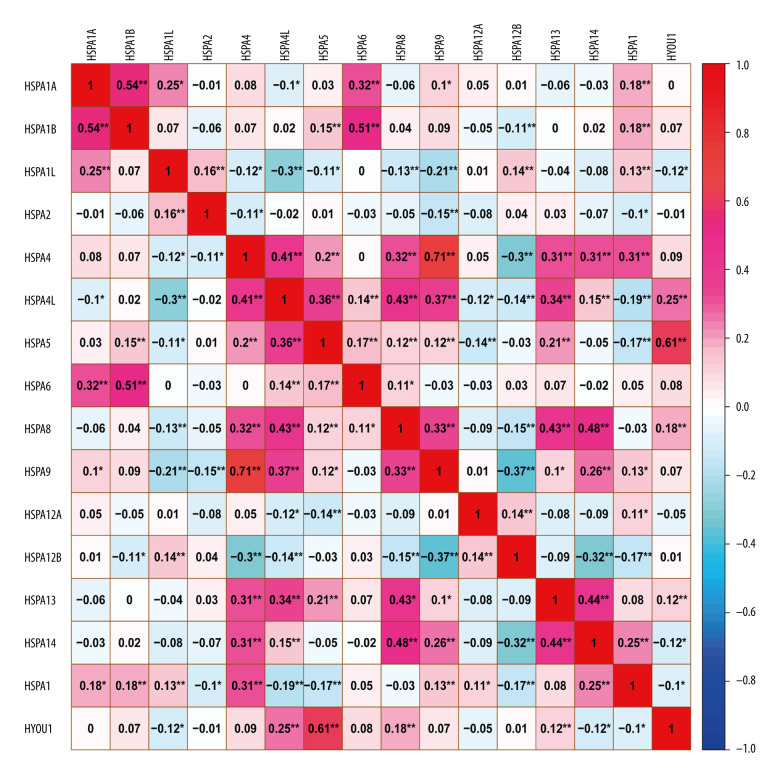
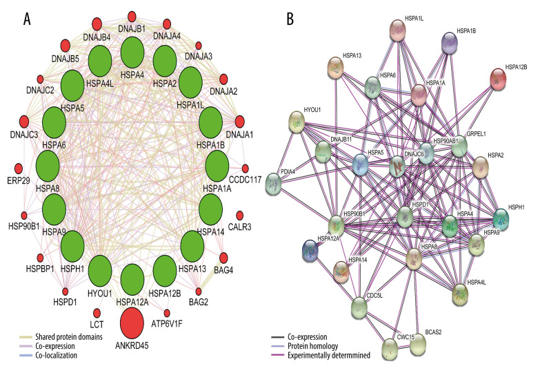
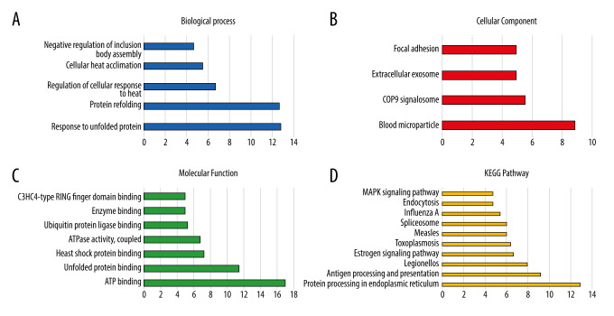
The Pearson correlation coefficient was used to analyze the correlation of Hsp70 genes (Figure 2). Figure 3 illustrates the gene-gene and protein–protein interaction network of the Hsp70 gene family. Figure 4 illustrates the GO pathway functional analysis and KEGG pathway functional analysis.
Figure 2.
Pearson’s correlation analysis for HSPA1A, HSPA1B, HSPA1L, HSPA2, HSPA4, HSPA4L, HSPA5, HSPA6, HSPA8, HSPA9, HSPA12A, HSPA12B, HSPA13, HSPA14, HSPH1, and HYOU1.
Figure 3.
Gene-gene and protein–protein interaction network for Hsp70 gene family. (A) Gene-gene interaction network; (B) Protein–protein interaction network.
Figure 4.
GO and KEGG pathway analysis of Hsp70 gene family carried out by the online tool DAVID. (A) Biological process; (B) cellular component; (C) molecular function; (D) KEGG pathway.
Survival and Joint effect Analysis of the Hsp70 Gene Family
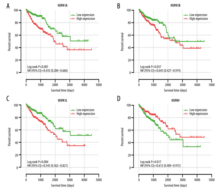
Table 2 summarizes the univariate and multivariate survival analyses of the Hsp70 genes. Low expression levels of HSPA1A, HSPA1B, and HSPA1L were associated with improved OS in univariate survival analysis. Meanwhile, the elevated expression level of HSPA9 was related to improved OS (Figure 5). Moreover, multivariate survival analysis demonstrated that lower expression levels of HSPA1A, HSPA1B, and HSPA1L were significantly associated with improved OS.
Table 2.
Prognostic survival analysis of Hsp70 family genes.
| Gene expression | Patients (n=438) | No. of events (%) | MST (days) | Crude HR (95% CI) | Crude P | Adjusted HR (95% CI) | Adjusted P* |
|---|---|---|---|---|---|---|---|
| HSPA1A | <0.001 | 0.004 | |||||
| Low | 219 | 85.8 | 2969 | Ref. | Ref. | ||
| High | 219 | 69.4 | 2412 | 0.435 (0.284–0.666) | 0.514 (0.327–0.808) | ||
| HSPA1B | 0.037 | 0.044 | |||||
| Low | 219 | 83.6 | 2966 | Ref. | Ref. | ||
| High | 219 | 71.7 | 2462 | 0.645 (0.427–0.974) | 0.643 (0.419–0.988) | ||
| HSPA1L | 0.004 | 0.046 | |||||
| Low | 219 | 83.1 | 3046 | Ref. | Ref. | ||
| High | 219 | 72.1 | 2247 | 0.545 (0.362–0.821) | 0.650 (0.425–0.993) | ||
| HSPA2 | |||||||
| Low | 219 | 76.7 | 2584 | Ref. | 0.776 | Ref. | 0.720 |
| High | 219 | 78.5 | 2810 | 0.944 (0.635–1.404) | 1.077 (0.717–1.619) | ||
| HSPA4 | |||||||
| Low | 219 | 76.3 | 2222 | Ref. | 0.103 | Ref. | 0.180 |
| High | 219 | 79.0 | 2852 | 0.717 (0.481–1.069) | 1.325 (0.879–1.998) | ||
| HSPA4L | |||||||
| Low | 219 | 76.3 | 2054 | Ref. | 0.134 | Ref. | 0.622 |
| High | 219 | 79.0 | 2908 | 0.734 (0.490–1.100) | 0.900 (0.591–1.370) | ||
| HSPA5 | |||||||
| Low | 219 | 79.0 | 2734 | Ref. | 0.656 | Ref. | 0.674 |
| High | 219 | 76.3 | 2594 | 1.095 (0.736–1.629) | 1.092 (0.725–1.644) | ||
| HSPA6 | |||||||
| Low | 219 | 80.8 | 2732 | Ref. | 0.067 | Ref. | 0.284 |
| High | 219 | 74.4 | 2591 | 1.455 (0.975–2.171) | 1.254 (0.829–1.899) | ||
| HSPA8 | |||||||
| Low | 219 | 73.5 | 2481 | Ref. | 0.050 | Ref. | 0.333 |
| High | 219 | 81.7 | 2790 | 0.668 (0.446–0.999) | 0.815 (0.538–1.233) | ||
| HSPA9 | |||||||
| Low | 219 | 74.0 | 2335 | Ref. | 0.017 | Ref. | 0.059 |
| High | 219 | 81.3 | 2962 | 0.612 (0.409–0.915) | 0.670 (0.442–1.016) | ||
| HSPA12A | |||||||
| Low | 219 | 78.1 | 2634 | Ref. | 0.863 | Ref. | 0.605 |
| High | 219 | 77.2 | 2688 | 1.035 (0.697–1.539) | 1.114 (0.741–1.674) | ||
| HSPA12B | |||||||
| Low | 219 | 76.3 | 2644 | Ref. | 0.935 | Ref. | 0.697 |
| High | 219 | 79.0 | 2712 | 0.984 (0.661–1.464) | 0.922 (0.612–1.388) | ||
| HSPA13 | |||||||
| Low | 219 | 74.0 | 2666 | Ref. | 0.189 | Ref. | 0.484 |
| High | 219 | 81.3 | 2570 | 0.763 (0.510–1.142) | 0.863 (0.571–1.305) | ||
| HSPA14 | |||||||
| Low | 219 | 76.7 | 2646 | Ref. | 0.388 | Ref. | 0.645 |
| High | 219 | 78.5 | 2666 | 0.840 (0.565–1.249) | 0.909 (0.604–1.366) | ||
| HSPH1 | |||||||
| Low | 219 | 78.5 | 2672 | Ref. | 0.836 | Ref. | 0.131 |
| High | 219 | 76.7 | 2661 | 1.043 (0.701–1.551) | 0.721 (0.472–1.102) | ||
| HYOU1 | |||||||
| Low | 219 | 74.4 | 2617 | Ref. | 0.244 | Ref. | 0.185 |
| High | 219 | 80.8 | 2692 | 0.788 (0.528–1.177) | 0.757 (0.501–1.143) | ||
Adjusted for TNM stage.
Hsp70 – heat shock protein 70; MST – median survival time; HR – hazard ratio; CI – confidence interval.
Figure 5.
The univariate survival analysis of HSPA1A, HSPA1B, HSPA1L, and HSPA9 (P<0.05). Kaplan-Meier survival curves concerning (A) HSPA1A; (B) HSPA1B; (C) HSPA1L; (D) HSPA9 expression.
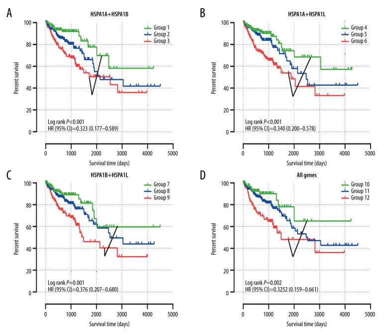
Based on the multivariate survival analysis of HSPA1A, HSPA1B, and HSPA1L, a joint-effects framework was performed with different groups (Table 3). As illustrated in Figure 6, low expression levels of HSPA1A, HSPA1B, and HSPA1L in Groups 1, 4, 7, and 10 were significantly correlated with improved OS.
Table 3.
Grouping according to HSPA1A, HSPA1B, and HSPA1L.
| Group | Composition | Group | Composition |
|---|---|---|---|
| 1 | Low HSPA1A +low HSPA1B | 10 | Low HSPA1A +low HSPA1B+ low HSPA1L |
| 2 | Low HSPA1A +high HSPA1B | 11 | Low HSPA1A +low HSPA1B+ high HSPA1L |
| High HSPA1A +low HSPA1B | Low HSPA1A +high HSPA1B+ low HSPA1L | ||
| 3 | High HSPA1A +high HSPA1B | High HSPA1A +low HSPA1B+ low HSPA1L | |
| 4 | Low HSPA1A +low HSPA1L | High HSPA1A +high HSPA1B+ low HSPA1L | |
| 5 | Low HSPA1A +high HSPA1L | High HSPA1A +low HSPA1B+ high HSPA1L | |
| High HSPA1A +low HSPA1L | Low HSPA1A +high HSPA1B+ high HSPA1L | ||
| 6 | High HSPA1A +high HSPA1L | 12 | High HSPA1A +high HSPA1B+ high HSPA1L |
| 7 | Low HSPA1B +low HSPA1L | ||
| 8 | Low HSPA1B +high HSPA1L | ||
| High HSPA1B +low HSPA1L | |||
| 9 | High HSPA1B +high HSPA1L |
Figure 6.
The joint-effects analysis of the influence of combined HSPA1A, HSPA1B, and HSPA1L. Kaplan-Meier survival curves concerning (A) HSPA1A+HSPA1B; (B) HSPA1A+HSPA1L; (C) HSPA1B+HSPA1L; (D) HSPA1A+HSPA1B+HSPA1L.
Nomogram
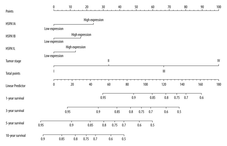
The nomogram was utilized for investigating the association between HSPA1A, HSPA1B, HSPA1L, and tumor stage in CRC. The points of each variable could be calculated. Figure 7 shows the prediction of the 1-, 3-, 5-, and 10-year survival rates.
Figure 7.
A nomogram model was performed to analyze the prognosis correlation of HSPA1A, HSPA1B, HSPA1L and tumor stage in CRC. The points of each variable were calculated at the top of the nomogram. A vertical line down to the 1-, 3-, 5-, and 10-year survival lines allowed for the determination of survival probabilities.
GSEA Analysis
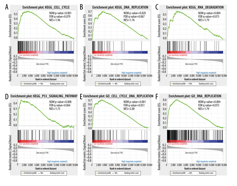
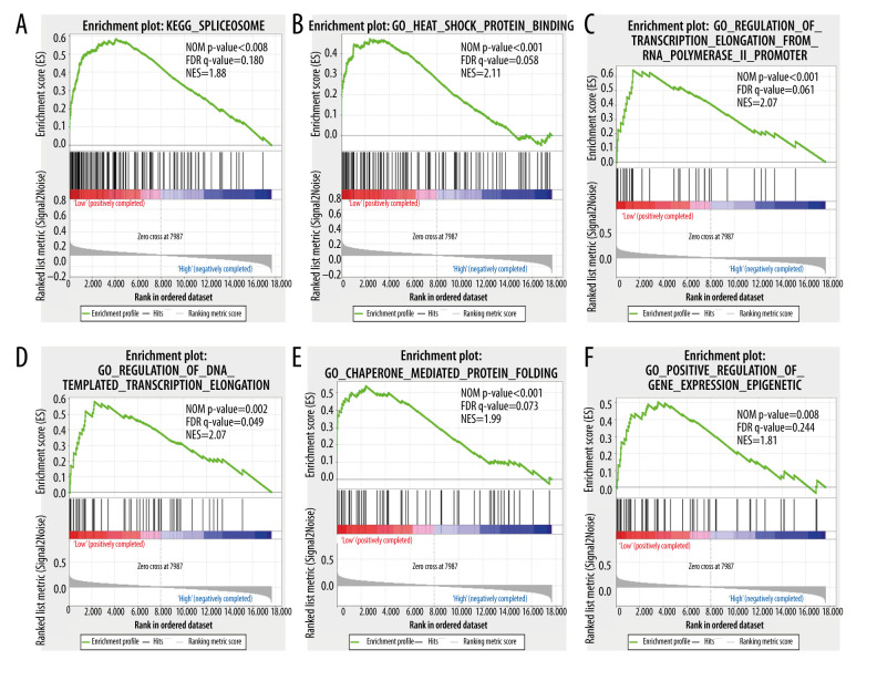
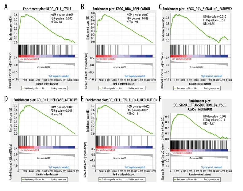
To investigate the enrichment pathway with HSPA1A, HSPA1B, and HSPA1L, GSEA analysis was conducted. As illustrated in Figure 8, according to the GSEA, the low expression of HSPA1A was positively correlated with the cell cycle, DNA replication, RNA degradation, and P53 pathway. As illustrated in Figure 9, the GSEA indicated that the high expression of HSPA1B was positively correlated with the spliceosome, heat shock protein binding, RNA polymerase II promoter transcription elongation, DNA-templated transcription elongation, chaperone-mediated protein folding, and positive regulation of gene-expression epigenetics. The GSEA also indicated that a low expression of HSPA1L was positively correlated with the cell cycle, DNA replication, DNA helicase activity, and P53 pathway (Figure 10).
Figure 8.
Gene set enrichment analysis shows the enrichment analysis of HSPA1A. (A–F) Statistical significance was implied by NOM P<0.05 and FDR<0.25. NOM – normalized; FDR – false discovery rate; NES – normalized enrichment score.
Figure 9.
Gene set enrichment analysis shows the enrichment analysis of HSPA1B. (A–F) Statistical significance was implied by NOM P<0.05 and FDR<0.25. NOM – normalized; FDR – false discovery rate; NES – normalized enrichment score.
Figure 10.
Gene set enrichment analysis shows the enrichment analysis of HSPA1L. (A–F) Statistical significance was implied by NOM P<0.05 and FDR<0.25. NOM – normalized; FDR – false discovery rate; NES – normalized enrichment score.
Discussion
The TCGA database was utilized to illustrate the importance of Hsp70 genes in predicting the prognosis of patients with CRC. We found that gene levels were statistically significantly higher in CRC tissue samples than in normal colon tissue samples for HSPA1A, HSPA1B, HSPA1L, HSPA2, HSPA4, HSPA4L, HSPA5, HSPA8, HSPA9, HSPA12B, HSPA14, HSPH1, and HYOU1. Furthermore, interaction and functional analyses were established in the investigation of the Hsp70 genes. According to the multivariate survival analysis and joint-effects analysis, low expression levels of HSPA1A, HSPA1B, and HSPA1L had a strong association with improved OS. Additionally, a nomogram based on HSPA1A, HSPA1B, HSPA1L, and tumor stage was formulated for predicting 1-, 3-, 5-, and 10-year survival rates in the patients with CRC. The investigation of potential molecular mechanisms with HSPA1A, HSPA1B, and HSPA1L was facilitated by GSEA analysis. Accordingly, low expression of HSPA1A exhibited a positive correlation with the cell cycle, DNA replication, RNA degradation, and P53 pathway. Furthermore, an elevated expression level in HSPA1B was positively correlated with the spliceosome, heat shock protein binding, RNA polymerase II promoter transcription elongation, DNA-templated transcription elongation, chaperone-mediated protein folding, and positive regulation of gene-expression epigenetics. In addition, the low expression of HSPA1L was positively correlated with the cell cycle, DNA replication, DNA helicase activity, and P53 pathway.
Since the Hsp70 genes are important members of the HSPs family, they were assumed to be responsible for multiple cellular developments and for responding to environmental changes [4]. The human Hsp70 is a multigene family which consists of 17 genes and 30 pseudogenes [7]. It also includes 1 putative gene, HSPA7 [17]. In the present study, we selected data from 16 Hsp70 genes to investigate the significance of Hsp70 genes in the prediction of prognosis in patients with CRC. According to the multivariate survival analysis, low expression levels of HSPA1A, HSPA1B, and HSPA1L were significantly related with improved OS. Previous studies have done detailed analysis of the evolutionary history of HSPA1A, HSPA1B, and HSPA1L [18], and these genes, respectively, encode 3 highly analogous Hsp70 proteins, namely, Hsp70-1, Hsp70-2, and Hsp70-hom, which are located on chromosome 6p21.3 [19]. The genes HSPA1A and HSPA1B have been studied extensively, and their coded proteins are thought to be completely interchangeable because only 2 amino acids are different [20]. In a majority of human tissues, the expression levels of HSPA1A and HSPA1B are expressed much more than are other Hsp70 family genes. Furthermore, HSPA1L is highly expressed in testis [7].
It has been demonstrated that HSPA1A plays an essential role in cancer development. Apparently, HSPA1A could be significant in the development of cancer cells, protecting them from oxidative stress, hypoxia, inflammatory cytokines, and the anti-apoptotic pathway [21]. It has been demonstrated that HSPA1A is essential to the survival of different cancer cells [22–24]. It has also been established that HSPA1A has a role on changes in the immune system [4]. Moreover, the HSPA1A and HSPA1L genes could be related to the prognosis in ovarian epithelial cancer [25].
Similar to HSPA1A, HSPA1B also assumes a vital role in cancer. It has been reported that HSPA1B variations are related to lung cancer risk and survival [19]. Numerous studies have shown that HSPA1B is related to the growth of tumors in colorectal and breast cancer [26,27]. Additionally, variant HSPA1L could be related to prostate cancer risk [28]. Hsp70 exhibits various anticancer therapies, including playing the role of lifeguard and having anti-apoptotic effects in cancer cells [6,29,30]. It also plays a role in the regulation of the intrinsic, extrinsic, and caspase-independent pathways [31,32]. GSEA was used to discover the potential underlying molecular mechanisms of HSPA1A, HSPA1B, and HSPA1L in CRC. It is likely that these genes possess anticancer effects by affecting the cell cycle, DNA replication, and P53 pathway.
This study has a number of limitations. First, the public databases lack detailed clinical information. Second, the patient data were obtained from a single source. To generalize the results, it will be necessary to validate the conclusions through the analysis of independent data in future studies. Finally, since this study is mainly a bioinformatics study using data from a public database, it lacks empirical conclusiveness. The anticancer properties of HSPA1A, HSPA1B, and HSPA1L in CRC should be tested through various in vitro and in vivo experiments. Studies have demonstrated that Hsp70 genes have prognostic significance in some common tumors [9,33,34]; however, the present study is the first to report on the significance of the Hsp70 family of genes in estimating the prognosis of patients with CRC.
Conclusions
Through comprehensive analysis, we identified the potential molecular mechanisms of HSPA1A, HSPA1B, and HSPA1L in CRC. Additionally, we discovered that low expression levels of HSPA1A, HSPA1B, and HSPA1L were significantly correlated with an improved prognosis in CRC. Importantly, HSPA1A, HSPA1B, and HSPA1L have potential value as prognostic biological markers in CRC.
Acknowledgements
The authors thank the TCGA for sharing public data.
Footnotes
Conflicts of Interest
None.
Source of support: Departmental sources
References
- 1.Siegel RL, Miller KD, Jemal A. Cancer statistics, 2020. Cancer J Clin. 2020;70:7–30. doi: 10.3322/caac.21590. [DOI] [PubMed] [Google Scholar]
- 2.Kannagi R, Izawa M, Koike T, et al. Carbohydrate-mediated cell adhesion in cancer metastasis and angiogenesis. Cancer Sci. 2004;95:377–84. doi: 10.1111/j.1349-7006.2004.tb03219.x. [DOI] [PMC free article] [PubMed] [Google Scholar]
- 3.Brodsky JL, Chiosis G. Hsp70 molecular chaperones: Emerging roles in human disease and identification of small molecule modulators. Curr Top Med Chem. 2006;6:1215–25. doi: 10.2174/156802606777811997. [DOI] [PubMed] [Google Scholar]
- 4.Daugaard M, Rohde M, Jaattela M. The heat shock protein 70 family: Highly homologous proteins with overlapping and distinct functions. FEBS Letters. 2007;581:3702–10. doi: 10.1016/j.febslet.2007.05.039. [DOI] [PubMed] [Google Scholar]
- 5.Nylandsted J, Brand K, Jäättelä M. Heat shock protein 70 is required for the survival of cancer cells. Ann N Y Acad Sci. 2000;926:122–25. doi: 10.1111/j.1749-6632.2000.tb05605.x. [DOI] [PubMed] [Google Scholar]
- 6.Kumar S, Stokes J, 3rd, Singh UP, et al. Targeting Hsp70: A possible therapy for cancer. Cancer Lett. 2016;374:156–66. doi: 10.1016/j.canlet.2016.01.056. [DOI] [PMC free article] [PubMed] [Google Scholar]
- 7.Radons J. The human HSP70 family of chaperones: Where do we stand? Cell Stress Chaperon. 2016;21:379–404. doi: 10.1007/s12192-016-0676-6. [DOI] [PMC free article] [PubMed] [Google Scholar]
- 8.Brocchieri L, Conway de Macario E, Macario AJ. Hsp70 genes in the human genome: Conservation and differentiation patterns predict a wide array of overlapping and specialized functions. BMC Evol Biol. 2008;8:19. doi: 10.1186/1471-2148-8-19. [DOI] [PMC free article] [PubMed] [Google Scholar]
- 9.Bauer K, Nitsche U, Slotta-Huspenina J, et al. High HSP27 and HSP70 expression levels are independent adverse prognostic factors in primary resected colon cancer. Cell Oncol (Dordrecht) 2012;35:197–205. doi: 10.1007/s13402-012-0079-3. [DOI] [PubMed] [Google Scholar]
- 10.Soleimani A, Zahiri E, Ehtiati S, et al. Therapeutic potency of heat-shock protein-70 in the pathogenesis of colorectal cancer: Current status and perspectives. Biochem Cell Biol. 2019;97:85–90. doi: 10.1139/bcb-2018-0177. [DOI] [PubMed] [Google Scholar]
- 11.Weinstein JN, Collisson EA, Mills GB, et al. The Cancer Genome Atlas Pan-Cancer analysis project. Nat Genet. 2013;45:1113–20. doi: 10.1038/ng.2764. [DOI] [PMC free article] [PubMed] [Google Scholar]
- 12.Warde-Farley D, Donaldson SL, Comes O, et al. The GeneMANIA prediction server: Biological network integration for gene prioritization and predicting gene function. Nucleic Acids Res. 2010;38:W214–20. doi: 10.1093/nar/gkq537. [DOI] [PMC free article] [PubMed] [Google Scholar]
- 13.Huang DW, Sherman BT, Lempicki RA. Systematic and integrative analysis of large gene lists using DAVID bioinformatics resources. Nat Protoc. 2009;4:44–57. doi: 10.1038/nprot.2008.211. [DOI] [PubMed] [Google Scholar]
- 14.Balachandran VP, Gonen M, Smith JJ, DeMatteo RP. Nomograms in oncology: More than meets the eye. Lancet Oncol. 2015;16:e173–80. doi: 10.1016/S1470-2045(14)71116-7. [DOI] [PMC free article] [PubMed] [Google Scholar]
- 15.Subramanian A, Tamayo P, Mootha VK, et al. Gene set enrichment analysis: A knowledge-based approach for interpreting genome-wide expression profiles. Proc Natl Acad Sci USA. 2005;102:15545–50. doi: 10.1073/pnas.0506580102. [DOI] [PMC free article] [PubMed] [Google Scholar]
- 16.Pan X, Wang Q, Xu C, et al. Prognostic value of chloride channel accessory mRNA expression in colon cancer. Oncol Lett. 2019;18:2967–76. doi: 10.3892/ol.2019.10615. [DOI] [PMC free article] [PubMed] [Google Scholar]
- 17.Leung TK, Hall C, Rajendran M, et al. The human heat-shock genes HSPA6 and HSPA7 are both expressed and localize to chromosome 1. Genomics. 1992;12:74–79. doi: 10.1016/0888-7543(92)90409-l. [DOI] [PubMed] [Google Scholar]
- 18.Kudla G, Helwak A, Lipinski L. Gene conversion and GC-content evolution in mammalian Hsp70. Mol Biol Evol. 2004;21:1438–44. doi: 10.1093/molbev/msh146. [DOI] [PubMed] [Google Scholar]
- 19.Guo H, Deng Q, Wu C, et al. Variations in HSPA1B at 6p21.3 are associated with lung cancer risk and prognosis in Chinese populations. Cancer Res. 2011;71:7576–86. doi: 10.1158/0008-5472.CAN-11-1409. [DOI] [PubMed] [Google Scholar]
- 20.Kampinga HH, Hageman J, Vos MJ, et al. Guidelines for the nomenclature of the human heat shock proteins. Cell Stress Chaperon. 2009;14:105–11. doi: 10.1007/s12192-008-0068-7. [DOI] [PMC free article] [PubMed] [Google Scholar]
- 21.Garrido C, Brunet M, Didelot C, et al. Heat shock proteins 27 and 70: Anti-apoptotic proteins with tumorigenic properties. Cell Cycle (Georgetown, Tex) 2006;5:2592–601. doi: 10.4161/cc.5.22.3448. [DOI] [PubMed] [Google Scholar]
- 22.Klink M, Nowak M, Kielbik M, et al. The interaction of HspA1A with TLR2 and TLR4 in the response of neutrophils induced by ovarian cancer cells in vitro. Cell Stress Chaperon. 2012;17:661–74. doi: 10.1007/s12192-012-0338-2. [DOI] [PMC free article] [PubMed] [Google Scholar]
- 23.Nadin SB, Cuello-Carrión FD, Sottile ML, et al. Effects of hyperthermia on Hsp27 (HSPB1), Hsp72 (HSPA1A) and DNA repair proteins hMLH1 and hMSH2 in human colorectal cancer hMLH1-deficient and hMLH1-proficient cell lines. Int J Hyperthermia. 2012;28:191–201. doi: 10.3109/02656736.2011.638962. [DOI] [PubMed] [Google Scholar]
- 24.Ban Y, Tan P, Cai J, et al. LNCAROD is stabilized by m6A methylation and promotes cancer progression via forming a ternary complex with HSPA1A and YBX1 in head and neck squamous cell carcinoma. Mol Oncol. 2020;14:1282–96. doi: 10.1002/1878-0261.12676. [DOI] [PMC free article] [PubMed] [Google Scholar]
- 25.De Andrade WP, Da Conceição Braga L, Gonçales NG, et al. HSPA1A, HSPA1L and TRAP1 heat shock genes may be associated with prognosis in ovarian epithelial cancer. Oncol Lett. 2020;19:359–67. doi: 10.3892/ol.2019.11095. [DOI] [PMC free article] [PubMed] [Google Scholar]
- 26.Jagadish N, Parashar D, Gupta N, et al. Heat shock protein 70-2 (HSP70-2) is a novel therapeutic target for colorectal cancer and is associated with tumor growth. BMC Cancer. 2016;16:561. doi: 10.1186/s12885-016-2592-7. [DOI] [PMC free article] [PubMed] [Google Scholar]
- 27.Jagadish N, Agarwal S, Gupta N, et al. Heat shock protein 70-2 (HSP70-2) overexpression in breast cancer. J Exp Clin Cancer Res. 2016;35:150. doi: 10.1186/s13046-016-0425-9. [DOI] [PMC free article] [PubMed] [Google Scholar]
- 28.Sfar S, Saad H, Mosbah F, Chouchane L. Association of HSP70-hom genetic variant with prostate cancer risk. Mol Biol Rep. 2008;35:459–64. doi: 10.1007/s11033-007-9107-1. [DOI] [PubMed] [Google Scholar]
- 29.Mosser DD, Caron AW, Bourget L, et al. The chaperone function of hsp70 is required for protection against stress-induced apoptosis. Mol Cell Biol. 2000;20:7146–59. doi: 10.1128/mcb.20.19.7146-7159.2000. [DOI] [PMC free article] [PubMed] [Google Scholar]
- 30.Nylandsted J, Rohde M, Brand K, et al. Selective depletion of heat shock protein 70 (Hsp70) activates a tumor-specific death program that is independent of caspases and bypasses Bcl-2. Proc Natl Acad Sci USA. 2000;97:7871–76. doi: 10.1073/pnas.97.14.7871. [DOI] [PMC free article] [PubMed] [Google Scholar]
- 31.Jäättelä M, Wissing D, Kokholm K, et al. Hsp70 exerts its anti-apoptotic function downstream of caspase-3-like proteases. EMBO J. 1998;17:6124–34. doi: 10.1093/emboj/17.21.6124. [DOI] [PMC free article] [PubMed] [Google Scholar]
- 32.Kalinowska M, Garncarz W, Pietrowska M, et al. Regulation of the human apoptotic DNase/RNase endonuclease G: Involvement of Hsp70 and ATP. Apoptosis. 2005;10:821–30. doi: 10.1007/s10495-005-0410-9. [DOI] [PubMed] [Google Scholar]
- 33.Kang Y, Jung WY, Lee H, et al. Prognostic significance of heat shock protein 70 expression in early gastric carcinoma. Korean J Pathol. 2013;47:219–26. doi: 10.4132/KoreanJPathol.2013.47.3.219. [DOI] [PMC free article] [PubMed] [Google Scholar]
- 34.Dimas DT, Perlepe CD, Sergentanis TN, et al. The prognostic significance of Hsp70/Hsp90 expression in breast cancer: A systematic review and meta-analysis. Anticancer Res. 2018;38:1551–62. doi: 10.21873/anticanres.12384. [DOI] [PubMed] [Google Scholar]


