

Abstract
In rare instances, calcific tendonitis may manifest in the pediatric population as inflammatory calcium hydroxyapatite deposition. To our knowledge, there have been no previous case reports involving the flexor pollicis longus tendon at the thumb interphalangeal joint. We present a 9-year-old boy with a painful mass at the right thumb interphalangeal joint. Initial radiographs revealed a 7-mm ovoid calcific mass along the volar soft tissues of the thumb interphalangeal joint. Subsequent ultrasound and magnetic resonance findings further confirmed calcification with surrounding edema. Because the pain was limiting the patient’s school activities, his family elected for excisional biopsy of the calcific mass. Pathology ultimately revealed prominent dystrophic calcifications with surrounding granulomatous inflammation, consistent with calcific tendonitis.
Keywords: Calcific tendonitis, flexor pollicis tendon, magnetic resonance imaging, pediatric, radiograph, ultrasound
Calcific tendonitis, a disease of calcium hydroxyapatite crystal deposition in tendons, most commonly presents as monoarticular pain in adults.1–4 Rarely, it may present in a child as acute digit pain with inflammatory features. It may be acute, chronic, or asymptomatic; however, acute calcific tendonitis is notable for swelling, erythema, pain, and potentially elevated laboratory inflammatory markers.3 The clinical presentation overlaps with other etiologies, such as infection, trauma, tenosynovitis, or septic arthritis.2,4–8 Appropriate workup and recognition of pediatric calcific tendonitis can allow for conservative management.
CASE DESCRIPTION
A 9-year-old boy with no significant past medical history presented to the emergency department with a painful mass at the right thumb interphalangeal joint. Physical exam demonstrated a small, tender, ball-like mass volar to the interphalangeal joint of the right thumb with normal range of motion and intact sensation. All labs were unremarkable, including complete blood count with differential, comprehensive metabolic panel, C-reactive protein, and blood cultures. Initial radiographs of the finger revealed soft tissue swelling with a 7-mm ovoid calcific soft tissue mass volar to the thumb interphalangeal joint (Figure 1). The patient was discharged with a plan to follow up with his pediatrician and obtain magnetic resonance imaging (MRI).
Figure 1.
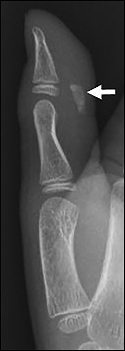
Lateral radiograph of the right thumb showing an ovoid calcific density (arrow) longitudinally oriented and superficial to the volar aspect of the interphalangeal joint.
At follow-up 2 days later, the pediatrician prescribed a 7-day course of clindamycin due to consideration of infection and requested an ultrasound. Ultrasound revealed a 7 × 6 × 3 mm hyperechoic hypovascular nodule with acoustic shadowing superficial to and intimately associated with the flexor tendon (Figure 2). There was soft tissue hyperemia. MRI was performed 4 days after initial presentation to rule out malignancy because calcific tendonitis is so rare at this age. MRI revealed a hypointense ovoid calcification and surrounding edema. There was no soft tissue mass, abnormal marrow signal, or abnormal tendon signal (Figure 3).
Figure 2.

Ultrasound of the calcific mass. (a) Longitudinal view demonstrating that the ovoid hyperechoic mass (between calipers) is superficial to the longitudinal fibers of the tendon (T). (b) Transverse image showing no color Doppler flow in the hyperechoic mass, although there is increased pericalcific flow reflecting inflammation.
Figure 3.

MRI of the right thumb. (a) Axial T2 fat-saturated image demonstrating the calcified mass lesion (arrow) markedly hypointense, consistent with calcification, with surrounding hyperintense edema. (b) Sagittal T1 fat-saturated postcontrast image showing the mass lesion (arrow) superficial to the tendon (T) with surrounding enhancement from edema and inflammation.
The patient was referred to pediatric orthopedic surgery following the MRI. Because persistent pain affected the patient’s daily activities, his family elected for excisional biopsy about 3 weeks after symptom presentation. At surgery there was a calcified lesion superficial to the flexor pollicis longus tendon. Pathology revealed dystrophic calcifications with surrounding granulomatous inflammation, consistent with calcific tendonitis. The patient fully recovered.
DISCUSSION
Acute digital pain with inflammatory features in a child is most often due to infection or trauma; however, in rare cases it reflects calcific tendonitis.2,4–8 Calcific tendonitis most commonly presents as painful rotator cuff tendonitis in women aged 40 to 60.1,2 Calcific tendonitis is rarely found in children, with a limited number of published cases.1,3–8 Our case involving the flexor pollicis longus tendon at the thumb interphalangeal joint is a unique location not previously described in the literature.
Current imaging modalities for diagnosing calcific tendonitis include conventional radiography, ultrasound, and MRI. Radiographic findings of calcium deposition range from fluffy, ill-defined densities (recent deposit) to homogenous, well-defined densities (chronic deposit).3,9,10 Ultrasound typically demonstrates an arc-shaped, nodular, fragmented, or globular hyperechoic focus with acoustic shadowing.9,10 MRI reveals a hypointense mass within or adjacent to a tendon, ligament, joint capsule, or bursa. A paucity of inflammatory changes surrounding the calcium deposit implies chronicity or a postcalcific stage, which is then typically termed calcific tendinosis if associated with a tendon. The imaging diagnosis of calcific tendonitis is given in the setting of pericalcific inflammatory changes on MRI or ultrasound that indicate edema and hyperemia involving the tendon and adjacent soft tissues and/or bone.3,9
This case report serves as a reminder to include calcific tendonitis in the differential diagnosis, even in children. Initial treatment for calcific tendonitis is generally conservative with splint immobilization, nonsteroidal anti-inflammatories, rest, physical therapy, and ice.1,4,9,11,12 Other treatment options include periarticular steroid injections and extracorporeal shockwave therapy.3,11,12 If conservative management fails, calcific tendonitis may be managed surgically.3,12 With recognition of typical radiographic and sonographic appearance and clinical characteristics, one may avoid advanced imaging, antibiotic therapy, and biopsy.
References
- 1.Walocko FM, Sando IC, Haase SC, Kozlow JH.. Acute calcific tendinitis of the index finger in a child. Hand (N Y). 2017;12:NP84–NP87. [DOI] [PMC free article] [PubMed] [Google Scholar]
- 2.De Carli A, Pulcinelli F, Rose GD, Pitino D, Ferretti A.. Calcific tendinitis of the shoulder. Joints. 2014;2:130–136. [DOI] [PMC free article] [PubMed] [Google Scholar]
- 3.Kheterpal A, Zoga A, McClure K.. Acute calcific tendinitis of the flexor pollicis longus in an 8-year-old boy. Skeletal Radiol. 2014;43:1471–1475. doi: 10.1007/s00256-014-1908-4. [DOI] [PubMed] [Google Scholar]
- 4.Hakozaki M, Iwabuchi M, Konno S, et al. Acute calcific tendinitis of the thumb in a child: a case report. Clin Rheumatol. 2007;26:841–844. doi: 10.1007/s10067-006-0384-1. [DOI] [PubMed] [Google Scholar]
- 5.Hansen U, Battista V.. Pediatric trigger finger from calcific tendonitis. J Hand Surg Am. 2007;32:1558–1559. doi: 10.1016/j.jhsa.2007.08.005. [DOI] [PubMed] [Google Scholar]
- 6.Bittmann S. Calcific tendinitis of the supraspinatus tendon in children. Klin Padiatr. 2006;218:45–46. doi: 10.1055/s-2005-836383. [DOI] [PubMed] [Google Scholar]
- 7.Millon SJ, Bush DC, Harrington TM.. Acute calcific tendinitis in a child: a case report. J Hand Surg Am. 1993;18:592–593. doi: 10.1016/0363-5023(93)90296-F. [DOI] [PubMed] [Google Scholar]
- 8.Fong CM. Calcific tendinitis of the supraspinatus tendon in a 7-year-old boy: diagnostic challenges. Hong Kong Med J. 2011;17:414–416. [PubMed] [Google Scholar]
- 9.Siegal DS, Wu JS, Newman JS, Del Cura JL, Hochman MG.. Calcific tendinitis: a pictorial review. Can Assoc Radiol J. 2009;60:263–272. doi: 10.1016/j.carj.2009.06.008. [DOI] [PubMed] [Google Scholar]
- 10.Beckmann NM. Calcium apatite deposition disease: diagnosis and treatment. Radiol Res Pract. 2016;1–16. doi: 10.1155/2016/4801474. [DOI] [PMC free article] [PubMed] [Google Scholar]
- 11.Sansone V, Maiorano E, Galluzzo A, Pascale V.. Calcific tendinopathy of the shoulder: clinical perspectives into the mechanisms, pathogenesis, and treatment. Orthop Res Rev. 2018;10:63–72. doi: 10.2147/ORR.S138225. [DOI] [PMC free article] [PubMed] [Google Scholar]
- 12.Chianca V, Albano D, Messina C, et al. Rotator cuff calcific tendinopathy: from diagnosis to treatment. Acta Biomed. 2018;89:186–196. doi: 10.23750/abm.v89i1-S.7022. [DOI] [PMC free article] [PubMed] [Google Scholar]