Abstract
Context
Field-based, portable motion-capture systems can be used to help identify individuals at greater risk of lower extremity injury. Microsoft Kinect-based markerless motion-capture systems meet these requirements; however, until recently, these systems were generally not automated, required substantial data postprocessing, and were not commercially available.
Objective
To validate the kinematic measures of a commercially available markerless motion-capture system.
Design
Descriptive laboratory study.
Setting
Laboratory.
Patients or Other Participants
A total of 20 healthy, physically active university students (10 males, 10 females; age = 20.50 ± 2.78 years, height = 170.36 ± 9.82 cm, mass = 68.38 ± 10.07 kg, body mass index = 23.50 ± 2.40 kg/m2).
Intervention(s)
Participants completed 5 jump-landing trials. Kinematic data were simultaneously recorded using Kinect-based markerless and stereophotogrammetric motion-capture systems.
Main Outcome Measure(s)
Sagittal- and frontal-plane trunk, hip-joint, and knee-joint angles were identified at initial ground contact of the jump landing (IC), for the maximum joint angle during the landing phase of the initial landing (MAX), and for the joint-angle displacement from IC to MAX (DSP). Outliers were removed, and data were averaged across trials. We used intraclass correlation coefficients (ICCs [2,1]) to assess intersystem reliability and the paired-samples t test to examine mean differences (α ≤ .05).
Results
Agreement existed between the systems (ICC range = −1.52 to 0.96; ICC average = 0.58), with 75.00% (n = 24/32) of the measures being validated (P ≤ .05). Agreement was better for sagittal- (ICC average = 0.84) than frontal- (ICC average = 0.35) plane measures. Agreement was best for MAX (ICC average = 0.77) compared with IC (ICC average = 0.56) and DSP (ICC average = 0.41) measures. Pairwise comparisons identified differences for 18.75% (6/32) of the measures. Fewer differences were observed for sagittal- (0.00%; 0/15) than for frontal- (35.29%; 6/17) plane measures. Between-systems differences were equivalent for MAX (18.18%; 2/11), DSP (18.18%; 2/11), and IC (20.00%; 2/10) measures. The markerless system underestimated sagittal-plane measures (86.67%; 13/15) and overestimated frontal-plane measures (76.47%; 13/17). No trends were observed for overestimating or underestimating IC, MAX, or DSP measures.
Conclusions
Moderate agreement existed between markerless and stereophotogrammetric motion-capture systems. Better agreement existed for larger (eg, sagittal-plane, MAX) than for smaller (eg, frontal-plane, IC) joint angles. The DSP angles had the worst agreement. Markerless motion-capture systems may help clinicians identify individuals at greater risk of lower extremity injury.
Keywords: movement assessment, motion analysis, biomechanics, injury screening
Key Points
Moderate agreement existed between markerless and stereophotogrammetric motion-capture systems for trunk and lower extremity kinematics during a jump-landing assessment.
The markerless motion-capture system was better at calculating larger (eg, sagittal-plane, maximum) than smaller (eg, frontal-plane, initial ground contact) joint angles.
The markerless motion-capture system can correctly identify gross movement-pattern differences but should be used with caution for identifying small differences in joint kinematics during a jump-landing assessment.
Laboratory-based1–4 and field-based1,2,5,6 jump-landing movement assessments can identify individuals at greater risk for lower extremity musculoskeletal injury. Laboratory-based movement assessments require expensive and cumbersome equipment to measure biomechanical patterns during movement assessments.1 These systems also typically require skilled technicians for optimal device operation.7 Therefore, laboratory-based movement assessments are not well suited for efficiently screening movement in large populations in a field (clinic) setting. Thus, a need exists for highly portable motion-capture systems that accurately calculate trunk and lower extremity kinematics so that movement assessments can be employed in field-based settings.
Markerless motion-capture systems using Kinect depth cameras (Microsoft Corp, Redmond, WA) to track trunk and lower extremity movement patterns have been developed.7–20 Overall, these systems provide valid measures of sagittal-plane8,9,13,16–18,20 and frontal-plane8–10,16–18 joint angles but are unable to provide valid measures of transverse-plane angles.16–19 These systems have demonstrated moderate-to-good validity and reliability during squatting9,13,16–18 and landing assessments.10,13,16,17,20 Compared with traditional laboratory-based, 3-dimensional (3D) motion-tracking systems, Kinect-based markerless motion-capture systems consistently provide the most valid measures for hip and knee sagittal-plane kinematics, but they provide only poor-to-moderately valid measures for hip and knee frontal-plane joint angles.13 The primary limitation of these previously proposed Kinect-based systems is that they still require substantial data postprocessing and specialized knowledge and software to complete the data processing.8,9,13,16–18,20
A commercially available markerless motion-capture system has been shown7,15 to reliably analyze qualitative movement patterns during jump-landing movement assessments. The findings of these studies are promising, because this system automates a valid and reliable clinical movement assessment that is capable of identifying individuals at greater risk of musculoskeletal injury.1,2 However, the joint angles reported by this commercially available markerless motion-capture system have yet to be validated against the current criterion standard of 3D motion assessment, marker-based stereophotogrammetry motion capture. Validation of this commercially available markerless motion-capture system is needed before widespread implementation can occur and aid clinicians in identifying lower extremity injury risks.
To the best of our knowledge, no researchers to date have validated a commercially available Kinect-based markerless motion-capture system. Therefore, the aim of our study was to validate the sagittal- and frontal-plane trunk and lower extremity joint angles reported by a commercially available Kinect-based markerless motion-capture system during a jump-landing assessment. We hypothesized that the markerless motion-capture system would provide valid calculations of trunk and lower extremity sagittal- and frontal-plane joint angles during a jump-landing assessment.
METHODS
Participants
A convenience sample of 20 participants (10 males, 10 females; age = 20.50 ± 2.78 years, height = 170.36 ± 9.82 cm, mass = 68.38 ± 10.07 kg, body mass index = 23.50 ± 2.40 kg/m2) was recruited from the general student body population of a large university. Participants were physically active for a minimum of 30 minutes, 3 times each week; were free of any lower extremity injury that required 3 consecutive days of missed physical activity for the 6 months preceding testing; and had no history of lower extremity or low back surgery. They reported to the motion-analysis laboratory for a single testing session. They wore standard nonreflective black spandex shorts and shirts and their own athletic shoes. All participants provided written informed consent, and the study was approved by the University of North Carolina Institutional Review Board.
Instrumentation
Markerless Motion-Capture System
A markerless motion-capture system using an Xbox Kinect camera (version 2; Microsoft Corp) and a laptop running proprietary software (version 2.11; PhysiMax Technologies Ltd, Tel Aviv, Israel) recorded all jump-landing movement assessments. The Kinect camera collected video depth data at 30 Hz. It was aligned 3.4 m in front of the participant on a tripod so that the camera was 0.84 cm off the ground. The markerless motion-capture system is capable of automatically capturing and calculating full-body kinematics without the use of reflective markers or electromagnetic sensors.7,15
Stereophotogrammetry Motion-Capture System
Participants were outfitted with 7 cluster sets containing 3 or 4 reflective markers each. The 7 clusters were placed over the sacrum (1), thighs (2), shanks (2), and feet (2). We placed 21 additional individual reflective markers over the sternal notch (1) and bilaterally over the acromioclavicular joints (2), anterior-superior iliac spines (2), greater trochanters (2), medial and lateral femoral epicondyles (4), medial and lateral malleoli (4), calcanei (2), first metatarsophalangeal joints (2), and fifth metatarsophalangeal joints (2). After the static calibration trial data were collected, the following markers were removed before the biomechanical assessment: greater trochanters, medial and lateral femoral epicondyles, and medial and lateral malleoli.
Marker trajectories were tracked via a 10-camera (Bonita 10 system; Vicon Motion Systems, Ltd, Oxford, United Kingdom) stereophotogrammetry motion-capture system (Vicon Motion Systems, Ltd). A right-handed global reference system was defined with the positive x-axis in the anterior direction, positive y-axis to the left of each participant, and positive z-axis in the superior direction. Marker trajectory data, sampled at 200 Hz, and force platform (model 4060-NC; Bertec Corp, Columbus, OH) data, sampled at 1200 Hz, were collected and time synchronized using Nexus software (version 1.8.5; Vicon Motion Systems, Ltd).
Data Collection
Participants warmed up on a stationary bicycle at a self-selected pace for 5 minutes. A static trial was then collected and served as the template for the stereophotogrammetric system to calculate trunk and lower extremity joint centers. Participants completed 5 jump-landing trials. They jumped from a 30-cm-tall box to the force platforms located 0.9 m in front of the box. They were instructed to complete a vertical jump for maximum height immediately after landing on the force platforms. Participants did not receive feedback or coaching concerning technique other than a definition of what constituted a successful trial. A trial was deemed successful if participants (1) jumped off the box with both feet leaving the box at the same time, (2) jumped forward and not vertically to reach the force platforms, (3) landed with each foot on its force platform, and (4) completed the movement fluidly.1 Data were simultaneously recorded using the markerless and stereophotogrammetric motion-capture systems.
Data Reduction
Markerless Motion-Capture System
The biomechanical data collected using the markerless motion-capture system were assessed using PhysiMax software via secondary data analyses. PhysiMax software processes the depth-camera data via proprietary kinematic machine learning algorithms. The algorithms extract, track, and dynamically refine virtual markers on the individual's body to assess dynamic motion. The algorithms are capable of calculating kinematic variables, including joint angles, ranges, velocities, and accelerations.15 Sagittal- and frontal- plane trunk, hip-joint, and knee-joint angles were reported at initial ground contact of the jump landing (IC; the frame before the entire foot was in contact with the ground), for the maximum joint angle during the landing phase (MAX), and for the joint-angle displacement (DSP) from IC to MAX during the landing phase of the initial landing DSP. The landing phase was defined as the time from IC to the point of greatest knee flexion during the initial landing from the box.
Stereophotogrammetry Motion-Capture System
The kinematic and kinetic data collected using the stereophotogrammetric system were imported into The MotionMonitor software (Innovative Sports Training, Inc, Chicago, IL). The locations of the hip-joint centers were approximated using the method of Bell et al,21 and the knee-joint centers were defined as the midpoints of the femoral epicondyles. Trunk and lower extremity joint angles were calculated using Euler angles with the following orders of rotation: Y (+ flexion), X (+ knee varus, hip adduction), and Z (+ internal rotation). Motion about the hip was defined as that of the thigh relative to the pelvis, and motion about the knee was defined as that of the shank relative to the thigh. Trunk motion was calculated relative to the global reference frame. Full extension of the trunk, hip, and knee was defined as 0° when the individual was standing in an erect, neutral position. All kinematic and kinetic data were filtered in The MotionMonitor software using a fourth-order, low-pass Butterworth filter and a cutoff frequency of 12.0 Hz.
The MotionMonitor data were further reduced via custom MATLAB software (version 2013a; The MathWorks, Inc, Natick, MA). Sagittal- and frontal-plane trunk, hip-joint, and knee-joint angles were reported for IC (vertical ground reaction forces > 10 N), the MAX during the landing phase, and the DSP between IC and the MAX during the landing phase. The landing phase was defined as the time from IC to the point of greatest knee flexion during the initial landing from the box.
General
Individual jump-landing kinematic data were examined for statistical outliers (>3 standard deviations from the individual participant's mean value); statistical outliers were removed from the dataset before statistical analyses. The markerless and stereophotogrammetric data were averaged for each time point of interest (IC, MAX, DSP) across all trials collected using each motion-capture system. Two participants had data missing from the markerless motion-capture system and were excluded from further analyses.
Data Analyses
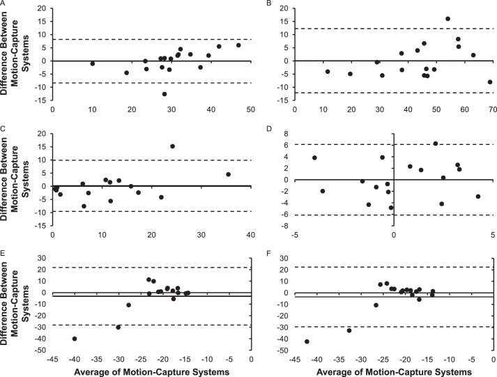
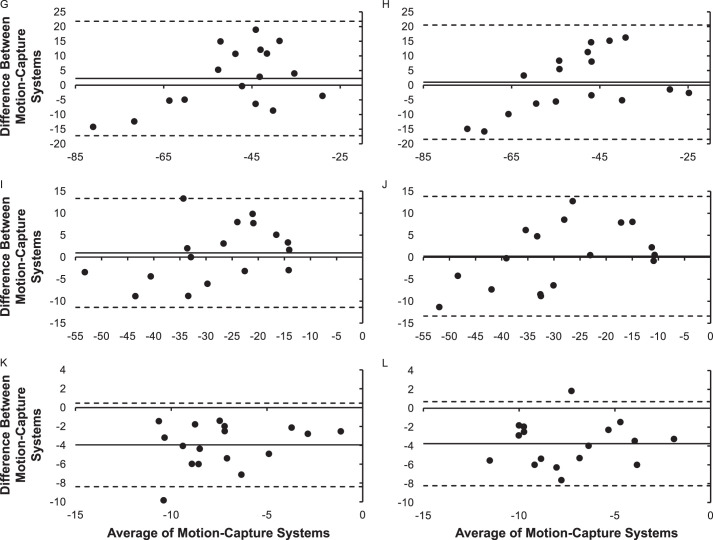
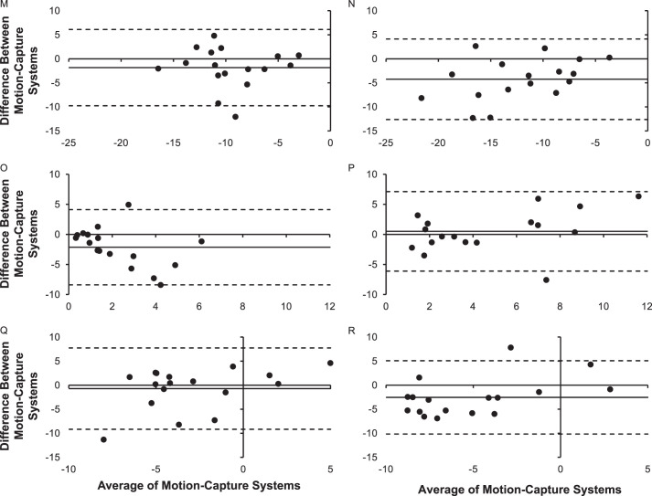
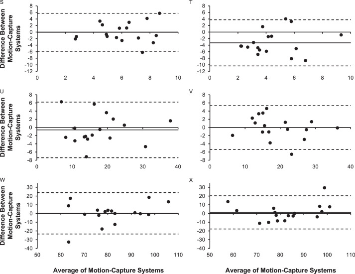
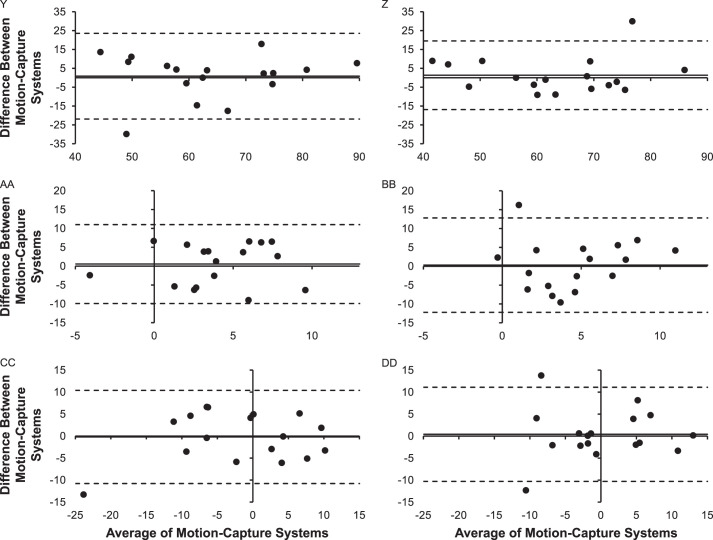
We assessed intersystem reliability via intraclass correlation coefficients (ICCs; model 2,1). The ICC values were interpreted as follows: poor, <0.50; moderate, 0.50–0.75; good, 0.76–0.90; or excellent, 0.91–1.00.22 Paired-samples t tests were used to identify between-systems differences for the mean joint angles of each outcome of interest. For both analyses, the α level was set a priori at ≤.05. Additionally, we calculated Bland-Altman plots with corresponding 95% limits of agreement to give a visual representation of intersystem agreement (Appendix). We used SPSS (version 21.0; IBM Corp, Armonk, NY) for all statistical analyses.
RESULTS
Overall, we observed moderate agreement between the markerless and stereophotogrammetry motion-capture systems (ICC average = 0.58; ICC range = −1.52 to 0.96), with most variables (24/32, 75.00%) demonstrating agreement between systems. Only 18.75% (6/32) of variable mean measurements were different between the systems (Table 1).
Table 1.
Summary of Results by Movement Plane and Variable Categories
| Category |
Intraclass Correlation Coefficient |
Paired-Samples t-Test Difference, n/N (%) |
|||
| Average |
Maximum |
Minimum |
Difference, n/N (%) |
||
| Sagittal plane | 0.84 | 0.96 | 0.50 | 14/15 (93.33) | 0/15 (0.00) |
| Frontal plane | 0.35 | 0.92 | −1.52 | 10/17 (58.82) | 6/17 (35.29) |
| Initial ground contact of jump landing | 0.56 | 0.96 | −0.19 | 6/10 (60.00) | 2/10 (20.00) |
| Maximum joint anglea | 0.77 | 0.96 | 0.55 | 11/11 (100.00) | 2/11 (18.18) |
| Joint-angle displacementb | 0.41 | 0.95 | −1.52 | 7/11 (63.64) | 2/11 (18.18) |
| Overall | 0.58 | 0.96 | −1.52 | 24/32 (75.00) | 6/32 (18.75) |
Maximum joint angle during the landing phase of the initial landing.
Joint-angle displacement from initial ground contact to the maximum joint angle during the landing phase of the initial landing.
Specifically, we noted excellent agreement for 8 variables, good agreement for 7 variables, moderate agreement for 10 variables, and poor agreement for 7 variables. Better agreement existed between motion-capture systems for sagittal-plane (poor = 0, moderate = 3, good = 5, excellent = 7) than frontal-plane (poor = 7, moderate = 7, good = 2, excellent = 1) variables. Agreement was also better between systems for MAX (poor = 0, moderate = 5, good = 4, excellent = 2) than either IC angle (poor = 3, moderate = 4, good = 0, excellent = 3) or DSP (poor = 4, moderate = 1, good = 3, excellent = 3). These findings are reported in Tables 2, 3, and 4 for the trunk, hip, and knee, respectively.
Table 2.
Trunk Joint-Angle Means, 95% CI, Intraclass Correlation Coefficients, and Paired-Samples t-Tests
| Variable |
Motion-Capture System |
Mean (95% CI) |
Intraclass Correlation Coefficient (2,1) |
Paired-Samples t-Test |
||
| Value |
P Value |
Value |
P Value |
|||
| Trunk flexion | ||||||
| Initial ground contact of jump landing | Stereophotogrammetric | 30.07 (25.55, 34.59) | 0.94 | <.001d | −0.09 | .93 |
| Kinectc-based markerless | 30.12 (26.62, 33.69) | |||||
| Maximum joint anglea | Stereophotogrammetric | 43.55 (36.20, 50.91) | 0.96 | <.001d | 0.18 | .86 |
| Kinect-based markerless | 42.39 (36.78, 49.80) | |||||
| Joint-angle displacementb | Stereophotogrammetric | 13.49 (7.55, 19.42) | 0.95 | <.001d | 0.30 | .77 |
| Kinect-based markerless | 13.13 (8.33, 17.94) | |||||
| Lateral trunk flexion | ||||||
| Initial ground contact of jump landing | Stereophotogrammetric | 0.26 (−1.10, 1.63) | 0.53 | .07 | −0.20 | .84 |
| Kinect-based markerless | 0.41 (−0.83, 1.66) | |||||
Maximum joint angle during the landing phase of the initial landing.
Joint-angle displacement from initial ground contact to the maximum joint angle during the landing phase of the initial landing.
Microsoft Corp, Redmond, WA.
Indicates difference (P ≤ .05).
Table 3.
Hip-Joint Angle Means, 95% CI, Intraclass Correlation Coefficients, and Paired-Samples t-Tests
| Variable |
Motion-Capture System |
Mean (95% CI) |
Intraclass Correlation Coefficient (2,1) |
Paired-Samples t-Test |
||
| Value |
P Value |
Value |
P Value |
|||
| Hip flexion | ||||||
| Initial ground contact of jump landing, right side | Stereophotogrammetric | −18.09 (−20.61, −15.58) | 0.50 | .10 | 1.17 | .26 |
| Kinectc-based markerless | −19.70 (−21.84, −17.56) | |||||
| Initial ground contact of jump landing, left side | Stereophotogrammetric | −19.34 (−21.40, −17.29) | 0.63 | .03d | 1.08 | .30 |
| Kinect-based markerless | −20.60 (−22.91, −18.29) | |||||
| Maximum joint angle, right sidea | Stereophotogrammetric | −47.68 (−54.98, −39.98) | 0.86 | <.001d | 1.06 | .30 |
| Kinect-based markerless | −49.98 (−55.25, −44.71) | |||||
| Maximum joint angle, left sidea | Stereophotogrammetric | −49.65 (−57.22, −42.07) | 0.86 | <.001d | 0.55 | .59 |
| Kinect-based markerless | −50.94 (−56.36, −45.52) | |||||
| Joint-angle displacement, right sideb | Stereophotogrammetric | −27.50 (−33.26, −21.73) | 0.91 | <.001d | 0.50 | .62 |
| Kinect-based markerless | −28.26 (−32.91, −23.60) | |||||
| Joint-angle displacement, left sideb | Stereophotogrammetric | −28.28 (−34.99, −21.59) | 0.92 | <.001d | 0.17 | .87 |
| Kinect-based markerless | −28.56 (−33.70, −23.42) | |||||
| Hip frontal | ||||||
| Initial ground contact of jump landing, right side | Stereophotogrammetric | −9.06 (−10.38, −7.74) | 0.47 | .01d | −5.16 | <.001d |
| Kinect-based markerless | −5.68 (−6.94, −4.41) | |||||
| Initial ground contact of jump landing, left side | Stereophotogrammetric | −10.01 (−11.62, −8.41) | 0.61 | <.001d | −7.83 | <.001d |
| Kinect-based markerless | −5.94 (−7.27, −4.60) | |||||
| Hip adduction | ||||||
| Maximum joint angle, right sidea | Stereophotogrammetric | −2.99 (−4.96, −1.01) | 0.55 | .050d | −0.02 | .99 |
| Kinect-based markerless | −3.01 (−4.51, −1.50) | |||||
| Maximum joint angle, left sidea | Stereophotogrammetric | −6.94 (−9.18, −4.70) | 0.68 | .002d | −3.27 | .004d |
| Kinect-based markerless | −4.17 (−5.60, −2.73) | |||||
| Joint-angle displacement, right sideb | Stereophotogrammetric | 6.07 (4.92, 7.23) | 0.02 | .49 | −0.05 | .96 |
| Kinect-based markerless | 6.03 (5.09, 6.98) | |||||
| Joint-angle displacement, left sideb | Stereophotogrammetric | 3.10 (1.84, 4.37) | −0.04 | .55 | −3.58 | .002d |
| Kinect-based markerless | 5.71 (4.80, 6.62) | |||||
| Hip abduction | ||||||
| Maximum joint angle, right sidea | Stereophotogrammetric | −10.38 (−12.02, −8.74) | 0.60 | .02d | −1.38 | .18 |
| Kinect-based markerless | −9.04 (−10.98, −7.09) | |||||
| Maximum joint angle, left sidea | Stereophotogrammetric | −15.08 (−18.00, −12.17) | 0.63 | .001d | −4.785 | <.001d |
| Kinect-based markerless | −9.77 (−11.55, −8.00) | |||||
| Joint-angle displacement, right sideb | Stereophotogrammetric | 1.32 (0.56, 2.08) | 0.17 | .30 | −2.97 | .008d |
| Kinect-based markerless | 3.36 (2.14, 4.57) | |||||
| Joint-angle displacement, left sideb | Stereophotogrammetric | 5.15 (3.35, 6.96) | 0.67 | .01d | 1.11 | .28 |
| Kinect-based markerless | 3.85 (2.70, 5.00) | |||||
Maximum joint angle during the landing phase of the initial landing.
Joint-angle displacement from initial ground contact to the maximum joint angle during the landing phase of the initial landing.
Microsoft Corp, Redmond, WA.
Indicates difference (P ≤ .05).
Table 4.
Knee-Joint Angle Means, 95% CIs, Intraclass Correlation Coefficients, and Paired-Samples t-Tests
| Variable |
Motion-Capture System |
Mean (95% CI) |
Intraclass Correlation Coefficient (2,1) |
Paired-Samples t-Test |
||
| Value |
P Value |
Value |
P Value |
|||
| Knee flexion | ||||||
| Initial ground contact of jump landing, right side | Stereophotogrammetric | 16.74 (12.94, 20.53) | 0.95 | <.001d | −0.97 | .35 |
| Kinectc-based markerless | 17.56 (13.88, 21.24) | |||||
| Initial ground contact of jump landing, left side | Stereophotogrammetric | 19.14 (15.87, 22.42) | 0.96 | <.001d | 0.14 | .89 |
| Kinectc-based markerless | 19.05 (15.50, 22.60) | |||||
| Maximum joint angle, right sidea | Stereophotogrammetric | 81.57 (74.53, 88.60) | 0.75 | .003d | 0.21 | .84 |
| Kinectc-based markerless | 80.97 (75.47, 86.46) | |||||
| Maximum joint angle, left sidea | Stereophotogrammetric | 83.03 (76.40, 89.66) | 0.85 | <.001d | 0.60 | .56 |
| Kinectc-based markerless | 81.67 (76.09, 87.24) | |||||
| Joint-angle displacement, right sideb | Stereophotogrammetric | 64.83 (58.28, 71.37) | 0.76 | .003d | 0.51 | .62 |
| Kinectc-based markerless | 63.41 (57.48, 69.34) | |||||
| Joint-angle displacement, left sideb | Stereophotogrammetric | 63.89 (57.87, 69.91) | 0.85 | <.001d | 0.58 | .57 |
| Kinectc-based markerless | 62.62 (56.72, 68.53) | |||||
| Knee valgus | ||||||
| Initial ground contact of jump landing, right side | Stereophotogrammetric | 3.96 (1.87, 6.05) | 0.21 | .31 | 0.06 | .95 |
| Kinectc-based markerless | 3.87 (2.00, 5.74) | |||||
| Initial ground contact of jump landing, left side | Stereophotogrammetric | 4.33 (2.06, 6.60) | −0.19 | .64 | −0.07 | .95 |
| Kinectc-based markerless | 4.80 (2.58, 6.29) | |||||
| Maximum joint angle, right sidea | Stereophotogrammetric | −3.20 (−8.32, 1.92) | 0.92 | <.001d | −0.45 | .66 |
| Kinectc-based markerless | −2.60 (−6.97, 1.77) | |||||
| Maximum joint angle, left sidea | Stereophotogrammetric | −0.38 (−4.19, 3.43) | 0.83 | <.001d | −0.11 | .92 |
| Kinectc-based markerless | −0.23 (−3.56, 3.10) | |||||
| Joint-angle displacement, right sideb | Stereophotogrammetric | −7.16 (−10.72, −3.60) | 0.80 | .001d | −0.67 | .51 |
| Kinectc-based markerless | −5.96 (−10.84, −1.07) | |||||
| Joint-angle displacement, left sideb | Stereophotogrammetric | −4.71 (−6.99, −2.44) | −1.52 | .97 | −0.06 | .95 |
| Kinectc-based markerless | −4.57 (−7.83, −1.31) | |||||
Maximum joint angle during the landing phase of the initial landing.
Joint-angle displacement from initial ground contact to the maximum joint angle during the landing phase of the initial landing.
Microsoft Corp, Redmond, WA.
Indicates difference (P ≤ .05).
The joint quantitative and qualitative assessment of the mean comparisons and Bland-Altman plots (Appendix) identified the following trends for the markerless motion-capture system: underestimated sagittal-plane measures (13/15; 86.67%) and overestimated frontal-plane measures (13/17; 76.47%). No trends were present for IC measures (underestimate: 4/10, 40.00%; overestimate: 6/10, 60.00%), MAX measurements (underestimate: 6/11, 54.55%; overestimate: 5/11, 45.45%), or DSP measures (underestimate: 7/11, 63.64%; overestimate: 4/11, 36.36%). The Bland-Altman plots also showed that, in general, the mean difference between the 2 motion-capture systems was more closely centered on zero for sagittal- than frontal-plane variables.
Trunk
Agreement was excellent for sagittal-plane trunk angles between the markerless and stereophotogrammetric motion-capture systems. Moderate agreement was evident for lateral trunk flexion at IC between the systems. No mean trunk-angle differences were observed between systems. Trunk-angle means with 95% confidence intervals (CIs), ICCs, and paired-samples t-test statistics are reported in Table 2.
Hip
Moderate-to-excellent agreement was present for sagittal-plane hip angles between the markerless and stereophotogrammetric motion-capture systems. Poor-to-moderate agreement was demonstrated for frontal-plane hip-joint angles between the systems. Intraclass correlations were significant for all hip-joint angle measurements except right hip flexion at IC, right and left hip-adduction DSP, and right hip-abduction DSP. Mean hip-joint angle differences were seen for right and left hip frontal-plane joint angles at IC, left hip-adduction and -abduction MAX, left hip-adduction DSP, and right hip-abduction DSP. Hip-joint angle means with 95% CIs, ICCs, and paired-samples t-test statistics are reported in Table 3.
Knee
Agreement was moderate to excellent for sagittal-plane knee-joint angles between the markerless and stereophotogrammetric motion-capture systems. Poor-to-excellent agreement was found for frontal-plane knee-joint angles between the systems. Significant ICCs were present for all knee-joint angles except right and left knee-valgus angle at IC and left knee-valgus angle DSP. No mean knee-joint angle differences existed between systems. Knee-joint angle means with 95% CIs, ICCs, and paired-samples t-test statistics are reported in Table 4.
DISCUSSION
Overall, moderate agreement was observed between the markerless motion-capture system and the criterion standard stereophotogrammetric system. In general, agreement was better between sagittal-plane kinematic measures than between frontal-plane measures, as well as between maximum joint-angle outcomes, than between IC joint angles or DSP outcomes. Our findings are consistent with previous work in which researchers9,10,13 compared markerless and stereophotogrammetric motion-capture systems. To our knowledge, we are the first to validate a commercially available Kinect-based markerless motion-capture system that can readily provide clinicians with quantitative data for use in clinical assessments.
The differences in sagittal- and frontal-plane levels of agreement in our study were similar to those reported earlier.8–10 These findings were not surprising but were counterintuitive to what would be expected. The Microsoft Kinect camera is aligned perpendicular to the frontal plane, so one would expect the camera to be better able to detect frontal- than sagittal-plane joint angles. However, sagittal-plane joint angles are typically larger than frontal-plane angles, especially for MAX, so any limitations in the markerless motion-capture system's ability to detect minute changes in joint angles may be minimized because of the larger overall joint angles in the sagittal plane. Similar findings have been noted among validated 3D motion-capture systems.23
Our results are comparable with those reported by Mauntel et al15 and Dar et al,7 who compared a markerless motion-capture system with the criterion standard (ie, expert raters) for qualitative analysis of trunk and lower extremity movement patterns during a jump-landing. Both groups7,15 validated the ability of a Kinect-based markerless motion-capture system to accurately assess the Landing Error Scoring System.1,24 In these studies,7,15 the markerless motion-capture system reliably identified trunk and lower extremity movement errors during a jump-landing, with most Landing Error Scoring System items demonstrating almost perfect agreement.
In the Landing Error Scoring System, gross movement quality is visually scored and, thus, minute changes in joint angles are less important. As such, Mauntel et al15 reported better agreement between the markerless motion-capture system and expert raters for MAX and DSP movement errors than for movement errors identified at IC. Collectively, these findings suggest that markerless motion-capture systems are limited in their ability to identify small differences in trunk and lower extremity kinematics. However, markerless motion-capture systems can effectively identify larger movement patterns and may be useful in automating and objectively quantifying clinical movement screenings that have previously involved visual identification of gross movement patterns.1,24
The inherent limitations of Kinect-based markerless motion-capture systems affect their ability to consistently and accurately calculate trunk and lower extremity joint angles during jump-landing assessments. The markerless motion-capture system was limited in its ability to calculate hip frontal-plane angles because individuals landing from a jump commonly exhibit deep knee flexion, and the knees can block the Kinect camera from visualizing the hip joints. Therefore, the markerless motion-capture system may be unable to track the virtual hip-joint markers. Overall, the markerless motion-capture system is limited in its ability to accurately calculate frontal-plane hip angles.
Additionally, the markerless motion-capture system was limited in its ability to identify trunk and lower extremity joint angles at IC. Microsoft Kinect depth cameras collect video data at 30 Hz, whereas we sampled the force-platform data at 1200 Hz. Fewer data points (frames) inhibit the Microsoft Kinect's ability to accurately identify IC, and the actual frame in which ground contact occurs may be missed by the camera. The markerless motion-capture system software attempts to correct for this limitation by identifying IC and the frames immediately preceding and following that frame. The software then averages the trunk and hip-joint angles across those 3 frames.
Also, the markerless motion-capture and stereophotogrammetric systems defined initial ground contact differently. The markerless motion-capture system defined initial ground contact as the frame before the entire foot was in contact with the ground. The stereophotogrammetric system identified initial ground contact as the point when the vertical ground reaction forces exceeded 10 N. This difference in definitions could have led to some of the discrepancies observed between the systems for trunk and lower extremity kinematics at IC.
Poor agreement was demonstrated for the DSP measures (Table 1). This is likely the result of these measurements being derived from 2 directly measured joint angles: the joint angle at IC and the MAX during the initial landing phase of the jump landing. As such, more noise, and subsequently error, may be introduced into the measure; this is true for both the stereophotogrammetric and markerless motion-capture systems. The additional error in these measures may have reduced the agreement between systems.
Microsoft Kinect-based motion-capture systems are highly efficient for assessing many individuals in a short time.7,15 Whereas the Kinect-based system may be limited in its ability to identify small differences in trunk and lower extremity joint angles, it can provide reliable and valid identification of gross movement-pattern differences.7,15 Objective identification of gross movement-pattern differences may be useful as a preliminary screening tool when assessing many individuals (eg, during preparticipation physical examinations) or in the rehabilitation setting when assessing individuals for movement alterations after a musculoskeletal injury. In both cases, individuals may benefit from undergoing more precise testing to further quantify their movement quality. Therefore, the Kinect-based motion-capture system can aid clinical practice by efficiently identifying primary and secondary biomechanical injury risk factors, as well as changes in movement quality over time when more advanced measures (eg, 3D motion-capture systems) are unavailable or not feasible within the clinical setting.
The following limitations should be considered when interpreting our findings. Only 1 movement assessment was examined; additional movements should be assessed in order to develop this markerless motion-capture system into a more robust system. Our study sample consisted solely of healthy individuals. Therefore, the system must be validated in individuals with previous lower extremity injuries because they are at the greatest risk of future injury. We did not evaluate transverse-plane joint angles. However, previous researchers8,16–19 who examined the ability of Microsoft Kinect markerless motion-capture systems to accurately calculate transverse-plane joint angles demonstrated poor agreement with stereophotogrammetric systems. Similar findings regarding worse agreement for transverse-plane measures have been seen between validated 3D motion-capture systems.23 Finally, ankle-joint kinematics were not included in our analyses. Future researchers should evaluate the validity of Microsoft Kinect-based markerless motion-capture systems for ankle-joint kinematics, as the ankle plantar-flexion angle may influence the lower extremity injury risk.3,6,25,26
CONCLUSIONS
Moderate agreement existed between the markerless and stereophotogrammetric motion-capture systems for trunk and lower extremity kinematics during a jump-landing assessment. The markerless motion-capture system was better at calculating sagittal- than frontal-plane joint angles. Furthermore, the markerless motion-capture system was limited in its ability to accurately calculate joint angles at IC and frontal-plane joint angles, which may have important implications for injury risk. For these reasons, until further refinement occurs, markerless motion-capture systems should be used with caution for identifying small differences in joint kinematics during high-velocity functional assessments. However, the Microsoft Kinect-based markerless motion-capture system correctly identified differences in gross movement patterns and thus may aid clinicians in identifying individuals at increased risk of injury. The system can be used to efficiently screen many individuals and identify individuals with gross movement errors who may benefit from a more robust and in-depth biomechanical screening assessment, including the use of a stereophotogrammetric motion-capture system.
ACKNOWLEDGMENTS
The views expressed in this paper are those of the authors and do not reflect the official policy of the Department of the Army/Navy/Air Force, Department of Defense, Uniformed Services University of the Health Sciences, or US government.
No financial assistance was provided for this research. PhysiMax Technologies Ltd provided their software to the research laboratory at no cost. Dr Padua has served as a member of the PhysiMax Scientific Advisory Board. However, he and members of his family do not have any financial interest in and receive no form of financial compensation from PhysiMax.
Appendix.
A, Trunk flexion, IC; B, Trunk flexion, MAX; C, Trunk flexion, DSP; D, Lateral trunk flexion at IC; E, Right hip flexion, IC; F, Left hip flexion, IC; G, Right hip flexion, MAX; H, Left hip flexion, MAX; I, Right hip flexion, DSP; J, Left hip flexion, DSP; K, Right hip abduction or adduction, IC; L, Left hip abduction or adduction, IC; M, Right hip abduction, MAX; N, Left hip abduction, MAX; O, Right hip-abduction, DSP; P, Left hip-abduction, DSP; Q, Right hip adduction, MAX; R, Left hip adduction, MAX; S, Right hip adduction, DSP; T, Left hip adduction, DSP; U, Right knee flexion, IC; V, Left knee flexion, IC; W, Right knee flexion, MAX; X, Left knee flexion, MAX; Y, Right knee flexion, DSP; Z, Left knee flexion, DSP; AA, Right knee valgus or varus, IC; BB, Left knee valgus or varus, IC; CC, Right knee valgus or varus, MAX; DD, Left knee valgus or varus, MAX; EE, Right knee valgus or varus, DSP; and FF, Left knee valgus or varus, DSP. Abbreviations: IC, initial ground contact of the jump landing; MAX, maximum joint angle during the landing phase of the initial landing; DSP, joint-angle displacement from initial ground contact to maximum joint angle during the landing phase of the initial landing. Continued on next page.
Appendix.
Continued from previous page. Continued on next page.
Appendix.
Continued from previous page. Continued on next page.
Appendix.
Continued from previous page. Continued on next page.
Appendix.
Continued from previous page. Continued on next page.
Appendix.
Continued from previous page.
REFERENCES
- 1. .Padua DA, Marshall SW, Boling MC, Thigpen CA, Garrett WE, Jr, Beutler AI. The Landing Error Scoring System (LESS) is a valid and reliable clinical assessment tool of jump-landing biomechanics: the JUMP-ACL study. Am J Sports Med. 2009;37(10):1996–2002. doi: 10.1177/0363546509343200. [DOI] [PubMed] [Google Scholar]
- 2. .Padua DA, DiStefano LJ, Beutler AI, de la Motte SJ, DiStefano MJ, Marshall SW. The Landing Error Scoring System as a screening tool for an anterior cruciate ligament injury-prevention program in elite-youth soccer athletes. J Athl Train. 2015;50(6):589–595. doi: 10.4085/1062-6050-50.1.10. [DOI] [PMC free article] [PubMed] [Google Scholar]
- 3. .Cameron KL, Peck KY, Owens BD, et al. Biomechanical risk factors for lower extremity stress fracture. Orthop J Sports Med. 2013;1(suppl 4) doi: 10.1177/2325967113S00019. [DOI] [Google Scholar]
- 4. .Hewett TE, Myer GD, Ford KR, et al. Biomechanical measures of neuromuscular control and valgus loading of the knee predict anterior cruciate ligament injury risk in female athletes: a prospective study. Am J Sports Med. 2005;33(4):492–501. doi: 10.1177/0363546504269591. [DOI] [PubMed] [Google Scholar]
- 5. .Teyhen D, Bergeron MF, Deuster P, et al. Consortium for health and military performance and American College of Sports Medicine Summit: utility of functional movement assessment in identifying musculoskeletal injury risk. Curr Sports Med Rep. 2014;13(1):52–63. doi: 10.1249/JSR.0000000000000023. [DOI] [PubMed] [Google Scholar]
- 6. .Cameron KL, Peck KY, Owens BD, et al. Landing Error Scoring System (LESS) items are associated with the incidence rate of lower extremity stress fractures. Orthop J Sports Med. 2014;2(7) doi: 10.1177/2325967114S00080. (suppl 2) [DOI] [PMC free article] [PubMed] [Google Scholar]
- 7. .Dar G, Yehiel A, Cale' Benzoor M. Concurrent criterion validity of a novel portable motion analysis system for assessing the landing error scoring system (LESS) test. Sports Biomech. 2019;18(4):426–436. doi: 10.1080/14763141.2017.1412495. [DOI] [PubMed] [Google Scholar]
- 8. .Schmitz A, Ye M, Shapiro R, Yang R, Noehren B. Accuracy and repeatability of joint angles measured using a single camera markerless motion capture system. J Biomech. 2014;47(2):587–591. doi: 10.1016/j.jbiomech.2013.11.031. [DOI] [PubMed] [Google Scholar]
- 9. .Schmitz A, Ye M, Boggess G, Shapiro R, Yang R, Noehren B. The measurement of in vivo joint angles during a squat using a single camera markerless motion capture system as compared to a marker based system. Gait Posture. 2015;41(2):694–698. doi: 10.1016/j.gaitpost.2015.01.028. [DOI] [PubMed] [Google Scholar]
- 10. .Gray AD, Marks JM, Stone EE, Butler MC, Skubic M, Sherman SL. Validation of the Microsoft Kinect as a portable and inexpensive screening tool for identifying ACL injury risk. Orthop J Sports Med. 2014;2(7)(suppl 2) doi:org/10.1177/2325967114S00106. [Google Scholar]
- 11. .Clark RA, Pua YH, Oliveira CC, et al. Reliability and concurrent validity of the Microsoft Xbox One Kinect for assessment of standing balance and postural control. Gait Posture. 2015;42(2):210–213. doi: 10.1016/j.gaitpost.2015.03.005. [DOI] [PubMed] [Google Scholar]
- 12. .Clark RA, Pua YH, Fortin K, et al. Validity of the Microsoft Kinect for assessment of postural control. Gait Posture. 2012;36(3):372–377. doi: 10.1016/j.gaitpost.2012.03.033. [DOI] [PubMed] [Google Scholar]
- 13. .Eltoukhy M, Kelly A, Kim CY, Jun HP, Campbell R, Kuenze C. Validation of the Microsoft Kinect® camera system for measurement of lower extremity jump landing and squatting kinematics. Sports Biomech. 2016;15(1):89–102. doi: 10.1080/14763141.2015.1123766. [DOI] [PubMed] [Google Scholar]
- 14. .Bonnechere B, Jansen B, Salvia P, et al. Validity and reliability of the Kinect within functional assessment activities: comparison with standard stereophotogrammetry. Gait Posture. 2014;39(1):593–598. doi: 10.1016/j.gaitpost.2013.09.018. [DOI] [PubMed] [Google Scholar]
- 15. .Mauntel TC, Padua DA, Stanley LE, et al. Automated quantification of the Landing Error Scoring System with a markerless motion-capture system. J Athl Train. 2017;52(11):1002–1009. doi: 10.4085/1062-6050-52.10.12. [DOI] [PMC free article] [PubMed] [Google Scholar]
- 16. .Mentiplay BF, Hasanki K, Perraton LG, Pua YH, Charlton PC, Clark RA. Three-dimensional assessment of squats and drop jumps using the Microsoft Xbox One Kinect: reliability and validity. J Sports Sci. 2018;36(19):2202–2209. doi: 10.1080/02640414.2018.1445439. [DOI] [PubMed] [Google Scholar]
- 17. .Kotsifaki A, Whiteley R, Hansen C. Dual Kinect v2 system can capture lower limb kinematics reasonably well in a clinical setting: concurrent validity of a dual camera markerless motion capture system in professional football players. BMJ Open Sport Exerc Med. 2018;4(1):e000441. doi: 10.1136/bmjsem-2018-000441. [DOI] [PMC free article] [PubMed] [Google Scholar]
- 18. .Eltoukhy M, Kuenze C, Oh J, Wooten S, Signorile J. Kinect-based assessment of lower limb kinematics and dynamic postural control during the Star Excursion Balance Test. Gait Posture. 2017;58:421–427. doi: 10.1016/j.gaitpost.2017.09.010. [DOI] [PubMed] [Google Scholar]
- 19. .Choppin S, Lane B, Wheat J. The accuracy of the Microsoft Kinect in joint angle measurement. Sports Technol. 2014;7(1–2):98–105. doi: 10.1080/19346182.2014.968165. [DOI] [Google Scholar]
- 20. .Guess TM, Razu S, Jahandar A, Skubic M, Huo Z. Comparison of 3D joint angles measured with the Kinect 2.0 skeletal tracker versus a marker-based motion capture system. J Appl Biomech. 2017;33(2):176–181. doi: 10.1123/jab.2016-0107. [DOI] [PubMed] [Google Scholar]
- 21. .Bell AL, Pedersen DR, Brand RA. A comparison of the accuracy of several hip center location prediction methods. J Biomech. 1990;23(6):617–621. doi: 10.1016/0021-9290(90)90054-7. [DOI] [PubMed] [Google Scholar]
- 22. .Portney LG, Watkins MP. Foundations of Clinical Research Applications to Practice 3rd ed. Upper Saddle River, NJ: Pearson/Prentice Hall; 2009. [Google Scholar]
- 23. .Hewett TE, Roewer B, Ford K, Myer G. Multicenter trial of motion analysis for injury risk prediction: lessons learned from prospective longitudinal large cohort combined biomechanical-epidemiological studies. Braz J Phys Ther. 2015;19(5):398–409. doi: 10.1590/bjpt-rbf.2014.0121. [DOI] [PMC free article] [PubMed] [Google Scholar]
- 24. .Root H, Trojian T, Martinez J, Kraemer W, DiStefano LJ. Landing technique and performance in youth athletes after a single injury-prevention program session. J Athl Train. 2015;50(11):1149–1157. doi: 10.4085/1062-6050-50.11.01. [DOI] [PMC free article] [PubMed] [Google Scholar]
- 25. .Kovacs I, Tihanyi J, Devita P, Racz L, Barrier J, Hortobagyi T. Foot placement modifies kinematics and kinetics during drop jumping. Med Sci Sports Exerc. 1999;31(5):708–716. doi: 10.1097/00005768-199905000-00014. [DOI] [PubMed] [Google Scholar]
- 26. .van der Worp H, Vrielink JW, Bredeweg SW. Do runners who suffer injuries have higher vertical ground reaction forces that those who remain injury-free? A systematic review and meta-analysis. Br J Sports Med. 2016;50:450–457. doi: 10.1136/bjsports-2015-094924. [DOI] [PubMed] [Google Scholar]


