Abstract
A 9-year-old male patient, with an ultrasonography diagnosis of horseshoe kidney condition, was referred to our hospital for recurrent urinary tract infection. He was submitted to 99mTc-diethylenetriamine pentaacetic acid sequential scintigraphy that demonstrated urine stasis in the calyces of both kidneys and was also suggestive for 8-shaped right-to-left crossed-fused renal ectopia. A subsequently performed 99mTc-dimercaptosuccinic acid scan confirmed the diagnosis of crossed renal ectopia, also disclosing, through single-photon emission computed tomography (SPECT) acquisition, cortical uptake defects in both kidneys, and indicative of renal scarring due to recurrent pyelonephritis. Combined scintigraphy and appropriate technological approaches (SPECT/3D volume rendering) may be useful in selected patients with congenital anomalies.
Keywords: 99mTc-diethylenetriamine pentaacetic acid, 99mTc-dimercaptosuccinic acid, congenital abnormalities, renal scintigraphy, single-photon emission computed tomography
Congenital anomalies of the kidneys include a wide range of abnormalities, from substantially asymptomatic conditions to severe and life-threatening pathologies, and constitute about 10%–12% of the malformations identified in the adults.[1] As concerns the positional abnormalities, fused crossed ectopia is the rarest one, occurring in 1/1000 live births, and is defined as a condition in which one of the kidneys is positioned across the midline and merged with the contralateral one.
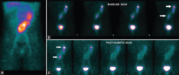
Here, we report a case of a 9-year-old male patient affected by congenital external auditory canal atresia, who was admitted to our attention for recurrent urinary tract infections and pain in the left flank. He had previously undergone an abdominal ultrasound that had diagnosed a horseshoe condition, without evidence of calculi or ectasia. A 9-month follow-up showed normal glomerular and tubular function (glomerular filtration rate 116 ml/min/1.73 m2, EFNa 0.8%, and TRP 90%) and the absence of significant proteinuria (Urinary Proteine/creatinine ratio 0.15). Due to the recurrent fever and pain in the left flank, obstruction was suspected and for further examination, he was submitted to sequential scintigraphy with 99mTc-diethylenetriamine pentaacetic acid (99mTc-DTPA) acquired through both anterior and posterior detectors, according to Santa Fe Consensus Report.[2] A baseline 20-min acquisition was performed: the phase of parenchymal uptake showed an image suggestive for 8-shaped right-to-left crossed fused renal ectopia, furthermore at the excretion phase urine stasis in the calyces of both kidneys was registered. At the conclusion of the baseline scan, furosemide (1 mg/kg) was intravenously administered and a second 20-min dynamic study gave evidence of prompt resolution of the calyceal stasis [Figure 1], thus excluding the clinical suspect of urinary tract obstruction (T1/2, calculated as the time necessary for the activity in the kidney reducing to 50% of its maximum value, was 2 min).
Figure 1.
Sequential scintigraphy with 99mTc-diethylenetriamine pentaacetic acid, anterior view. Parenchymal phase was suggestive for 8-shaped right-to-left crossed-fused renal ectopia (a). The excretory phase showed stasis of urine in the calyceal system of both kidneys (b, white arrows). Post diuretic scan demonstrated prompt resolution of the stasis in the calyceal system (c, white arrows)
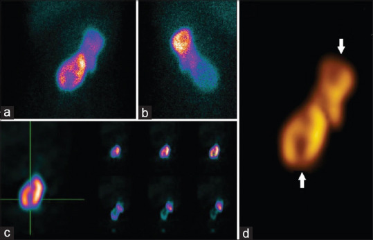
One week later, the patient was submitted to scintigraphy with Tc99 m dimercaptosuccinic acid (99mTc-DMSA), that showed an empty right renal fossa and left crossed renal ectopia with fusion, but also disclosed cortical uptake defects borne by both kidneys, as well demonstrated by planars, single-photon emission computed tomography (SPECT) examination and 3D volume rendering, suggestive of renal scarring due to recurrent pyelonephritis [Figure 2].
Figure 2.

Scintigraphy with 99mTc-dimercaptosuccinic acid. Planar anterior (a) and posterior (b) confirmed the scintigraphic findings of right-to-left crossed fused renal ectopia. Cortical defects, suggestive of scarring due to recurrent pyelonephritis, are visible in both kidneys, as well evident at single-photon emission computed tomography examination (c, triangulation) and at the 3D volume rendering (d, white arrow)
Among the possible presentations of crossed fused renal ectopia, the case we describe is the rarest variant since left to right is more common than right to left with a 3:1 ratio. Fused crossed ectopia can be accidentally diagnosed in asymptomatic children during occasional clinical investigations, but in some patients, it can cause complications such as recurrent infections and renal obstruction. The diagnosis of crossed fused renal ectopia may be challenging, especially in children, since ultrasound examination presents several limitations. Computed tomography (CT) or intravenous urography, although more sensitive and accurate, are characterized by a significant radiation burden delivered to the patients.[3]
Sequential scintigraphy with 99mTc-DTPA is extensively used in pediatric clinical practice, for the estimation of different features of the renal function, such as clearance and excretion. On the other hand, imaging with 99mTc-DMSA provides crucial information for the diagnosis of cortical defects in children with vesicoureteric reflux and urinary tract infections, as demonstrated by Ditchfield and Nadel in a large cohort of 129 children.[4] In the previously cited article, the authors reported that cortical defects represent common findings at 99mTc-DSMA scan in pediatric subjects with urinary infection, frequently occurring even in the absence of vesicoureteric reflux. Therefore, the combined use of 99mTc-DTPA and 99mTc-DMSA has been introduced in pediatric nephrology for the simultaneous evaluation of clearance/excretion and cortical scars. From a technical point of view, it has been demonstrated that the tomographic technique (i.e., SPECT) may be of great value for the detection of small cortical defects.[5] In this regard, it has to be underlined that hybrid SPECT/CT has been more recently introduced for further improving SPECT accuracy.[6] In a recently published article by Dehesa et al., SPECT/CT resulted in value for the correct characterization of crossed renal ectopia in an 11-year-old boy.[7] Of note, some concerns have to be raised due to dosimetric issues. Although Dehesa et al. calculated the additional radiation burden due to the CT component of the SPECT/CT system as very low (i.e., 3 mSv), in the pediatric setting, the hybrid approach should be reserved only to well-selected cases. In our specific case, the activity to be administered was calculated, both for 99mTc-DMSA and 99mTc-DTPA radiopharmaceuticals, according to the international guidelines that were demonstrated to provide very low dosimetry to children.[8,9]
Our case illustrates the clinical importance of the combined use of 99mTc-DTPA/ 99mTc-DMSA scintigraphy, also through the utilization of some appropriate technological approaches (i.e., SPECT/3D rendering), for the morpho-functional imaging of the kidney malformations in children. In addition, we suggest that combined scintigraphy may be useful in selected patients with congenital anomalies, together with laboratory tests and ambulatory blood pressure monitoring, to carefully monitor possible complications due to recurrent pyelonephritis.
Declaration of patient consent
The authors certify that they have obtained all appropriate patient consent forms.
Financial support and sponsorship
Nil.
Conflicts of interest
There are no conflicts of interest.
References
- 1.Mudoni A, Caccetta F, Caroppo M, Musio F, Accogli A, Zacheo MD, et al. Crossed fused renal ectopia: Case report and review of the literature. J Ultrasound. 2017;20:333–7. doi: 10.1007/s40477-017-0245-6. [DOI] [PMC free article] [PubMed] [Google Scholar]
- 2.O'Reilly P, Aurell M, Britton K, Kletter K, Rosenthal L, Testa T Consensus committee on diuresis renography. Consensus on diuresis renography for investigating the dilated upper urinary tract Radionuclides in nephrourology group Consensus committee on diuresis renography. J Nucl Med. 1996;37:1872–6. [PubMed] [Google Scholar]
- 3.Loganathan AK, Bal HS. Crossed fused renal ectopia in children: A review of clinical profile, surgical challenges, and outcome. J Pediatr Urol. 2019;15:315–21. doi: 10.1016/j.jpurol.2019.06.019. [DOI] [PubMed] [Google Scholar]
- 4.Ditchfield MR, Nadel HR. The DMSA scan in paediatric urinary tract infection. Australas Radiol. 1998;42:318–20. doi: 10.1111/j.1440-1673.1998.tb00530.x. [DOI] [PubMed] [Google Scholar]
- 5.Sheehy N, Tetrault TA, Zurakowski D, Vija AH, Fahey FH, Treves ST. Pediatric 99mTc-DMSA SPECT performed by using iterative reconstruction with isotropic resolution recovery: Improved image quality and reduced radiopharmaceutical activity. Radiology. 2009;251:511–6. doi: 10.1148/radiol.2512081440. [DOI] [PubMed] [Google Scholar]
- 6.Filippi L, Biancone L, Petruzziello C, Schillaci O. Tc-99m HMPAO-labeled leukocyte scintigraphy with hybrid SPECT/CT detects perianal fistulas in Crohn disease. Clin Nucl Med. 2006;31:541–2. doi: 10.1097/01.rlu.0000233082.89996.3a. [DOI] [PubMed] [Google Scholar]
- 7.Dehesa A, Zugazaga A, De Miguel MB, Biassoni L. Evaluation of a crossed fused renal ectopia in a paediatric patient using 99m Tc-DMSA SPECT/CT. Rev Esp Med Nucl Imagen Mol. 2016;35:406–8. doi: 10.1016/j.remn.2016.01.003. [DOI] [PubMed] [Google Scholar]
- 8.Taylor AT, Brandon DC, de Palma D, Blaufox MD, Durand E, Erbas B, et al. SNMMI procedure standard/EANM practice guideline for diuretic renal scintigraphy in adults with suspected upper urinary tract obstruction 1.0. Semin Nucl Med. 2018;48:377–90. doi: 10.1053/j.semnuclmed.2018.02.010. [DOI] [PMC free article] [PubMed] [Google Scholar]
- 9.Arteaga MV, Caballero VM, Rengifo KM. Dosimetry of 99mTc (DTPA, DMSA and MAG3) used in renal function studies of newborns and children. Appl Radiat Isot. 2018;138:25–8. doi: 10.1016/j.apradiso.2017.07.054. [DOI] [PubMed] [Google Scholar]


