Abstract
Background
The asymmetric medial and lateral condyles of the distal femur and proximal tibia have a direct influence on the biomechanics of knee joint and prostheses design. This study aimed to determine the morphologic data, that is., anteroposterior (AP) and mediolateral (ML) widths, and the radius of curvature (ROC) of the geometric arcs of the distal femur and proximal tibia.
Methods
One hundred and seventeen adult dry bones (57 femurs and 60 tibias) were studied. Aspect ratios (AP/ML) were calculated. The AP and ML widths were measured using digital Vernier Caliper with a measuring range of 0-150 mm, resolution of 0.01 mm, and accuracy ± 0.02 mm. The geometric arcs of femoral and tibial condyles were divided into three parts namely anterior 1/3rd, distal (femur) or middle (tibia) 1/3rd and posterior 1/3rd and were estimated in the sagittal plane for the femur and transverse plane for tibia using the ROC gauges.
Results
For the femur, the mean AP length for medial and lateral condyles was 55.62 mm and 57.93 mm, respectively, while the mean ML width was 73.45 mm. For the tibia, the mean AP length for medial condyle (MC) and lateral condyle (LC) was 47.74 mm and 43.46 mm, respectively. The mean aspect ratios for the distal femur and proximal tibia were 1.26 and 1.45, respectively. The mean aspect ratios for MC and LC of the femur were 0.50 and 0.52, respectively, whereas, for tibia, they were 0.61 and 0.71, respectively. The mean ROC for femoral MC - 20.77 mm, 31.42 mm, and 19.68 mm and for LC - 21.48 mm, 64.40 mm and 19.06 mm for the anterior, distal and posterior arcs, respectively. The mean ROC for tibial MC - 22.42 mm, 22.49 mm and 19.94 mm, and LC - 19.92 mm, 21.79 mm and 20.95 mm for the anterior, middle and posterior arcs, respectively.
Conclusions
The morphologic data accumulated in this study for both the distal femur as well as the proximal tibia would provide guidelines and help the manufacturers of joint prostheses to address the potential for compromised implant fit and re-design and make available ‘anatomic’ knee prostheses appropriate for the local population which would not only improve function but also prolong the longevity of the prostheses.
Keywords: : anatomic, asian, femoral condyle, knee, morphometric analysis, tibial condyle
Introduction
Knowledge of the three-dimensional geometry of the knee joint - the distal end of the femur and the proximal end of the tibia - is important as this joint is frequently affected in trauma, primary tumours of bone, metabolic bone disorders and degenerative joint diseases.
The distal end of the femur is widely expanded as a bearing surface for transmission of weight to the tibia. The medial and lateral condyles are significantly asymmetric in their morphology, the lateral condyle being longer anteroposteriorly, flatter and lying at a higher plane. These differences are important determinants of knee joint motion. The proximal surface of the tibia, also called the tibial plateau, is reciprocally expanded and presents medial and lateral articular surfaces for corresponding femoral condyles. The plateau also slopes posteriorly and downwards relative to the long axis of the tibia. The articular surface of the medial condyle (MC) is oval with longer anteroposterior (AP) length while that of lateral condyle (LC) is circular. The menisci which overlie the proximal tibia serve to widen and deepen the tibial articular surfaces that receive the femoral condyles thereby improving the tibiofemoral congruence [1].
Arthroplasty is an effective and routinely performed procedure to relieve pain and improve function in patients with osteoarthritis. Unicondylar knee arthroplasty (UKA) for the treatment of localized symptomatic osteoarthritis [2] while total knee arthroplasty (TKA) for more extensive joint involvement are routine procedures now. Soft-tissue balancing along with optimum coverage of the resected bone surfaces is of prime importance to achieve implant longevity in these procedures. The key dimensions for selecting a suitable implant are based on the patient’s unique anatomy - AP length and mediolateral (ML) width [3,4]. Implant incompatibility has been suggested as a possible reason for poor outcomes following knee replacement surgery [5]. Studies done in the Asian countries have shown differences in morphometric measurements from the Caucasian knees [6-10]. The existing prostheses for these surgeries are designed based on Caucasian morphometric analyses [3,11] and are not always appropriate for the Asian population who have a smaller build and stature when compared with their Western counterparts [6-11] leading to problems of implant size mismatch.
The knowledge of normal morphometry of the femoral and tibial condyles is therefore indispensable in effectively designing these replacement prostheses to be used on the local populations. There are not many published data available on the morphometry of distal femur and proximal tibia from India. Hence, the authors felt the need for a study on the morphometry of the distal femur and proximal tibia.
Materials and methods
One hundred and seventeen dry adult bones available at two medical institutions in central India were included for the study. Institutional human ethics committee clearances (ref: IHEC-LOP/2014/STS0027) were duly obtained before commencing the study. The bones which exhibited gross pathological changes and manual damages due to storage were excluded. The right and the left-sided bones were identified and sorted. Morphometric features of the distal femur and proximal tibia were measured. The measurement definitions are described in Table 1.
Table 1. Measurement definitions for distal femur and proximal tibia.
ML: mediolateral; AP: anteroposterior
| Measurement definitions for the distal femur | |
| Bicondylar width (ML width) | Distance between medial and lateral epicondyle |
| Medial condyle AP length | Distance between most anterior and posterior aspects of the medial condyle |
| Lateral condyle AP length | Distance between most anterior and posterior aspects of the lateral condyle |
| Measurement definitions for the proximal tibia | |
| Bicondylar width (ML width) | Maximum distance between medial and lateral condyles |
| Medial condyle AP length | Maximum distance between most anterior and posterior aspects of the medial condyle |
| Lateral condyle AP length | Maximum distance between most anterior and posterior aspects of the lateral condyle |
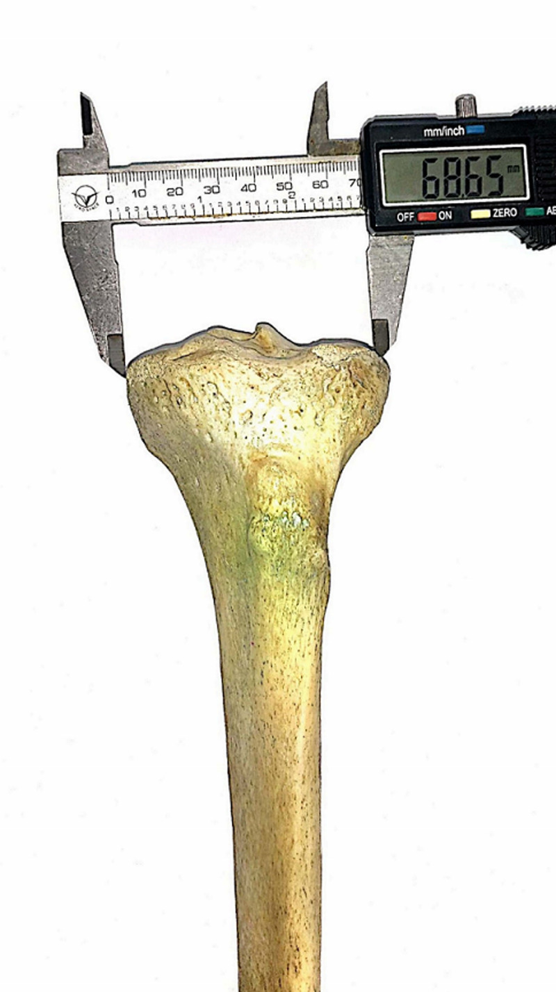
All measurements were performed using a digital Vernier Caliper with a measuring range of 0-150 mm, resolution of 0.01 mm, and accuracy ± 0.02 mm (Figure 1). The measurements were made three times by the two of the authors and the mean of the measurements was recorded.
Figure 1. Measurement of bicondylar width of proximal tibia.
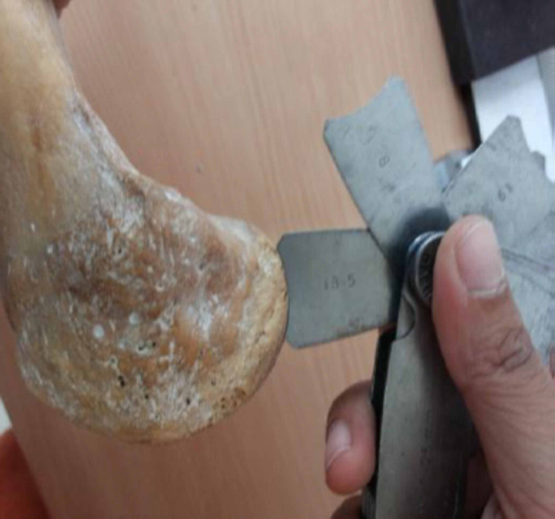
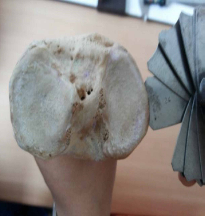
The geometric arcs of femoral and tibial condyles were divided into three parts namely anterior 1/3rd, distal (femur) or middle (tibia) 1/3rd and posterior 1/3rd and were estimated in the sagittal plane for the femur and transverse plane for tibia using the radius of curvature (ROC) gauges of ranges 7.5-15 mm, 15.5-25 mm, 25.5-35 mm and 35-70 mm (Figure 2, Figure 3).
Figure 2. Measurement of the posterior arc of the lateral femoral condyle using the radius of curvature gauge.
Figure 3. Measurement of middle third arc of tibia.
The measurements were tabulated, and the data were analyzed using the Excel 2010 program (Microsoft®, Redmond, Washington, USA). Mean values of AP length and ML width of femoral and tibial condyles; anterior, distal/ middle and posterior geometric arcs of the same were calculated. Aspect ratios (ratio of ML width to the AP length) for the distal femur and proximal tibia were calculated for each bone and for MC and LC individually.
Results
Twenty-four femurs (n = 57) and 29 tibias (n = 60) belonged to the right side. Table 2 and Table 3 show the various morphometric measurements of the distal femur and proximal tibia. The mean aspect ratios for the distal femur and proximal tibia were 1.26 and 1.45, respectively. The average aspect ratios for MC and LC of the femur were 0.50 and 0.52, respectively, whereas, for tibia, they were 0.61 and 0.71, respectively.
Table 2. Morphometric analysis of the distal femur (n = 57).
| Femur | Mean | SD |
| Length (cm) | 43.1 | 5.1 |
| Antero-Posterior diameter | ||
| Medial condyle (mm) | 55.62 | 4.20 |
| Lateral condyle (mm) | 57.93 | 4.20 |
| Medio-Lateral diameter | ||
| Medial condyle (mm) | 28.02 | 2.77 |
| Lateral condyle (mm) | 30.32 | 2.75 |
| Intercondylar width (mm) | 73.45 | 11.18 |
| Geometric arcs | ||
| Medial condyle | ||
| Anterior – third (mm) | 20.77 | 1.99 |
| Distal – third (mm) | 31.42 | 6.02 |
| Posterior – third (mm) | 19.68 | 1.40 |
| Lateral condyle | ||
| Anterior – third (mm) | 21.48 | 1.49 |
| Distal – third (mm) | 64.40 | 5.88 |
| Posterior – third (mm) | 19.06 | 1.56 |
Table 3. Morphometric analysis of proximal tibia (n = 60).
| Tibia | Mean | SD |
| Length (cm) | 37.0 | 2.5 |
| Antero-Posterior diameter | ||
| Medial condyle (mm) | 47.74 | 3.34 |
| Lateral condyle(mm) | 43.46 | 2.64 |
| Medio-Lateral diameter | ||
| Medial condyle(mm) | 30.09 | 1.81 |
| Lateral condyle(mm) | 31.10 | 2.11 |
| Intercondylar width(mm) | 70.66 | 4.77 |
| Geometric arcs | ||
| Medial condyle | ||
| Anterior – third (mm) | 22.42 | 1.84 |
| Middle – third (mm) | 22.49 | 2.39 |
| Posterior – third (mm) | 19.94 | 4.19 |
| Lateral condyle | ||
| Anterior – third (mm) | 19.92 | 2.65 |
| Middle – third (mm) | 21.79 | 2.17 |
| Posterior – third (mm) | 20.95 | 2.15 |
Discussion
Knee replacement is considered a precision surgery, which requires accurate soft tissue balancing and resection of bone thickness equal to the thickness of the prosthetic component implanted, thereby ensuring that the flexion-extension spacing is equal while allowing joint stability throughout the range of motion resulting in a successful outcome [5]. Problems of implant size mismatch are frequently encountered with the existing prostheses as they are designed based on Caucasian morphometric analyses [12] and are not always appropriate for the Asian population who have a smaller build and stature when compared with their Western counterparts [6-10].
The morphometric data obtained in the present study were compared to other studies from Asia retrieved from the PubMed database [5-10,12-17]. Vaidya and colleagues [5] studied computed tomography (CT) of 86 osteoarthritic and 25 dry femurs. In their study, based on CT measurements, the mean AP length was 61.09 mm and 55.58 mm for men and women respectively, while, it was 55.26 mm for the dry femurs. The mean AP length for MC and LC separately were 55.62 mm and 57.93 mm, respectively, in the present study. The mean ML width in their study was 64.84 whereas it was 73.45 mm in the present study. They did not study the proximal tibia. Awasthi and colleagues [12] studied the distal femur in 62 patients presenting with osteoarthritis and trauma of the knee using helical CT, and they reported, the ML width to be 72.74 ± 4.45 mm in males and 63.59 ± 2.61 mm in females, and the AP length to be 49.62 ± 3.86 mm in males and 45.11 ± 4.4 mm in females. Aspect ratio measurements for the distal femur in various studies from Asian countries have ranged from 1.14 to 1.49 [3,6,9,10,15,18]. The mean aspect ratio for distal femur in the present study was 1.26 which is comparable to other studies.
The geometry of proximal tibia has a direct influence on the biomechanics of knee joint [7] and the tibial component is recognized to be more prone to complications compared to the femoral component [2,4]. The aspect ratio of the proximal tibia is an important parameter that helps to anticipate the shape of the tibial component. The aspect ratio in most studies from Asian countries has ranged from 1.33 to 1.8 [7,16,19]. The mean aspect ratio for proximal tibia in the present study was 1.45 which is comparable to other authors as are the aspect ratios for the MC and LC, which were 0.61 and 0.71, respectively [14,20].
This asymmetry of the proximal tibia needs to be incorporated in the TKA prosthesis design as an asymmetric tibial component would be more anatomical, provide correct rotational alignment leading to better survivorship, patellar tracking and function [17,21]. The conventional UKA implants are designed with an asymmetric femoral component and none have an asymmetric tibial component. The present study agrees with Servien and colleagues [20] that the shape of the medial tibial condyle differs from that of the lateral condyle. This difference can lead to ML overhang for medial UKA if the surgeon aims for optimal AP coverage. Custom/patient-specific implants are suggested as they would provide the potential for complete cortical rim coverage better anatomical fit to maximize bone coverage and bone preservation [22,23].
Most authors have only looked at the AP and ML dimensions for the distal femur and proximal tibia [5,6,12,24]. The ROC of the femoral condyles and proximal tibia are not uniform and are integral in dictating normal knee motion as the curvature of the condyles is one of the main factors affecting knee kinematics. In general, a more curved knee would have a higher range of motion [19]. Measurement of the ROC of the femoral condyles is important as the Asian TKA as well as UKA prostheses need to be designed keeping the Asian patient in mind. The ROC of the condyles also has implications for osteochondral allografting procedures [25]. The social and religious needs of prospective patients to keep the knees flexed or for low-sitting, which is a common practice in most Asian cultures, must be incorporated in the prosthesis design [26]. This information is also required for planning the patellar component to avoid problems associated with patellar maltracking.
Various authors have studied the ROC of distal femoral condyles using a variety of techniques. Kapandji [27] divided the medial and lateral femoral condyles into multiple arcs; he noted that posteroanterior the ROC for the medial femoral condyle ranged from 17 to 38 mm and lateral femoral condyle ranged from 12 to 60 mm. Other authors have divided the geometric arcs of the femoral condyles into posterior, distal and anterior arcs. Nuno and Ahmed [28] from Canada studied the profile measurements of the articular surfaces of the femoral condyles in 12 fresh-frozen knees using the two-circular-arc model. They found the posterior arc of the medial and lateral condyle to be 18.9 mm and 19.6 mm respectively and the distal arc measurements as 35 mm and 36.6 mm respectively; in the present study, the mean measurement of the medial and lateral condyle, the posterior arc was 19.68 mm and 19.06 mm respectively and the distal arc was 31.42 mm and 64.4 mm respectively. A smaller ROC was found for the medial condyle than for the lateral condyle; the lateral condyle was much flatter compared to the medial condyle. These observations are comparable to other studies which have noted that the ROC of the posterior arc of lateral condyle is smaller than the anterior and distal arcs [11,19,29].
The measurements of the ROC of the arcs of the proximal tibia would be of immense help in designing tibial components for TKA as well as for UKA; we could not find other researchers looking at this important anthropometric parameter. In the present study, the mean ROC for tibial MC was 22.42 mm, 22.49 mm and 19.94 mm, and for LC 19.92 mm, 21.79 mm and 20.95 mm for the anterior, middle and posterior arcs, respectively.
Some previous studies have used specimen resected during surgery for anthropometric measurements [18,24]. The specimens obtained during knee arthroplasty surgery are obviously arthritic and may not be suitable for basing implant design. We have used dry bones most of which were not arthritic. This makes our data suitable for designing replacement prostheses.
The study has a few limitations. Since we were studying dry bones we were not able to calculate the dimensions for male and female gender separately. Bones in females are generally smaller in dimensions compared to males, and the smaller sized prostheses are generally used in females. Also, the bones were devoid of hyaline cartilage. It has been suggested in previous studies, that the articular cartilage follows the surface topography of subchondral bone, and measurements of the sagittal radii can be extrapolated from the bone measurements [30]. Hence, it may be assumed that the shape and dimension of the distal femur and proximal tibia measured along the periphery are impacted little by the thickness of the articular cartilage [19]. The bone samples, in the present study, were only from central India. To make this study pan-Asian, we would suggest researchers in other countries to undertake similar anthropometric measurements so that we get reliable data available from all over Asia. Such collated data would help in designing UKA and TKA prostheses for our patients.
Conclusions
The morphologic data accumulated in this study for both the distal femur as well as proximal tibia would provide guidelines and help the manufacturers of joint prostheses to address the potential for compromised implant fit and re-design and make available ‘anatomic’ knee prostheses appropriate for the Asian knee which would not only improve function but also prolong the longevity of the prostheses.
Acknowledgments
The authors wish to acknowledge the support of the department of Anatomy, Gandhi Medical College, Bhopal, MP, India for making available the dry bones for the study.
The content published in Cureus is the result of clinical experience and/or research by independent individuals or organizations. Cureus is not responsible for the scientific accuracy or reliability of data or conclusions published herein. All content published within Cureus is intended only for educational, research and reference purposes. Additionally, articles published within Cureus should not be deemed a suitable substitute for the advice of a qualified health care professional. Do not disregard or avoid professional medical advice due to content published within Cureus.
The authors have declared that no competing interests exist.
Human Ethics
Consent was obtained or waived by all participants in this study. Institutional Human Ethics Committee, All India Institute of Medical Sciences, Bhopal issued approval IHEC-LOP/2014/STS0027. Approved (Exempted from review)
Animal Ethics
Animal subjects: All authors have confirmed that this study did not involve animal subjects or tissue.
References
- 1.Standring S. Gray’s Anatomy: The Anatomical Basis of Clinical Practice. 41st ed. China: Churchill Livingstone Elsevier; 2011. Gray’s Anatomy: The Anatomical Basis of Clinical Practice. 41st ed. [Google Scholar]
- 2.Unicondylar knee arthroplasty: key concepts. Halawi MJ, Barsoum WK. J Clin Orthop Trauma. 2017;8:11–13. doi: 10.1016/j.jcot.2016.08.010. [DOI] [PMC free article] [PubMed] [Google Scholar]
- 3.What differences in morphologic features of the knee exist among patients of various races? A systematic review. Kim TK, Phillips M, Bhandari M, et al. Clin Orthop Rel Res. 2017;475:170–182. doi: 10.1007/s11999-016-5097-4. [DOI] [PMC free article] [PubMed] [Google Scholar]
- 4.Anthropometric measurements of tibial plateau and correlation with the current tibial implants. Erkocak OF, Kucukdurmaz F, Sayar S, Erdil ME, Ceylan HH, Tuncay I. Knee Surg Sports Traumatol Arthrosc. 2016;24:2990–2997. doi: 10.1007/s00167-015-3609-5. [DOI] [PubMed] [Google Scholar]
- 5.Anthropometric measurements to design total knee prostheses for the Indian population. Vaidya SV, Ranawat CS, Aroojis A, Laud NS. J Arthroplasty. 2000;15:79–85. doi: 10.1016/s0883-5403(00)91285-3. [DOI] [PubMed] [Google Scholar]
- 6.Anthropometric measurements of the human distal femur: a study of the adult Malay population. Hussain F, Abdul Kadir MR, Zulkifly AH, et al. BioMed Res Int. 2013;2013:175056. doi: 10.1155/2013/175056. [DOI] [PMC free article] [PubMed] [Google Scholar]
- 7.Differences of knee anthropometry between Chinese and white men and women. Yue B, Varadarajan KM, Ai S, Tang T, Rubash HE, Li G. J Arthroplasty. 2011;26:124–130. doi: 10.1016/j.arth.2009.11.020. [DOI] [PMC free article] [PubMed] [Google Scholar]
- 8.Anthropometry of the proximal tibia of patients with knee arthritis in Shanghai. Liu Z, Yuan G, Zhang W, Shen Y, Deng L. J Arthroplasty. 2013;28:778–783. doi: 10.1016/j.arth.2012.12.006. [DOI] [PubMed] [Google Scholar]
- 9.Resected femoral anthropometry for design of the femoral component of the total knee prosthesis in a Korean population. Kwak DS, Han S, Han CW, Han S-H. Anat Cell Biol. 2010;43:252. doi: 10.5115/acb.2010.43.3.252. [DOI] [PMC free article] [PubMed] [Google Scholar]
- 10.The correctness of fit of current total knee prostheses compared with intra-operative anthropometric measurements in Korean knees. Ha CW, Na SE. J Bone Joint Surg Br. 2012;94:638–641. doi: 10.1302/0301-620X.94B5.28824. [DOI] [PubMed] [Google Scholar]
- 11.Knee morphology as a guide to knee replacement. Mensch JS, Amstutz HC. https://pubmed.ncbi.nlm.nih.gov/1192638/ Clin Orthop Relat Res. 1975;112:231–241. [PubMed] [Google Scholar]
- 12.Morphometric study of lower end femur by using helical computed tomography. Awasthi B, Raina SK, Negi V, et al. Indian J Orthop. 2019;53:304–308. doi: 10.4103/ortho.IJOrtho_136_17. [DOI] [PMC free article] [PubMed] [Google Scholar]
- 13.Morphometric analysis of upper end of tibia. Gandhi S, Singla RK, Kullar JS, Suri RK, Mehta V. J Clin Diagn Res. 2014;8:0. doi: 10.7860/JCDR/2014/8973.4736. [DOI] [PMC free article] [PubMed] [Google Scholar]
- 14.Anatomical morphometry of the tibial plateau in South Indian population. Murlimanju BV, Purushothama C, Srivastava A, et al. Ital J Anat Embryol. 2017;121:258–264. [Google Scholar]
- 15.Morphologic features of the distal femur and tibia plateau in Southeastern Chinese population: a cross-sectional study. Fan L, Xu T, Li X, Zan P, Li G. Medicine. 2017;96:0. doi: 10.1097/MD.0000000000008524. [DOI] [PMC free article] [PubMed] [Google Scholar]
- 16.Correlation of anthropometric measurements of proximal tibia in Iranian knees with size of current tibial implants. Karimi E, Zandi R, Norouzian M, Birjandinejad A. https://www.ncbi.nlm.nih.gov/pmc/articles/PMC6686072/ Arch Bone Jt Surg. 2019;7:339–345. [PMC free article] [PubMed] [Google Scholar]
- 17.Comparison of intraoperative anthropometric measurements of the proximal tibia and tibial component in total knee arthroplasty. Miyatake N, Sugita T, Aizawa T, et al. J Orthop Sci. 2016;21:635–639. doi: 10.1016/j.jos.2016.06.003. [DOI] [PubMed] [Google Scholar]
- 18.A new approach of designing the tibial baseplate of total knee prostheses. Cheng CK, Lung CY, Lee YM, Huang CH. Clin Biomech. 1999;14:112–117. doi: 10.1016/s0268-0033(98)00054-0. [DOI] [PubMed] [Google Scholar]
- 19.Three-dimensional morphology of the knee reveals ethnic differences. Mahfouz M, Abdel Fatah EE, Bowers LS, Scuderi G. Clin Orthop Relat Res. 2012;470:172–185. doi: 10.1007/s11999-011-2089-2. [DOI] [PMC free article] [PubMed] [Google Scholar]
- 20.Lateral versus medial tibial plateau: morphometric analysis and adaptability with current tibial component design. Servien E, Saffarini M, Lustig S, Chomel S, Neyret P. Knee Surg Sports Traumatol Arthrosc. 2008;16:1141–1145. doi: 10.1007/s00167-008-0620-0. [DOI] [PubMed] [Google Scholar]
- 21.Maximizing tibial coverage is detrimental to proper rotational alignment. Martin S, Saurez A, Ismaily S, Ashfaq K, Noble P, Incavo SJ. Clin Orthop Relat Res. 2014;472:121–125. doi: 10.1007/s11999-013-3047-y. [DOI] [PMC free article] [PubMed] [Google Scholar]
- 22.Varadarajan KM, Porteous A, Freiberg AA. Partial Knee Arthroplasty. Cham: Springer International Publishing; 2019. Tibiofemoral partial knee arthroplasty implant designs; pp. 133–146. [Google Scholar]
- 23.Influence of tibiofemoral congruency design on the wear of patient-specific unicompartmental knee arthroplasty using finite element analysis. Koh YG, Park KM, Lee HY, Kang KT. Bone Joint Res. 2019;8:156–164. doi: 10.1302/2046-3758.83.BJR-2018-0193.R1. [DOI] [PMC free article] [PubMed] [Google Scholar]
- 24.Anthropometric study of the knee in patients with osteoarthritis: intraoperative measurement versus magnetic resonance imaging. Loures FB, Carrara RJ, Góes RF de A, et al. Radiol Bras. 2017;50:170–175. doi: 10.1590/0100-3984.2016.0007. [DOI] [PMC free article] [PubMed] [Google Scholar]
- 25.Differences in the radius of curvature between femoral condyles: implications for osteochondral allograft matching. Du PZ, Markolf KL, Levine BD, McAllister DR, Jones KJ. J Bone Jt Surg. 2018;100:1326–1331. doi: 10.2106/JBJS.17.01509. [DOI] [PubMed] [Google Scholar]
- 26.Sina A, Hosseinzadeh Hosseinzadeh, Hossein G, et al. 2013. Special Considerations in Asian Knee Arthroplasty. [Google Scholar]
- 27.Kapandji Kapandji, I. A. Edinburgh. Churchill Livingstone/Elsevier; 2011. The Physiology of the Joints. Vol. 2: Lower Limb; p. 70203942. [Google Scholar]
- 28.Three-dimensional morphometry of the femoral condyles. Nuño N, Ahmed AM. Clin Biomech Bristol Avon. 2003;18:924–932. doi: 10.1016/s0268-0033(03)00172-4. [DOI] [PubMed] [Google Scholar]
- 29.A correlative study of the geometry and anatomy of the distal femur. Elias SG, Freeman R, Gokcay EI. https://pubmed.ncbi.nlm.nih.gov/2225651/ Clin Orthop Relat Res. 1990;260:98–103. [PubMed] [Google Scholar]
- 30.Knee cartilage topography, thickness, and contact areas from MRI: in-vitro calibration and in-vivo measurements. Cohen ZA, McCarthy DM, Kwak SD, et al. Osteoarthritis Cartilage. 1999;7:95–109. doi: 10.1053/joca.1998.0165. [DOI] [PubMed] [Google Scholar]


