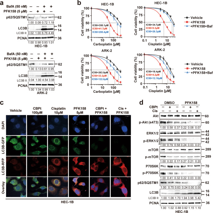
Fig. 4. Combined effects of PFK158 and carboplatin/cisplatin on autophagy flux and the Akt/mTOR signaling pathway.
a Western blot analysis of autophagy-related proteins (p62/SQSTM1 and LC3B) in HEC-1B and ARK-2 treated with PFK158 (5 μM) for 24 h in the presence or absence of bafilomycin A (50 nM) treatment for the last 12 h. PCNA was used as the loading control. b Cell viability assays were performed with a combination of increasing concentrations of CBPt/Cis with 5 μM PFK158 with and without bafilomycin A (BafA) pretreatment. Cells were pretreated with 50 nM BafA for 2 h followed by drug treatment. Cell viability was assessed by MTT assays 48 h later. The data are presented as mean ± SD. A minimum of three independent experiments were performed. c After transient expression of Cherry-GFP-LC3B (48 h), HEC-1B cells were treated with PFK158 (5 μM), CBPt (100 μM)/Cis (10 μM), or their combination for 24 h. Autophagic flux after treatment was investigated by confocal microscopy. Scale bar, 10 μm. d HEC-1B cells were treated with PFK158 (5 μM), CBPt (100 μM)/Cis (10 μM) ± for 24 h. Then, the cells were collected to assess the expression levels of autophagy-related proteins (Akt, p-Akt, mTOR, p-mTOR, p62 and LC3B). PCNA was used as the loading control.

