Abstract
Simple Summary
Melyridae, in a broad sense including Dasytinae and Malachiinae, is the largest family of Cleroidea distributed worldwide. However, the former two subfamilies are always treated as independent families by the European coleopterists. Opposite results have been produced by the two latest molecular phylogenetic works, so the development of reliable markers for reconstructing the phylogenetic relationships between the above taxa is of great importance. Here, we present the annotated complete mitogenome of Cordylepherus sp., which is the first complete mitogenome in Melyridae. The mitogenome of Cordylepherus sp. presents the typical organization of an insect mitochondrion. Comparisons of the newly generated mitogenome of Cordylepherus sp. to all available mitochondrial genomes of other Melyridae revealed no significant differences among them in terms of the length of each protein-coding gene, AT content of different genome regions, amino acid composition and relative synonymous codon usage. Phylogenetic analyses based on 13 protein-coding genes of mitogenomes show that the monophyly of Melyridae sensu lato is not supported, and Malachiinae and Dasytinae are suggested to be independent families, which are sister groups of Prionoceridae and Cleridae, respectively. Large-scale analyses with denser locus and taxon sampling are needed to confirm the present results.
Abstract
To explore the characteristics of the mitogenome of Melyridae and reveal phylogenetic relationships, the mitogenome of Cordylepherus sp. was sequenced and annotated. This is the first time a complete mitochondrial genome has been generated in this family. Consistent with previous observations of Cleroidea species, the mitogenome of Cordylepherus sp. is highly conserved in gene size, organization and codon usage, and secondary structures of tRNAs. All protein-coding genes (PCGs) initiate with the standard start codon ATN, except ND1, which starts with TTG, and terminate with the complete stop codons of TAA and TAG, or incomplete forms, TA- and T-. Most tRNAs have the typical clover-leaf structure, except trnS1 (Ser, AGN), whose dihydrouridine (DHU) arm is reduced. In the A+T-rich region, three types of tandem repeat sequence units are found, including a 115 bp sequence tandemly repeated twice, a 16 bp sequence tandemly repeated three times with a partial third repeat and a 10 bp sequence tandemly repeated seven times. Phylogenetic analyses based on 13 protein-coding genes by both Bayesian inference (BI) and maximum likelihood (ML) methods suggest that Melyridae sensu lato is polyphyletic, and Dasytinae and Malchiinae are supported as independent families.
Keywords: Melyridae, mitochondrial genome, annotation, phylogenetic analysis
1. Introduction
The soft-winged flower beetles belong to the family Melyridae, which is the largest group of superfamily Cleroidea, with about 300 genera and 5500 species occurring in all major regions of the world [1]. The adults are commonly found on flowers where they feed on pollen or flower-visiting insects [2]. There has been inconsistency in its family definition in the history.
Most of the earlier authors considered the family Melyridae in the broad sense, including Prionocerinae, Rhadalinae, Malachiinae and Dasytinae [3,4,5]. Majer [6] split the former Melyridae sensu lato into several families, with Prionoceridae, Melyridae, Dasytidae and Malachiidae treated as independent families. This classification was followed by Mayor [7] in the latest catalogue of Palaearctic Coleoptera, also in agreement with Bocakova et al. [1] based on a four-gene (18S, 28S, 16S and cox1) phylogenetic study.
However, the modern coleopterological community [2,8,9] considers Prionoceridae and Melyridae as distinct families, while Dasytinae, Malachiinae and Rhadalinae are subfamilies of the latter. Most recently, Gimmel et al. [10] reconstructed the most comprehensive phylogeny of Cleroidea based on a four-gene dataset (18S, 28S, cox1 and cytb), and redefined Melyridae, including Dasytinae and Malachiinae but excluding Rhadalinae.
At the moment, the phylogenetic relationships of Melyridae, Dasytidae (Dasytinae) and Malachiidae (Malachiinae) remain controversial, and since the two most recent molecular phylogenetic studies, based on a few genes, produced conflicting results [2,10], the development of reliable markers for reconstructing the phylogenetic relationships for the above taxa is of great importance.
The mitochondrial genome has become a powerful tool for metazoan phylogenetic and evolutionary analysis [11,12,13,14,15,16] because of its small size, the presence of high copy numbers, strict orthologous genes, rare recombination and high evolutionary rate [12,17,18]. To date, only four mitochondrial genomes are available for Melyridae in GenBank, with two belonging to species for both Dasytinae and Malachiinae. However, none is a complete mitochondrial genome sequence. Deficiency of mitogenome data for Melyridae is an obstacle for a comprehensive phylogenetic analysis.
In the present study, the first complete mitochondrial genome for Melyridae, the complete mitogenome of Cordylepherus sp., was sequenced and analyzed. In addition, phylogenetic analyses based on two different methods were carried out to assess the phylogenetic position of Cordylepherus sp. in Cleroidea, and to contribute to further understanding the mitogenome evolution and phylogeny of the Cleroidea.
2. Materials and Methods
2.1. Sample Preparation and DNA Extraction
The material of Cordylepherus sp. was collected from Saihanba, Chengde, Hebei Province, China, on 29 May 2019. Specimens were preserved in 100% ethanol and deposited in the Museum of Hebei University, Baoding, China (MHBU, accession number CAN0146). Species identification was carried out one by one under a stereomicroscope. Total genomic DNAs were extracted using a DNeasy Blood & Tissue kit (QIAGEN, Beijing, China), according to the manufacturer’s instructions. DNAs were stored at −20 °C for long-term storage and further molecular analyses.
2.2. DNA Sequencing and Assembly
Whole mitochondrial genome sequencing was performed using an Illumina Novaseq 6000 platform (Illumina, Alameda, CA, USA) with 150 bp paired-end reads at Berry Genomics, Beijing, China. The sequence reads were first filtered following Zhou et al. [19] and then assembled using IDBA-UD [20] under similarity threshold 98% and k values of a minimum of 40 and a maximum of 160 bp. The gene cox1 was amplified by polymerase chain reaction (PCR) using universal primers as “reference sequences” to acquire the best-fit. The PCR cycling conditions comprised a predenaturation at 94 °C for 5 min and 35 cycles of denaturation at 94 °C for 50 s, annealing at 48 °C for 45 s and elongation at 72 °C for 8 min at the end of all cycles. Geneious 2019.2 [21] software was used to manually map the clean readings on the obtained mitochondrial scaffolds to check the accuracy of the assembly.
2.3. Sequence Annotation and Analyses
Gene annotation was performed by Geneious 2019.2 software [21] and MITOS Web Server (http://mitos.bioinf.uni-leipzig.de/index.py) [22], with Idgia oculata as reference [23]. The tRNA Scan-SE server v. 1.21 [24] was used to re-identify the 22 tRNAs as well as to reconfirm their secondary structures, and the secondary structures were plotted with Adobe Photoshop 7.0. The mitogenome map was illustrated using CG View (http://stothard.afns.ualberta.ca/cgview_server) [25]. Tandem repeat elements in the A + T-rich region were identified using the Tandem Repeats Finder program (http://tandem.bu.edu/trf/trf.html) [26]. Nucleotide composition, base composition skewness, codon usage and relative synonymous codon usage (RSCU) of protein-coding genes were analyzed using PhyloSuitev 1.2.2 [27]. Strand asymmetry was calculated according to the formulas: GC skew = [G − C]/[G + C] and AT skew = [A − T]/[A + T] [28]. The nucleotide diversity (Pi) and nonsynonymous (Ka)/synonymous (Ks) mutation rate ratios were calculated by DnaSP v5.10.01 [29].
2.4. Phylogenetic Analysis
Mitochondrial genomes of 13 species from 7different families of Cleroidea were selected as ingroups, and mitochondrial genomes of 2 Cucujoidea species were chosen as outgroups (details in Table 1). All mitochondrial genomes (except the one sequenced in this study) were obtained from GenBank (accession numbers given in Table 1). Data standardization and information extraction were performed by PhyloSuite v 1.2.2 [27]. The nucleotide sequences of the 13 protein-coding genes were analyzed. The protein-coding genes were aligned using the MAFFT v 7.313 plugins [30], optimized using MACES in PhyloSuite v 1.2.2 [27]. Intergenic gaps and ambiguous sites were removed using Gblocks v 0.91b [31], then all protein-coding genes were concatenated in PhyloSuitev 1.2.2 [27]. The optimal partition schemes and the best-fit replacement models were selected by Model Finder [32], and the results are presented in Supplementary Tables S1 and S2. The “greedy” algorithm with branch lengths estimated as “linked” and the Bayesian information criterion were used.
Table 1.
Summary of the representative species and their mitogenome information in this study.
| Superfamily | Family/Subfamily | Species | GenBankNo. | References |
|---|---|---|---|---|
| Cleroidea | Phloiophilidae | Phloiophilus edwardsi | JX412815.1 | Unpublished |
| (Ingroup) | Melyridae/ | Malachiinae sp. | JX412799.1 | Unpublished |
| Malachiinae | Clanoptilus assimilis | JX412833.1 | Unpublished | |
| Cordylepherus sp. | MW365444 | This study | ||
| Melyridae/ | Psilothrix sp. | JX412801.1 | [33] | |
| Dasytinae | Dasytinae sp. | JX412765.1 | Unpublished | |
| Trogossitidae | Temnoscheila virescens | JX412752.1 | Unpublished | |
| Prionoceridae | Idgia oculata | NC_044896.1 | [23] | |
| Byturidae | Byturus ochraceus | NC_036267.1 | [34] | |
| Cleridae | Hydnocerini sp. | KX035157.1 | Unpublished | |
| Trichodes sinae | NC_033340.1 | [35] | ||
| Cleridae sp. | MH789728.1 | [36] | ||
| Chaetosomatidae | Chaetosomas caritides | NC_011324.1 | [37] | |
| Cucujoidea | Coccinellidae | Coccidophilus cariba | MN447521.1 | [38] |
| (Outgroup) | Hippodamia variegata | MK334129.1 | [39] |
Phylogenetic trees were constructed based on both maximum likelihood (ML) and Bayesian inference (BI). The ML phylogenies were inferred using IQ-TREE.v.1.6.8 [40] and BI phylogenies using MrBayes 3.2.6 [41], respectively. The ML phylogenetic analyses were performed with the ultrafast bootstrap (UFboot) algorithm with 1000 replicates. In BI phylogenetic analyses, 5 × 10⁶ Markov Chain Monte Carlo (MCMC) generations after reaching stationarity (average standard deviation of split frequencies < 0.01) were used as the default settings, with estimated sample size >200, and potential scale reduction factor ≈1 [41]. Interactive Tree of Life (iTOL, http://itol.embl.de) was used to display, annotate and manage the phylogenetic tree.
3. Results and Discussion
3.1. Mitogenome Organization and Base Composition
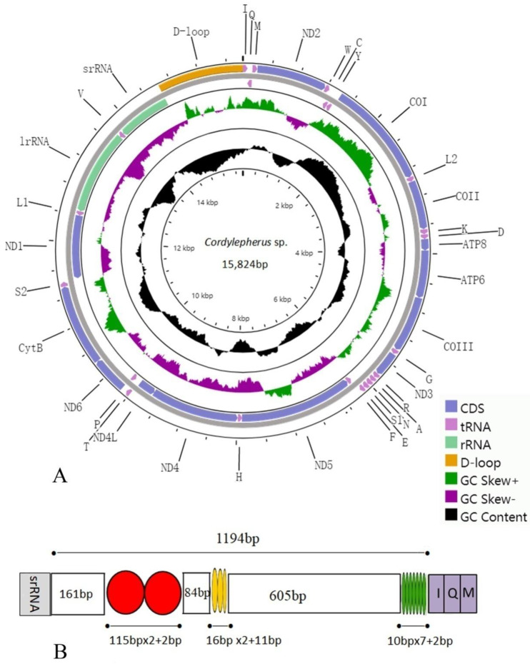
Sequence generated in this study is deposited in GenBank with accession number (MW365444). The complete mitogenome sequence of Cordylepherus sp. is 15,824 bp in length. It is a circular, double-stranded ring which includes 13 protein-coding genes (PCGs), 22 tRNA genes, 2 rRNA genes and an A+T-rich region (control region) (Figure 1A). The gene organization is the same as the hypothetical ancestral insect mitogenome as direction and order [42]: 15 genes (8 tRNAs, 4 PCGs and 2 rRNAs) are encoded on the minority strand (N-strand), and the others (14 tRNAs and 9 PCGs) are transcribed from the majority strand (J-strand) (Table S3). The mitogenome contains 7 overlapped genes (13 bp in total), and the longest overlap is 4 bp. Most of the gene overlaps occur in tRNA genes, which is possibly due to the lower evolutionary constraints of these genes [37]. Intergenic spacers in the Cordylepherus sp. mitogenome have 12 regions ranging from 1 to 34 bp (a total of 98bp), with the longest region detected between tRNA-Tyr and cox1.
Figure 1.
(A) Mitochondrial genome map of Cordylepherus sp.; (B) organization of the A + T-rich regions in the mitochondrial genomes of Cordylepherus sp.
The base composition of Cordylepherus sp. mitogenome is A (41.90%), T (39.10%), C (11.20%) and G (7.80%). The A/T nucleotide composition is 81.00%, thus exhibiting a high A/T bias, as other insects [13,14,15,16,33,35]. The AT-skew of the full mitogenome is positive (0.03), while the GC skew is negative (−0.18). This indicates that the content of bases G is higher than that of C, and A is higher than T in the whole (Table S4).
3.2. Protein-CodingGenes
The total length of the 13 PCGs in Cordylepherus sp. is 10,996 bp, approximately accounting for 69.5% of the whole mitogenome (Table S4). Nine of the 13 PCGs are coded on the J-strand (cox1, cox2, cox3, cytb, nad2, nad3, nad6, atp6, atp8), and the other four (nad1, nad4, nad4l, nad5) are located on N-strand (Figure 1, Table S3). The A/T nucleotide composition is 79.70%, also exhibiting a highly A/T bias (Table S4). Most of the 13 PCGs initiate with the standard start codon ATN, common among metazoan [43], except nad1, which starts with TTG. There are four types of stop codons, TAA (nad6, cox2, atp6, atp8, cox3, nad4l) and TAG (nad1), as well as TA- (nad4) and T- (nad2, cox1, nad3, nad5, cytb). It is common that incomplete stop codons occur in insects; this is believed to be completed by the rough polyadenylation processes and polycistronic transcription cleavage, and it may emerge as functional stop codons or serve to minimize gene overlap and spacer [44].
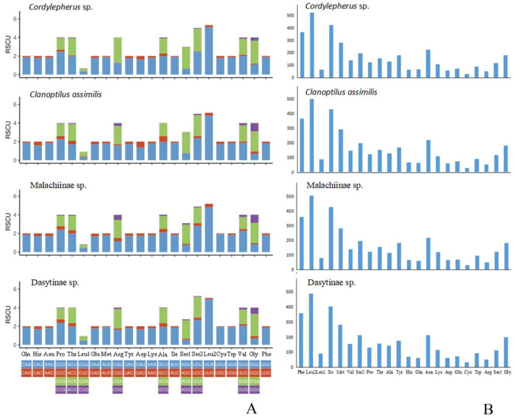
To characterize codon frequencies across Cordylepherus sp., relative synonymous codon usage (RSCU) was calculated and drawn, as shown in the Figure 2 and Table S5. In total, it contains 3656 codons excluding stop codons, and the most frequently used codons are UUA (502), AUU (405), UUU (343) and AUA (265). Accordingly, Leu, Ile, Phe and Met are the most frequently used amino acids, accounting for 16.11%, 11.57%, 9.98% and 7.66% of the total amino acids, respectively. In these most frequently used codons, A and U are the components which contribute to the high A + T bias of the full mitogenome. Additionally, the RSCU values of NNU and NNA are greater than 1, indicating that the third position of the codon is rich in A and T. Besides, PCGs shows positive AT skew (0.04) and negative GC skew (−0.16) (Table S4).
Figure 2.
(A) Relative synonymous codon usage (RSCU) in the Melyridae species’ mitogenomes; (B) amino acid composition in the Melyridae species’ mitogenomes.
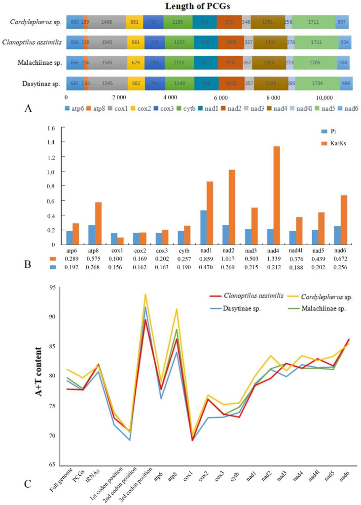
In order to illustrate differences between Malachiinae and Dasytinae, we compared the length of the 13 PCGs (Table S6), AT content (Table S7), RSCU and amino acid composition (Table S4) in different regions, as shown in the Figure 2 and Figure 3. The length of atp6, cox2, cytb, nad2, nad4, nad4l and nad5 is higher in Dasytinae than Malachinae, but nad6 is the opposite (Figure 3A). The AT contents of mitogenomes are lower in Dasytinae than Malachiinae in most regions, except for the full genome, 3rd codon position, nad2, nad4, nad5 and cytb (Figure 3C). Among the RSCU, the CUG codon used for Leu is absent in Dasytinae (Figure 2A). No differences were observed between them in the amino acid composition and proportion (Figure 2B).
Figure 3.
(A) Comparison of 13 PCGs in lengths of Melyridae species; (B) nucleotide diversity (Pi) and nonsynonymous (Ka) to synonymous (Ks) substitution rate ratios of 13 PCGs of Melyridae species (the Pi and Ka/Ks values of each PCGs shown under the gene name); (C) comparison of AT contents in different regions of mitogenomes of Melyridae species.
The nucleotide diversity of the Melyridae is shown in Figure 3B; it is highly variable among the 13 PCGs, with values ranging from 0.156 (cox1) to 0.470 (nad1). The gene nad1 (Pi = 0.470) has the most diverse nucleotide among all PCGs, followed by nad2 (Pi = 0.269), atp8 (Pi = 0.268), nad6 (Pi = 0.256) and nad4 (Pi = 0.212). In contrast, cox1 (Pi = 0.156), cox2 (Pi = 0.162) and cox3 (Pi = 0.163) have relatively low values of nucleotide diversity and are the most conserved genes.
Average non-synonymous (Ka)/synonymous (Ks) substitution rate ratios can be used to estimate the evolutionary rate [45]. The Ka/Ks ratios of nad2 (1.017) and nad4 (1.339) genes are greater than one, indicating that they are under positive selection [46], while the other genes are under purifying selection, with ratios ranging from 0.100 to 0.859 (all less than 1) [46]. The genes nad2 and nad4 have relatively high evolutionary rates, while cox1 (0.100) and cox2 (0.169) have comparatively low Ka/Ks ratios (Figure 3B). Like other insects [47,48], the genes with lower evolutionary rate could be used as barcodes for inferring the phylogenetic relationships, such as cox1 and cox2 in Melyridae, while those evolving more quickly are more suitable for species identification, especially for nad2 and nad4 in Melyridae.
3.3. Transfer and Ribosomal RNA Genes
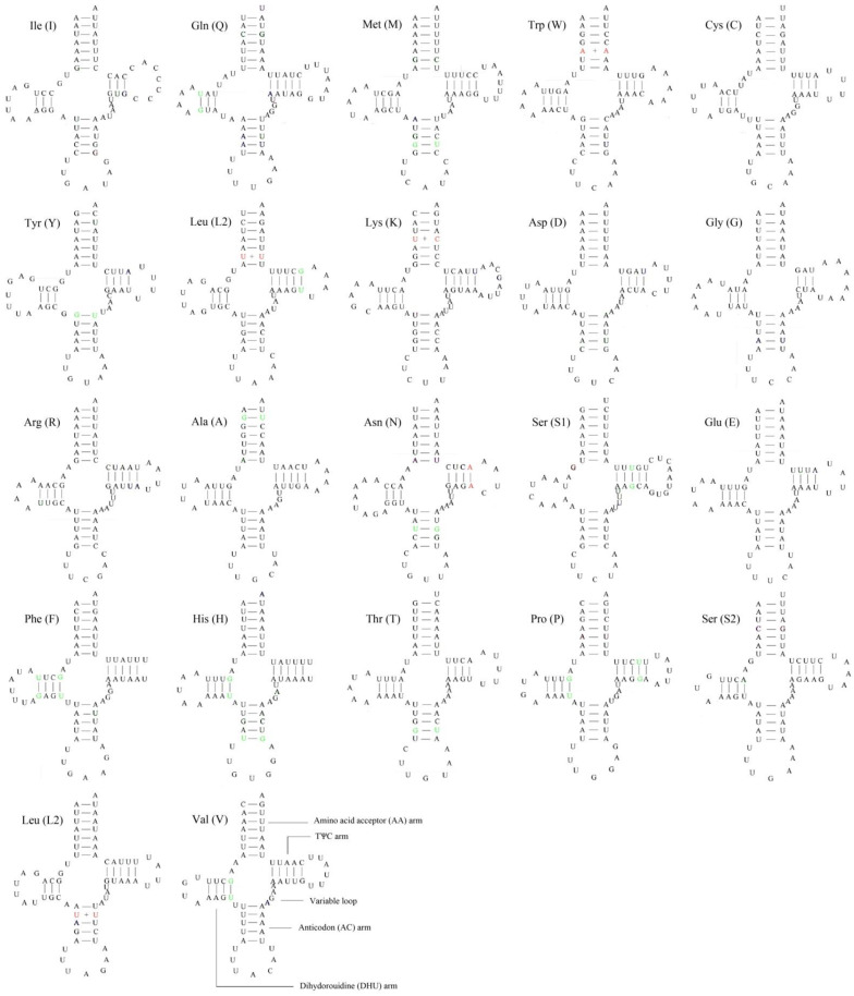
As other insects [14,15,16,33,35], the Cordylepherus sp. mitogenome contains 22 tRNAs genes, which range from 63 to 71 bp in length; the total length of tRNAs is 1149 bp (Table S3). Most tRNAs genes could be folded into the typical clover-leaf secondary structure, while trnS1 (AGN) lacked a dihydrouridine (DHU) arm (Figure 4), as observed in many other insect mitogenomes [12,43]. In general, the secondary structure is conserved in the length of the acceptor and anticodon arms (7 bp in the former and 5 bp in the latter), while variable in that of the DHU and TΨC arms [49,50,51]. In the secondary structures of all tRNAs of Cordylepherus sp., 3 or 4 base pairs in the DHU arms, and 3, 4 or 5 base pairs in the TΨC arms are found. Except the classic base pairs (A-U and C-G), 15 noncanonical base pairings (G–U and A–C) and 5 other mismatched base pairs (U-U and C-U) are found in the arms. The AT content is 81.70%, with a positive AT skew (0.07) and negative GC skew (−0.16) (Table S4).
Figure 4.
Putative secondary structures of tRNAs from Cordylepherus sp. mitogenome (noncanonical base pairings in green; mismatched base pairs in red).
There are two rRNAs, a 1258-bp 16SrRNA (rrnL) and an 815-bp 12SrRNA (rrnS) in Cordylepherus sp. (Table S3). It is hard to determine the boundaries of the rRNA genes, since that they do not have functional annotation features as PCGs [52,53]. Thus, the boundaries of flanking genes are decided by assuming no overlapping or gaps between adjacent genes, like that in the inferred insect mitogenome pattern. The 16S rRNA subunit is located between trnL1 and trnV; the 12S rRNA genes are located between trnV and the A+T-rich region. The AT content is 84.50%, with positive AT skew (0.02) and negative GC skew (−0.32) (Table S4).
3.4. A + T-RichRegion
The mitochondrial A+T-rich region (or control region, CR) acts on the initiation and regulation of insect replication and transcription [54,55]. This non-coding region is located between rrnS and trnI in the mitogenome of Cordylepherus sp., with a total length of 1194 bp (Table S3). The AT content is 90.00%, with both negative AT skew (−0.02) and GC skew (−0.09) (Table S4). Three tandem repeat sequence units are present; their positions and length are shown in the Figure 1B. They area 115 bp-sequence tandemly repeated twice, a 16 bp-sequence tandemly repeated three times with a partial third repeat and a 10 bp-sequence tandemly repeated seven times, respectively.
3.5. Phylogenetic Analysis
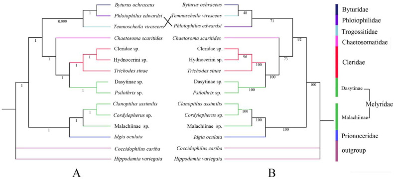
Similar topologies are yielded from ML (Figure 5A) and BI (Figure 5B) analyses with high nodal support values. The monophyly of Cleroidea is highly supported (PP = 1, BS = 100), with Byturidae included in it [56]. Within Cleroidea, it is divided into three branches. One clade consists of Malachiinae and Prionoceridae, with a high statistical support value (PP = 1, BS = 100). The other two clades (Chaetosomatidae (Dasytinae, Cleridae)) and (Trogossitidae, Phloiophilidae and Byturidae) seem more closely related in the tree and supported with a relatively high value (PP = 1, BS = 92). However, within each of the latter two clades, the interrelationships among the included taxa are not strongly supported in the BI analysis, except the sister relationship between Dasytinae and Cleridae (PP = 1, BS = 100).
Figure 5.
Phylogenetic tree of Cleroidea produced from maximum likelihood (ML) (A) and Bayesian inference (BI) (B) analyses based on 13 PCGs (posterior probabilities (PP) and bootstrap (BS) values indicated in each clade).
This result does not recover Melyridae sensu lato as monophyletic. Malachiinae, with three species included in the analyses, is resolved as monophyletic, and sister to Prionoceridae. Dasytinae, with two sampled species, formed a monophyletic clade sister to Cleridae. Both clades are strongly supported (PP = 1, BS = 100). Our analyses suggest that Malachiinae and Dasytinae should be treated as independent families, which agrees with the views of Majer [6] and Bocakova et al. [1], but is incongruent with the others [8,9,10]. It is necessary to further examine the phylogenetic relationships of Melyridae family groups or Cleroidea, if more molecular data (more representative species and more molecular markers) are available.
4. Conclusions
Consistent with previous observations of other Cleroidea species, the mitogenome sequences of Cordylepherus sp., which is the first complete mitochondrial genome of Melyridae, are highly conserved in gene size and organization, highly A + T biased base composition, codon usage of protein-coding genes and secondary structures of tRNAs. This provides the basic information to perform comparative analyses and further discussion of the mitogenome evolution of Cleroidea.
Phylogenetic analyses support Malachiinae and Dasytinaeas as independent families, instead of subfamilies of Melyridae sensu lato, which is suggested to be a polyphyletic group. Larger scale studies with more locus and taxon sampling are still needed to reconstruct more comprehensive phylogenies in order to achieve a better resolution of the relationships of Melyridae family groups or Cleroidea.
Acknowledgments
We are grateful to Hu Li and Ph.D. candidate Yunfei Wu (China Agricultural University, Beijing) for their valuable help in mitochondrial genomic assembling and phylogenetics. Thanks are also given to the anonymous reviewers for their wonderful suggestions in improving our original manuscript.
Supplementary Materials
The following are available online at https://www.mdpi.com/2075-4450/12/2/87/s1, Table S1: The optimal partition schemes and the best-fit replacement models for the Bayesian inference (BI) method; Table S2: The optimal partition schemes and the best-fit replacement models for the Maximum likelihood (ML) method; Table S3: Summary of the characteristics of the mitogenome of Cordylepherus sp.; Table S4: Nucleotide composition and skewness of the mitogenomes of Cordylepherus sp.; Table S5: Codon number and RSCU in the mitogenomes of Melyridae species (Cordylepherus sp./Clanoptilus assimilis/Malachiinae sp./Dasytinae sp.); Table S6: Length of PCGs in the mitogenomes of the Melyridae species; Table S7: AT content in the mitogenomes of Melyridae species.
Author Contributions
Conceptualization, Y.Y., H.L., and G.X.; Methodology, L.Y., X.G., and Y.Y.; Investigation, H.L.; Validation, L.Y. and Y.Y.; Writing—Original Draft Preparation, L.Y., X.G., and Y.Y.; Writing—Review and Editing, L.Y., H.L., and Y.Y.; Funding acquisition, Y.Y. and H.L. All authors have read and agreed to the published version of the manuscript.
Funding
This research was funded by the National Natural Science Foundation of China (Nos. 31772507, 41401064), the Natural Science Foundation of Hebei Province (No. C201720112) and the Science and Technology Project of Hebei Education Department (No. BJ2017030) to Y.Y., also the Biodiversity Survey and Assessment Project of the Ministry of Ecology and Environment, China (No. 2019HJ2096001006) and the Natural Science Foundation of Hebei Province (No. C2019201192) to H.L.
Data Availability Statement
The sequence generated in this study is deposited in GenBank with accession number (MW365444).
Conflicts of Interest
The authors declare no conflict of interest.
Footnotes
Publisher’s Note: MDPI stays neutral with regard to jurisdictional claims in published maps and institutional affiliations.
References
- 1.Bocakova M., Constantin R., Bocak L. Molecular phylogenetics of the melyrid lineage (Coleoptera: Cleroidea) Cladistics. 2011;28:117–129. doi: 10.1111/j.1096-0031.2011.00368.x. [DOI] [PubMed] [Google Scholar]
- 2.Lawrence J.F., Leschen R.A.B. 9.11. Melyridae Leach, 1815. In: Leschen R.A.B., Beutel R.G., Lawrence J.F., editors. Coleoptera, Beetles: Morphology and Systematics (Elateroidea, Bostrichiformia, Cucujiformiapartim) Volume 2. W. DeGruyter; Berlin, Germany: 2010. pp. 273–280. [Google Scholar]
- 3.Böving A.G., Craighead F.C. An illustrated synopsis ofthe principal larval forms of the order Coleoptera. Entomol. Am. 1931;11:1–351. [Google Scholar]
- 4.Crowson R.A. The Phylogeny of Coleoptera. Annu. Rev. Entomol. 1960;5:111–134. doi: 10.1146/annurev.en.05.010160.000551. [DOI] [Google Scholar]
- 5.Crowson R.A. A review of the classification of Cleroidea (Coleoptera), with descriptions of two new genera of Peltidae and of several new larval types. Ecol. Entomol. 2009;116:275–327. doi: 10.1111/j.1365-2311.1964.tb02298.x. [DOI] [Google Scholar]
- 6.Majer K. A review of the classification of the Melyridae and related families (Coleoptera, Cleroidea) Entomol. Basil. 1994;17:319–390. doi: 10.1023/A:1014214404699. [DOI] [Google Scholar]
- 7.Mayor A. Melyridae, Dasytidae, Malachiidae. In: Löbl I., Smetana A., editors. Catalogue of Palaearctic Coleoptera. Volume 4. Apollo Books; Stenstrup, Denmark: 2007. pp. 386–454. [Google Scholar]
- 8.Lawrence J.F., Newton A.F. Families and subfamilies of Coleoptera (with selected genera, notes, references and data on family-group names) In: Lawrence J., Pakaluk J.F., Slípínski S.A., editors. Biology, Phylogeny, and Classification of Coleoptera. Museum i Instytut Zoologii PAN; Warzawa, Poland: 1995. pp. 779–1092. [Google Scholar]
- 9.Bouchard P., Bousquet Y., Davies A.E., Alonso-Zarazaga M.A., Lawrence J.F., Lyal C.H.C., Newton A.F., Reid C.A.M., Schmitt M., Ślipiński S.A., et al. Family-Group Names In Coleoptera (Insecta) ZooKeys. 2011;88:1–972. doi: 10.3897/zookeys.88.807. [DOI] [PMC free article] [PubMed] [Google Scholar]
- 10.Gimmel M.L., Bocakova M., Gunter N., Leschen R.A.B. Comprehensive phylogeny of the Cleroidea (Coleoptera: Cucujiformia) Syst. Entomol. 2019;44:527–558. doi: 10.1111/syen.12338. [DOI] [Google Scholar]
- 11.Cameron S.L., Lambkin C.L., Barker S.C., Whiting M.F. A mitochondrial genome of Diptera: Whole genome sequence data accurately resolve relationships over broad timescales with high precision. Syst. Entomol. 2007;32:40–59. doi: 10.1111/j.1365-3113.2006.00355.x. [DOI] [Google Scholar]
- 12.Cameron S.L. Insect Mitochondrial Genomics: Implications for Evolution and Phylogeny. Annu. Rev. Entomol. 2014;59:95–117. doi: 10.1146/annurev-ento-011613-162007. [DOI] [PubMed] [Google Scholar]
- 13.Nie R., Andújar C., Gómez-Rodríguez C., Bai M., Xue H., Tang M., Yang C., Tang P., Yang X., Vogler A.P. The phylogeny of leaf beetles (Chrysomelidae) inferred from mitochondrial genomes. Syst. Entomol. 2019;45:188–204. doi: 10.1111/syen.12387. [DOI] [Google Scholar]
- 14.Liu Y., Song F., Jiang P., Wilson J.-J., Cai W., Li H. Compositional heterogeneity in true bug mitochondrial phylogenomics. Mol. Phylogenet. Evol. 2018;118:135–144. doi: 10.1016/j.ympev.2017.09.025. [DOI] [PubMed] [Google Scholar]
- 15.Liu Y., Li H., Song F., Zhao Y., Wilson J.J., Cai W. Higher-level phylogeny and evolutionary history of Pentatomorpha (Hemiptera: Heteroptera) inferred from mitochondrial genome sequences. Syst. Entomol. 2019;44:810–819. doi: 10.1111/syen.12357. [DOI] [Google Scholar]
- 16.Nie R., Vogler A.P., Yang X.-K., Lin M. Higher-level phylogeny of longhorn beetles (Coleoptera: Chrysomeloidea) inferred from mitochondrial genomes. Syst. Entomol. 2020 doi: 10.1111/syen.12387. [DOI] [Google Scholar]
- 17.Li H., Shao R., Song N., Song F., Jiang P., Li Z., Cai W. Higher-level phylogeny of paraneopteran insects inferred from mitochondrial genome sequences. Sci. Rep. 2015;5:8527. doi: 10.1038/srep08527. [DOI] [PMC free article] [PubMed] [Google Scholar]
- 18.Curole J.P., Kocher T.D. Mitogenomics: Digging deeper with complete mitochondrial genomes. Trends Ecol. Evol. 1999;14:394–398. doi: 10.1016/S0169-5347(99)01660-2. [DOI] [PubMed] [Google Scholar]
- 19.Zhou X., Li Y., Liu S., Yang Q., Su X., Zhou L., Tang M., Fu R., Li J., Huang Q. Ultra-deep sequencing enables high-fidelity recovery of biodiversity for bulk arthropod samples without PCR amplification. GigaScience. 2013;2:1–12. doi: 10.1186/2047-217X-2-4. [DOI] [PMC free article] [PubMed] [Google Scholar]
- 20.Peng Y., Leung H.C.M., Yiu S.M., Chin F.Y.L. IDBA-UD: A de novo assembler for single-cell and metagenomic sequencing data with highly uneven depth. Bioinformatics. 2012;28:1420–1428. doi: 10.1093/bioinformatics/bts174. [DOI] [PubMed] [Google Scholar]
- 21.Kearse M., Moir R., Wilson A., Stones-Havas S., Cheung M., Sturrock S., Buxton S., Cooper A., Markowitz S., Duran C., et al. Geneious Basic: An integrated and extendable desktop software platform for the organization and analysis of sequence data. Bioinformatics. 2012;28:1647–1649. doi: 10.1093/bioinformatics/bts199. [DOI] [PMC free article] [PubMed] [Google Scholar]
- 22.Bernt M., Donath A., Jühling F., Gärtner F., Florentz C., Fritzsch G., Pütz J., Middendorf M., Stadler P.F. MITOS: Improved de novo metazoan mitochondrial genome annotation. Mol. Phylogenetics Evol. 2013;69:313–319. doi: 10.1016/j.ympev.2012.08.023. [DOI] [PubMed] [Google Scholar]
- 23.Wu L., Nie R.E., Bai M., Yang Y.X. The complete mitochondrial genome of Idgia oculata (Coleoptera: Cleroidea: Prionoceridae) and a related phylogenetic analysis of Cleroidea. Mitochondrial DNA Part B. 2019;4:491–493. doi: 10.1080/23802359.2018.1545533. [DOI] [Google Scholar]
- 24.Lowe T.M., Eddy S.R. tRNAscan-SE.A program for improved detection of transfer RNA genes in genomic sequence. Nucleic Acids Res. 1997;25:955–964. doi: 10.1093/nar/25.5.955. [DOI] [PMC free article] [PubMed] [Google Scholar]
- 25.Grant J.R., Stothard P. The CG View Server: A comparative genomics tool for circular genomes. Nucleic Acids Res. 2008;36:181–184. doi: 10.1093/nar/gkn179. [DOI] [PMC free article] [PubMed] [Google Scholar]
- 26.Benson G. Tandem repeats finder: A program to analyze DNA sequences. Nucleic Acids Res. 1999;27:573–580. doi: 10.1093/nar/27.2.573. [DOI] [PMC free article] [PubMed] [Google Scholar]
- 27.Zhang D., Gao F., Jakovlić I., Zhou H., Zhang J., Li W.X., Wang G.T. PhyloSuite: An integrated and scalable desktop platform for streamlined molecular sequence data management and evolutionary phylogenetics studies. Mol. Ecol. Resour. 2019;20:348–355. doi: 10.1111/1755-0998.13096. [DOI] [PubMed] [Google Scholar]
- 28.Perna N.T., Kocher T.D. Patterns of nucleotide composition at fourfold degenerate sitesof animal mitochondrial genomes. J. Mol. Evol. 1995;41:353–358. doi: 10.1007/BF01215182. [DOI] [PubMed] [Google Scholar]
- 29.Librado P., Rozas J. DnaSPv5: A software for comprehensive analysis of DNA polymorphism data. Bioinformatics. 2009;25:1451–1452. doi: 10.1093/bioinformatics/btp187. [DOI] [PubMed] [Google Scholar]
- 30.Katoh K., Standley D.M. Mafft Multiple Sequence Alignment Software Version 7: Improvements in Performance and Usa-bility. Mol. Biol. Evol. 2013;4:772–780. doi: 10.1093/molbev/mst010. [DOI] [PMC free article] [PubMed] [Google Scholar]
- 31.Castresana J. Selection of Conserved Blocks from Multiple Alignments for Their Use in Phylogenetic Analysis. Mol. Biol. Evol. 2000;17:540–552. doi: 10.1093/oxfordjournals.molbev.a026334. [DOI] [PubMed] [Google Scholar]
- 32.Lanfear R., Frandsen P.B., Wright A.M., Senfeld T., Calcott B. PartitionFinder 2: New Methods for Selecting Partitioned Models of Evolution for Molecular and Morphological Phylogenetic Analyses. Mol. Biol. Evol. 2017;34:772–773. doi: 10.1093/molbev/msw260. [DOI] [PubMed] [Google Scholar]
- 33.Timmermans M.J.T.N., Barton C., Haran J., Ahrens D., Culverwell C.L., Ollikainen A., Dodsworth S., Foster P.G., Bocak L., Vogler A.P. Family-Level Sampling of Mitochondrial Genomes in Coleoptera: Compositional Heterogeneity and Phyloge-netics. Genome Biol. Evol. 2016;8:161–175. doi: 10.1093/gbe/evv241. [DOI] [PMC free article] [PubMed] [Google Scholar]
- 34.Linard B., Crampton-Platt A., Moriniere J., Timmermans M.J.T.N., Andújar C., Arribas P., Miller K.E., Lipecki J., Favreau E., Hunter A. The contribution of mitochondrial metagenomics to large-scale data mining and phylogenetic analysis of Coleoptera. Mol. Phylogenet. Evol. 2018;128:1–11. doi: 10.1016/j.ympev.2018.07.008. [DOI] [PubMed] [Google Scholar]
- 35.Tang P.A., Feng R.Q., Zhang L., Wang J., Wang X.T., Zhang L.J., Yuan M.L. Mitochondrial genomes of three Bostrichiformia species and phylogenetic analysis of Polyphaga (Insecta, Coleoptera) Genomics. 2020;112:2970–2977. doi: 10.1016/j.ygeno.2020.05.012. [DOI] [PubMed] [Google Scholar]
- 36.Crampton-Platt A., Timmermans M.J., Gimmel M.L., Kutty S.N., Cockerill T.D., Khen C.V., Vogler A.P. Soup to Tree: The Phylogeny of Beetles Inferred by Mitochondrial Metagenomics of a Bornean Rainforest Sample. Mol. Biol. Evol. 2015;32:2302–2316. doi: 10.1093/molbev/msv111. [DOI] [PMC free article] [PubMed] [Google Scholar]
- 37.Sheffield N.C., Song H., Cameron S.L., Whiting M.F. A Comparative Analysis of Mitochondrial Genomes in Coleoptera (Arthropoda: Insecta) and Genome Descriptions of Six New Beetles. Mol. Biol. Evol. 2008;25:2499–2509. doi: 10.1093/molbev/msn198. [DOI] [PMC free article] [PubMed] [Google Scholar]
- 38.Nattier R., Salazar K. Next-generation sequencing yields mitochondrial genome of Coccidophilus cariba Gordon (Coleoptera: Coccinellidae) from museum specimen. Mitochondrial DNA Part B. 2019;4:3780–3781. doi: 10.1080/23802359.2019.1679684. [DOI] [PMC free article] [PubMed] [Google Scholar]
- 39.Hao Y.-N., Liu C.-Z., Sun Y.-X. The complete mitochondrial genome of the Adonis ladybird, Hippodamia variegata (Coleoptera: Coccinellidae) Mitochondrial DNA Part B. 2019;4:1087–1088. doi: 10.1080/23802359.2019.1586474. [DOI] [Google Scholar]
- 40.Nguyen L.-T., Schmidt H.A., Von Haeseler A., Minh B.Q. IQ-TREE: A Fast and Effective Stochastic Algorithm for Estimating Maximum-Likelihood Phylogenies. Mol. Biol. Evol. 2015;32:268–274. doi: 10.1093/molbev/msu300. [DOI] [PMC free article] [PubMed] [Google Scholar]
- 41.Ronquist F., Huelsenbeck J.P. MrBayes 3: Bayesian phylogenetic inference under mixed models. Bioinformatics. 2003;19:1572–1574. doi: 10.1093/bioinformatics/btg180. [DOI] [PubMed] [Google Scholar]
- 42.Clary D.O., Wolstenholme D.R. The mitochondrial DNA molecule of Drosophila yakuba nucleotide sequence, gene organiza-tion, and genetic code. J. Mol. Phylogenet. Evol. 1985;22:252–271. doi: 10.1007/BF02099755. [DOI] [PubMed] [Google Scholar]
- 43.Wolstenholme D.R. Animal Mitochondrial DNA: Structure and Evolution. Adv. Virus Res. 1992;141:173–216. doi: 10.1016/s0074-7696(08)62066-5. [DOI] [PubMed] [Google Scholar]
- 44.Ojala D., Montoya J., Attardi G. tRNA punctuation model of RNA processing in human mitochondria. Nat. Cell Biol. 1981;290:470–474. doi: 10.1038/290470a0. [DOI] [PubMed] [Google Scholar]
- 45.Hurst L.D. The Ka/Ks ratio: Diagnosing the form of sequence evolution. Trends Genet. 2002;18:486–487. doi: 10.1016/S0168-9525(02)02722-1. [DOI] [PubMed] [Google Scholar]
- 46.Mori S., Matsunami M. Signature of positive selection in mitochondrial DNA in Cetartiodactyla. Genes Genet. Syst. 2018;93:65–73. doi: 10.1266/ggs.17-00015. [DOI] [PubMed] [Google Scholar]
- 47.Demari-Silva B., Foster P.G., Oliveira-de T.M.P., Bergo E.S., Sanabani S.S., Pessôa R., Sallum M.A.M. Mitochondrial ge-nomes and comparative analyses of Culex camposi, Culex coronator, Culex usquatus and Culex usquatissimus (Diptera: Culicidae), members of the Coronator group. BMC Genom. 2015;16:831. doi: 10.1186/s12864-015-1951-0. [DOI] [PMC free article] [PubMed] [Google Scholar]
- 48.Zhou X., Dietrich C.H., Huang M. Characterization of the complete mitochondrial genomes of two species with preliminary investigation on phylogenetic status of Zyginellini (Hemioptera: Cicadellidae: Typhlocybinae) Insects. 2020;11:684. doi: 10.3390/insects11100684. [DOI] [PMC free article] [PubMed] [Google Scholar]
- 49.Du C., He S., Song X., Liao Q., Zhang X., Yue B. The complete mitochondrial genome of Epicauta chinensis (Coleoptera: Meloidae) and phylogenetic analysis among Coleopteran insects. Gene. 2016;578:274–280. doi: 10.1016/j.gene.2015.12.036. [DOI] [PubMed] [Google Scholar]
- 50.Du C., Zhang L., Lu T., Ma J., Zeng C., Yue B., Zhang X. Mitochondrial genomes of blister beetles (Coleoptera, Meloidae) and two large intergenic spacers in Hycleus genera. BMC Genom. 2017;18:698. doi: 10.1186/s12864-017-4102-y. [DOI] [PMC free article] [PubMed] [Google Scholar]
- 51.Wang Q., Tang G. The mitochondrial genomes of two walnut pests, Gastrolina depressa depressa and G. depressa thoracica (Coleoptera: Chrysomelidae), and phylogenetic analyses. PeerJ. 2018;6:e4919. doi: 10.7717/peerj.4919. [DOI] [PMC free article] [PubMed] [Google Scholar]
- 52.Ballard J.W.O. Comparative Genomics of Mitochondrial DNA in Members of the Drosophila melanogaster Subgroup. J. Mol. Evol. 2000;51:48–63. doi: 10.1007/s002390010066. [DOI] [PubMed] [Google Scholar]
- 53.Boore J.L. Animal mitochondrial genomes. Nucleic Acids Res. 1999;27:1767–1780. doi: 10.1093/nar/27.8.1767. [DOI] [PMC free article] [PubMed] [Google Scholar]
- 54.Zhang D.-X., Szymura J.M., Hewitt G.M. Evolution and structural conservation of the control region of insect mitochondrial DNA. J. Mol. Evol. 1995;40:382–391. doi: 10.1007/BF00164024. [DOI] [PubMed] [Google Scholar]
- 55.Zhang D.-X., Hewitt G.M. Insect mitochondrial control region: A review of its structure, evolution and usefulness in evolutionary studies. Biochem. Syst. Ecol. 1997;25:99–120. doi: 10.1016/S0305-1978(96)00042-7. [DOI] [Google Scholar]
- 56.Robertson J.A., Ślipiński A., Moulton M., Shockley F.W., Giorgi A., Lord N.P., Mckenna D.D., Tomaszewska W., Forrester J., Miller K.B., et al. Phylogeny and classification of Cucujoidea and the recognition of a new super-family Coccinelloidea (Coleoptera: Cucujiformia) Syst. Entomol. 2015;40:745–778. doi: 10.1111/syen.12138. [DOI] [Google Scholar]
Associated Data
This section collects any data citations, data availability statements, or supplementary materials included in this article.
Supplementary Materials
Data Availability Statement
The sequence generated in this study is deposited in GenBank with accession number (MW365444).


