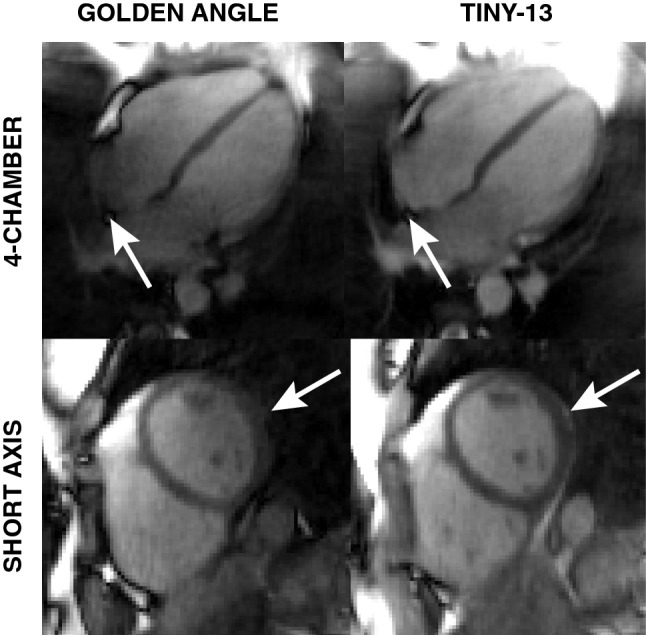
Fig. 7.

In vivo images show improved image quality with the reduced angular step, although the difference in eddy current-induced artifacts are not as pronounced as in the phantom experiments. With the conventional double golden-angle profile ordering there is an apparent signal loss in the right atrium and the epicardial fat (indicated by white arrows) which is not present with Tiny-13. Both images were acquired during free breathing and reconstructed from 126,478 spokes. The images represent the end-expiratory respiratory phase and the end-diastolic cardiac phase
