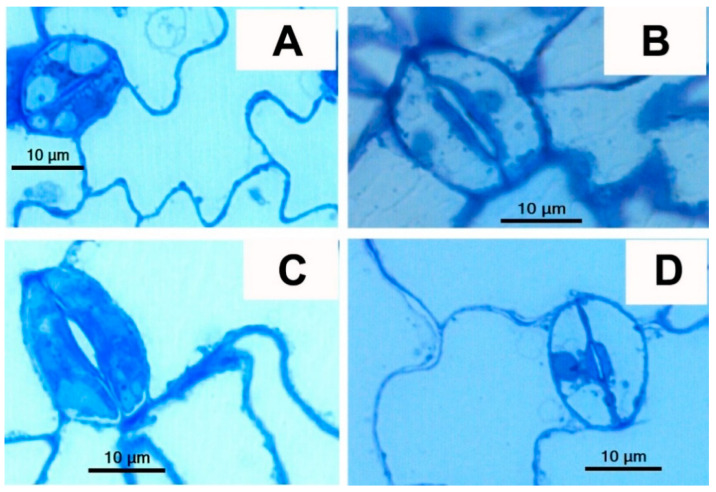
Figure 3.
LM images of stomata and GCs from leaf segments of semi-thin sections (longitudinal) of rosette leaves of WT, tgg single and double mutants of Arabidopsis stained with toluidine blue. (A). WT: GC where vacuoles showed toluidine staining, (B). tgg1 single mutant: GC where vacuoles lacked toluidine blue staining, (C). tgg2 single mutant: GC where vacuoles showed toluidine staining, (D). tgg1 tgg2 double mutant: GCs vacuoles showed no toluidine staining. (Scale bars = 10 μm).

